. 2023 Jul 10;6(1):704.
doi: 10.1038/s42003-023-05075-8.

Atypical functional connectivity hierarchy in Rolandic epilepsy

Affiliations

Atypical functional connectivity hierarchy in Rolandic epilepsy

Qirui Zhang et al. Commun Biol. .

Abstract

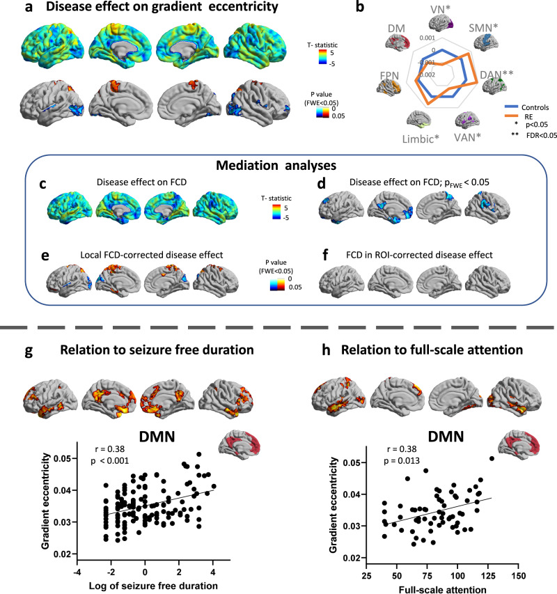
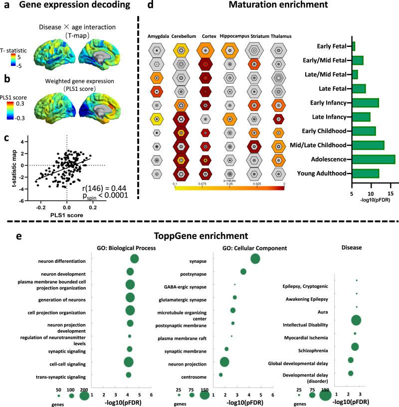
Functional connectivity hierarchy is an important principle in the process of brain functional organization and an important feature reflecting brain development. However, atypical brain network hierarchy organization in Rolandic epilepsy have not been systematically investigated. We examined connectivity alteration with age and its relation to epileptic incidence, cognition, or underlying genetic factors in 162 cases of Rolandic epilepsy and 117 typically developing children, by measuring fMRI multi-axis functional connectivity gradients. Rolandic epilepsy is characterized by contracting and slowing expansion of the functional connectivity gradients, highlighting the atypical age-related change of the connectivity hierarchy in segregation properties. The gradient alterations are relevant to seizure incidence, cognition, and connectivity deficit, and development-associated genetic basis. Collectively, our approach provides converging evidence for atypical connectivity hierarchy as a system-level substrate of Rolandic epilepsy, suggesting this is a disorder of information processing across multiple functional domains, and established a framework for large-scale brain hierarchical research.


Conflict of interest statement

The authors declare no competing interests.

Figures

Fig. 1
Fig. 1. Functional connectivity gradient eccentricity.
a Gradient eccentricity was calculated as the Euclidean distance between the centroid of all vertices and each vertex in an individual 3D gradient space. b Brain surfaces map showed mean gradient eccentricity values of RE (n = 140) and TDC (n = 91), joy plots showed relatively high eccentricity in VN, SMN, and DMN. TDC typically developing children, RE Rolandic epilepsy, VN visual network, SMN sensorimotor network, DAN dorsal attention network, VAN ventral attention network, FPN frontoparietal network, DMN default mode network.
Fig. 2
Fig. 2. Atypical age-related gradient eccentricity in Rolandic epilepsy.
a Surface-based age-related eccentricity changes in Rolandic epilepsy (n = 140) and TDC (n = 91). b Surface-based disease × age interaction analysis (n = 231), with significant decrease age-related change in superior parietal lobule, primary motor area and occipito-temporal area. c Surface-based disease × age interaction in each gradient (n = 231). d Community-based disease × age interaction of gradient eccentricity (n = 231) shows significant decrease age-re in the VN and DAN. e The relationship between typical and atypical age-related gradient eccentricity. A community-based scatterplot (n = 7 communities) of the age-related changes in TDC (x-axis) and disease × age interaction t-map (y-axis). TDC typically developing children, RE Rolandic epilepsy, VN visual network, SMN sensorimotor network, DAN dorsal attention network, VAN ventral attention network, FPN frontoparietal network, DMN default mode network.
Fig. 3
Fig. 3. Gradient eccentricity mapping in Rolandic epilepsy and TDC and relation to relation to cognitive and clinical variable.
a Surface-based case-control t-test between Rolandic epilepsy and TDC (n = 231), with significant increases in the superior parietal lobule, primary motor cortex, together with decreases in occipito-temporal and centro-temporal areas (Rolandic region). b Community-based z-score analysis (n = number of vertices in each community) of gradient eccentricity (with respect to controls) shows significant increases primarily in DAN, together with decreases in primarily in VN. c, d Compared to TDC, patients present decreased FCD in the precuneus, medial prefrontal lobe, temporal pole, superior parietal lobule, dorsal prefrontal lobe, and supramarginal gyrus. e Findings of eccentricity alterations remained robust after correcting for FCD in each vertex. f But the effect size of eccentricity alterations in Rolandic epilepsy was markedly reduced when analyses were repeated while controlling for mean FCD of the significant region in DMN (right bottom). g Surface-based and community-based (DMN) correlation between eccentricity and log of seizure free duration in RE (n = 140). h Surface-based and community-based (DMN) correlation between eccentricity and attention in RE (n = 65). TDC typically developing children, RE Rolandic epilepsy, VN visual network, SMN sensorimotor network, DAN dorsal attention network, VAN ventral attention network, FPN frontoparietal network, DMN default mode network, FCD functional connectivity density.
Fig. 4
Fig. 4. Transcriptomic analysis and development enrichment.
a Gene expression decoding of disease × age interaction. Statistic value in the left hemisphere. b A weighted gene expression map of regional PLS1 scores in the left hemisphere. c A scatterplot of regional PLS1 scores (a weighted sum of 10,027 gene expression scores) and disease × age interaction (Pearson’s r (146) = 0.44, pspin < 0.0001) (bottom). d We identified a gene set (n = 1627 genes) that showing consistent whole-brain expression pattern. These genes were input to a developmental enrichment analysis, showing strong associations with the cortex, thalamus, and cerebellum during the childhood-to-adolescence time window. e Enriched ontology terms in GO (gene ontology): biological process, GO: cellular component, and disease for the gene set. The size of the circle represents the number of genes involved in each term.

Similar articles

Cited by

References

    1. Specchio N, et al. International League Against Epilepsy classification and definition of epilepsy syndromes with onset in childhood: Position paper by the ILAE Task Force on Nosology and Definitions. Epilepsia. 2022;63:1398–1442. doi: 10.1111/epi.17241. - DOI - PubMed
    1. Panayiotopoulos CP, Michael M, Sanders S, Valeta T, Koutroumanidis M. Benign childhood focal epilepsies: assessment of established and newly recognized syndromes. Brain. 2008;131:2264–2286. doi: 10.1093/brain/awn162. - DOI - PubMed
    1. Bektas G, et al. Autism spectrum disorder and attention-deficit/hyperactivity disorder-related symptoms in benign childhood epilepsy with centrotemporal spikes: a prospective case-control study. Epilepsy Behav. 2019;95:61–64. doi: 10.1016/j.yebeh.2019.03.044. - DOI - PubMed
    1. Kim EH, Yum MS, Kim HW, Ko TS. Attention-deficit/hyperactivity disorder and attention impairment in children with benign childhood epilepsy with centrotemporal spikes. Epilepsy Behav. 2014;37:54–58. doi: 10.1016/j.yebeh.2014.05.030. - DOI - PubMed
    1. Smith SDW, Smith AB, Richardson MP, Pal DK. Neurodevelopmental origins of self-limiting rolandic epilepsy: systematic review of MR imaging studies. Epilepsia Open. 2021;6:310–322. doi: 10.1002/epi4.12468. - DOI - PMC - PubMed

Publication types