

Basilar artery diameter as neuroimaging biomarker in Chinese Fabry disease patients
- PMID: 37430370
- PMCID: PMC10334527
- DOI: 10.1186/s13023-023-02759-6
Basilar artery diameter as neuroimaging biomarker in Chinese Fabry disease patients
Abstract
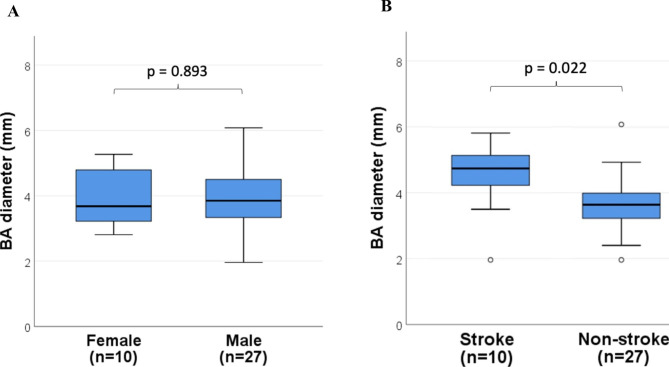
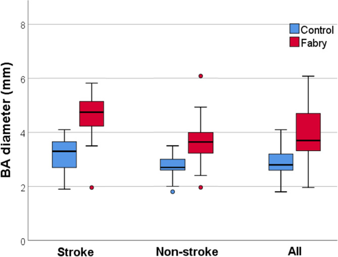
Background: Fabry disease (FD) is an X-linked lysosomal storage disease resulting from mutations of α-galactosidase A gene, and has been emphasized as one of the etiologies of young stroke and leukoencephalopathy. Vertebrobasilar dolichoectasia (VBD) is a highlighted finding in FD. We aim to examine the utility of VBD in Chinese FD by comparing the differences in basilar artery (BA) diameter of Chinese FD patients against age-matched controls with and without stroke.
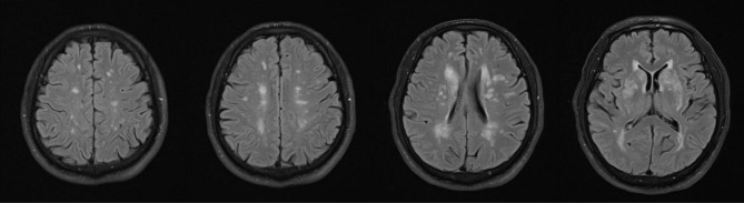
Methods: This was a matched case-control study involving 37 Chinese FD patients. The BA diameters were evaluated on axial T2-weighted magnetic resonance imaging and compared to two age-and-gender matched control groups, one with stroke and one without. The association between BA diameter and stroke occurrences and white matter hyperintensities (WMH) were analyzed among all FD patients.
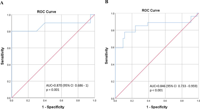
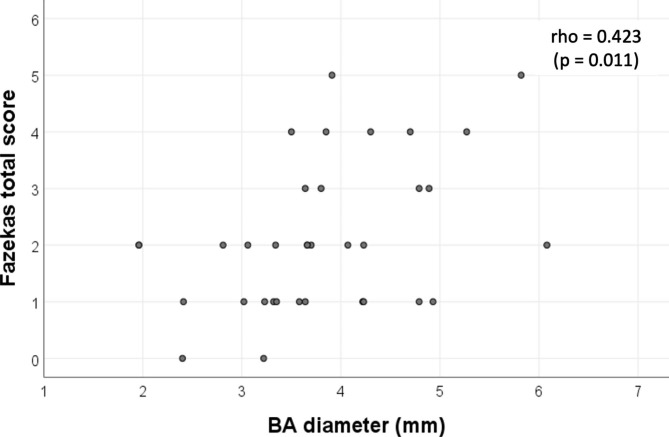
Results: Patients with FD had significantly increased BA diameter compared to controls with and without stroke (p < 0.001). A BA diameter of 4.16 mm could distinguish FD from controls in the stroke subgroup (ROC AUC 0.870, p = 0.001, sensitivity 80% specificity 100%), and with a cut-off of 3.21 mm in the non-stroke subgroup (ROC AUC 0.846, p < 0.001, sensitivity 77.8% specificity 88.9%). Larger BA diameter had more stroke occurrences and was moderately associated with heavier WMH load in terms of higher total FAZEKAS scores. (Spearman's rho = 0.423, p = 0.011).
Conclusion: VBD was also present in Chinese FD patients. BA diameter has high diagnostic utility in identifying FD from a mixed cohort of stroke and normal controls, and carried predictive value in evaluating neurological complications of FD.
Keywords: Basilar artery; Chinese; Fabry disease; IVS4; Vertebrobasilar dolichoectasia.
© 2023. The Author(s).
Conflict of interest statement
The authors declared there was no conflicts of interest.
Figures
Similar articles
-
Basilar artery diameter is a potential screening tool for Fabry disease in young stroke patients.Cerebrovasc Dis. 2011;31(3):294-9. doi: 10.1159/000322558. Epub 2010 Dec 22. Cerebrovasc Dis. 2011. PMID: 21196729
-
Studying the imaging features and infarction mechanism of vertebrobasilar dolichoectasia with high-resolution magnetic resonance imaging.Brain Pathol. 2023 Mar;33(2):e13135. doi: 10.1111/bpa.13135. Epub 2023 Jan 31. Brain Pathol. 2023. PMID: 36718993 Free PMC article.
-
Effect of Enzyme Replacement Therapy on Basilar Artery Diameter in Male Patients With Fabry Disease.Stroke. 2019 Apr;50(4):1010-1012. doi: 10.1161/STROKEAHA.118.024426. Stroke. 2019. PMID: 30852964
-
Vertebrobasilar dolichoectasia causing homonymous hemianopia: a case report and review of the literature.Acta Neurochir (Wien). 2018 Jan;160(1):161-164. doi: 10.1007/s00701-017-3367-x. Epub 2017 Oct 26. Acta Neurochir (Wien). 2018. PMID: 29075905 Review.
-
Development and clinical consequences of white matter lesions in Fabry disease: a systematic review.Mol Genet Metab. 2018 Nov;125(3):205-216. doi: 10.1016/j.ymgme.2018.08.014. Epub 2018 Sep 5. Mol Genet Metab. 2018. PMID: 30213639
Cited by
-
Expanding the Neurological Phenotype of Anderson-Fabry Disease: Proof of Concept for an Extrapyramidal Neurodegenerative Pattern and Comparison with Monogenic Vascular Parkinsonism.Cells. 2024 Jun 29;13(13):1131. doi: 10.3390/cells13131131. Cells. 2024. PMID: 38994983 Free PMC article. Review.
-
Clinical and Pathophysiologic Correlates of Basilar Artery Measurements in Fabry Disease.AJNR Am J Neuroradiol. 2024 Nov 7;45(11):1670-1677. doi: 10.3174/ajnr.A8403. AJNR Am J Neuroradiol. 2024. PMID: 38997124
References
-
- Sims K, Politei J, Banikazemi M, Lee P. Stroke in fabry disease frequently occurs before diagnosis and in the absence of other clinical events: natural history data from the Fabry Registry: natural history data from the Fabry Registry. Stroke. 2009;40(3):788–94. doi: 10.1161/STROKEAHA.108.526293. - DOI - PubMed
MeSH terms
Substances
LinkOut - more resources
Full Text Sources
Medical
