. 2023 Sep 1;325(3):G230-G238.
doi: 10.1152/ajpgi.00062.2023. Epub 2023 Jul 11.

Influenza A virus infection during pregnancy causes immunological changes in gut-associated lymphoid tissues of offspring mice

Affiliations

Influenza A virus infection during pregnancy causes immunological changes in gut-associated lymphoid tissues of offspring mice

Stella Liong et al. Am J Physiol Gastrointest Liver Physiol. .

Abstract

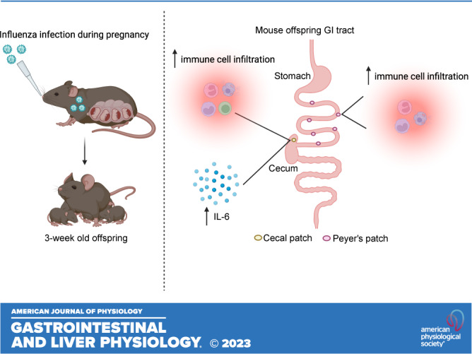
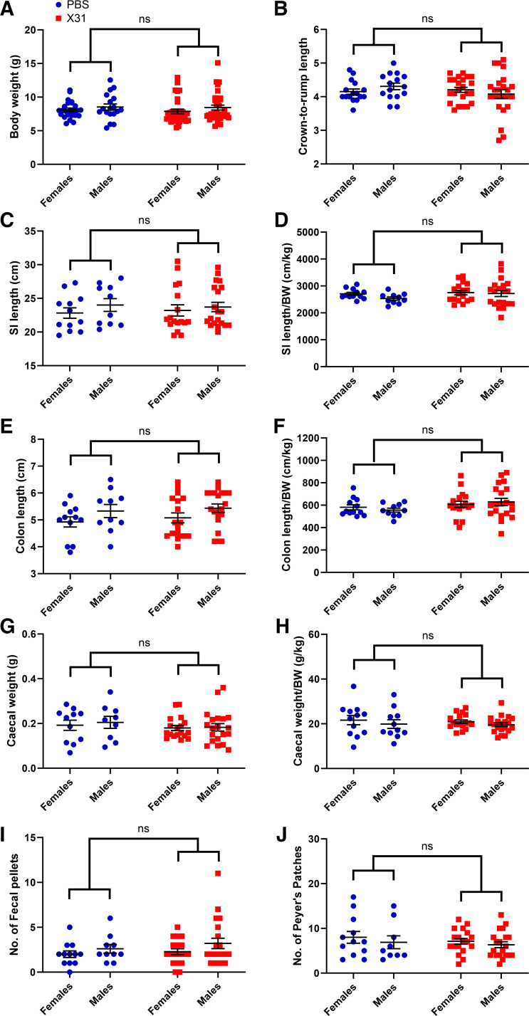
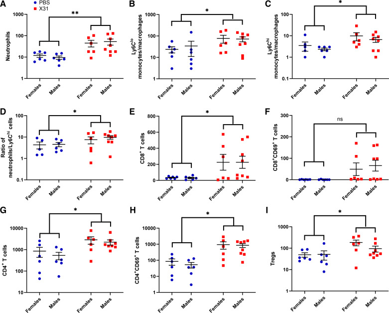
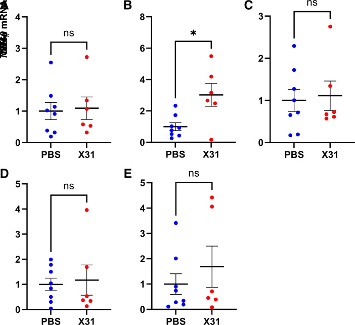
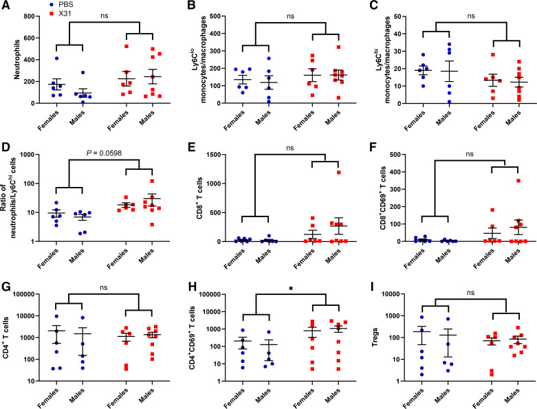
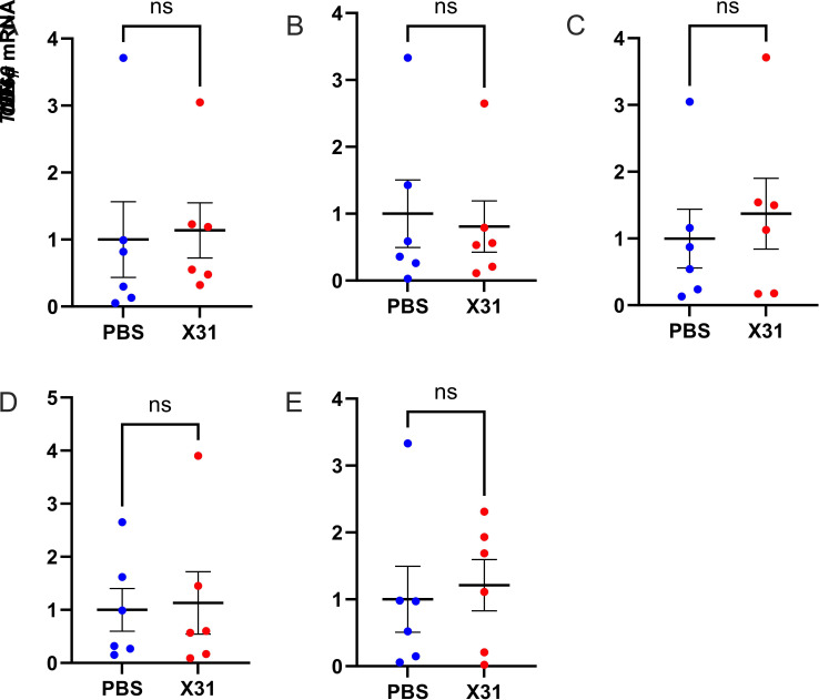
Maternal influenza A virus (IAV) infection during pregnancy can affect offspring immune programming and development. Offspring born from influenza-infected mothers are at increased risk of neurodevelopmental disorders and have impaired respiratory mucosal immunity against pathogens. The gut-associated lymphoid tissue (GALT) represents a large proportion of the immune system in the body and plays an important role in gastrointestinal (GI) homeostasis. This includes immune modulation to antigens derived from food or microbes, gut microbiota composition, and gut-brain axis signaling. Therefore, in this study, we investigated the effect of maternal IAV infection on mucosal immunity of the GI tract in the offspring. There were no major anatomical changes to the gastrointestinal tract of offspring born to influenza-infected dams. In contrast, maternal IAV did affect the mucosal immunity of offspring, showing regional differences in immune cell profiles within distinct GALT. Neutrophils, monocytes/macrophages, CD4+ and CD8+ T cells infiltration was increased in the cecal patch offspring from IAV-infected dams. In the Peyer's patches, only activated CD4+ T cells were increased in IAV offspring. IL-6 gene expression was also elevated in the cecal patch but not in the Peyer's patches of IAV offspring. These findings suggest that maternal IAV infection perturbs homeostatic mucosal immunity in the offspring gastrointestinal tract. This could have profound ramifications on the gut-brain axis and mucosal immunity in the lungs leading to increased susceptibility to respiratory infections and neurological disorders in the offspring later in life.NEW & NOTEWORTHY Influenza A virus (IAV) infection during pregnancy is associated with changes in gut-associated lymphoid tissue (GALT) in the offspring in a region-dependent manner. Neutrophils and monocytes/macrophages were elevated in the cecal patch of offspring from infected dams. This increase in innate immune cell infiltration was not observed in the Peyer's patches. T cells were also elevated in the cecal patch but not in the Peyer's patches.

Keywords: Peyer’s patches; cecal patch; inflammation; influenza A virus; offspring development.


Conflict of interest statement

No conflicts of interest, financial or otherwise, are declared by the authors.

Figures

None
Graphical abstract
Figure 1.
Figure 1.
Body weight and gastrointestinal (GI) tract anatomy of 3-wk-old mice offspring. The body weights (n = 24–32 females/group; n = 18–33 males/group) (A), crown-to-rump lengths (B), small intestine (SI) lengths (C), small intestine (SI) length/body weight (D), colon lengths (E), colon lengths/body weight (F), cecal weights (G), cecal weigh/body weight (H), number of fecal pellets (I), and number of Peyer’s patches (J) of 3-wk-old offspring born to influenza A virus (IAV)-infected (X31) or PBS-mock-infected dams. Body weights and crown-to-rump lengths were collected from n = 24 PBS females, n = 32 IAV (X31) females, n = 18 PBS males and n = 33 IAV (X31) males. All other GI assessments were collected from n = 12 PBS females, 17 IAV (X31) females, n = 10 PBS males, and n = 20 IAV (X31) males. Two-way ANOVA analysis identified no sex interaction. ns, not significant.
Figure 2.
Figure 2.
Gestational influenza A virus (IAV) infection increases immune cell infiltration into the cecal patches of 3-wk-old offspring. Neutrophils (A), patrolling Ly6Clo monocytes/macrophages (B), proinflammatory Ly6Chi monocytes/macrophages (C), neutrophil-to-Ly6Chi ratio (D), CD8+ T cells (E), CD69+-activated CD8+ T cells (F), CD4+ T cells (G), CD69+-activated CD4+ T cells (H), and CD4+ regulatory T cells (Tregs) (I) present in the cecal patches of 3-wk-old offspring born from IAV-infected (X31) or PBS mock-infected dams were quantified by flow cytometry (n = 6 or 7 animals/group). Two-way ANOVA analysis identified no sex interaction and thus male and female offspring were subsequently combined for Student’s t test analysis. *P < 0.05; **P < 0.01.
Figure 3.
Figure 3.
Gestational influenza A virus (IAV) infection increased IL-6 gene expression in the cecal patches of 3-wk-old offspring. IL-1β (A), IL-6 (B), TNF-α (C), IFN-γ (D), and CD69 mRNA expression (E) in the cecal patches of offspring born from IAV-infected (X31) or PBS mock-infected dams was assessed by qPCR (n = 6–8 animals/group). Student’s t test analysis. *P < 0.05. ns, Not significant.
Figure 4.
Figure 4.
Gestational influenza A virus (IAV) infection increases immune cell infiltration into the Peyer’s patches of 3-wk-old offspring. Neutrophils (A), patrolling Ly6Clo monocytes/macrophages (B), proinflammatory Ly6Chi monocytes/macrophages (C), neutrophil-to-Ly6Chi ratio (D), CD8+ T cells (E), CD69+-activated CD8+ T cells (F), CD4+ T cells (G), CD69+-activated CD4+ T cells (H), and CD4+ regulatory T cells (Tregs) (I) present in the Peyer’s patches of 3-wk-old offspring born from IAV-infected (X31) or PBS mock-infected dams were quantified by flow cytometry (n = 6 or 7 animals/group). Two-way ANOVA analysis identified no interaction sex and thus male and female offspring were subsequently combined for Student’s t test analysis. *P < 0.05. ns, not significant.
Figure 5.
Figure 5.
Gestational influenza A virus (IAV) infection had no effect on proinflammatory gene expression in the Peyer’s patches of 3-wk-old offspring. IL-1β (A), IL-6 (B), TNF-α (C), IFN-γ (D), and CD69 mRNA expression (E) in the cecal patches of offspring born from IAV-infected (X31) or PBS mock-infected dams was assessed by qPCR (n = 6 or 7 animals/group). Student’s t test analysis. ns, not significant.

References

    1. Rasmussen SA, Jamieson DJ, Uyeki TM. Effects of influenza on pregnant women and infants. Am J Obstet Gynecol 207: S3–S8, 2012. doi: 10.1016/j.ajog.2012.06.068. - DOI - PubMed
    1. Jacobsen H, Walendy-Gnirß K, Tekin-Bubenheim N, Kouassi NM, Ben-Batalla I, Berenbrok N, Wolff M, Dos Reis VP, Zickler M, Scholl L, Gries A, Jania H, Kloetgen A, Düsedau A, Pilnitz-Stolze G, Jeridi A, Yildirim AÖ, Fuchs H, Gailus-Durner V, Stoeger C, de Angelis MH, Manuylova T, Klingel K, Culley FJ, Behrends J, Loges S, Schneider B, Krauss-Etschmann S, Openshaw P, Gabriel G. Offspring born to influenza A virus infected pregnant mice have increased susceptibility to viral and bacterial infections in early life. Nat Commun 12: 4957, 2021. doi: 10.1038/s41467-021-25220-3. - DOI - PMC - PubMed
    1. Shi L, Fatemi SH, Sidwell RW, Patterson PH. Maternal influenza infection causes marked behavioral and pharmacological changes in the offspring. J Neurosci 23: 297–302, 2003. doi: 10.1523/JNEUROSCI.23-01-00297.2003. - DOI - PMC - PubMed
    1. Liong S, Oseghale O, To EE, Brassington K, Erlich JR, Luong R, Liong F, Brooks R, Martin C, O'Toole S, Vinh A, O'Neill LAJ, Bozinovski S, Vlahos R, Papagianis PC, O'Leary JJ, Brooks DA, Selemidis S. Influenza A virus causes maternal and fetal pathology via innate and adaptive vascular inflammation in mice. Proc Natl Acad Sci USA 117: 24964–24973, 2020. doi: 10.1073/pnas.2006905117. - DOI - PMC - PubMed
    1. Manresa MC, Taylor CT. Hypoxia inducible factor (HIF) hydroxylases as regulators of intestinal epithelial barrier function. Cell Mol Gastroenterol Hepatol 3: 303–315, 2017. doi: 10.1016/j.jcmgh.2017.02.004. - DOI - PMC - PubMed

Publication types