. 2023 Jul;16(7):523-535.
doi: 10.1161/CIRCIMAGING.123.015259. Epub 2023 Jul 11.

Rare Forms of Cardiac Amyloidosis: Diagnostic Clues and Phenotype in Apo AI and AIV Amyloidosis

Affiliations

Rare Forms of Cardiac Amyloidosis: Diagnostic Clues and Phenotype in Apo AI and AIV Amyloidosis

Adam Ioannou et al. Circ Cardiovasc Imaging. 2023 Jul.

Abstract

Background: Apo AI amyloidosis (AApoAI) and Apo AIV amyloidosis (AApoAIV) are rare but increasingly recognized causes of cardiac amyloidosis (CA). We sought to define the cardiac phenotype in AApoAI and AApoAIV using multimodality imaging.

Methods: We identified all patients with AApoAI and AApoAIV assessed at our center between 2000 and 2021, and 2 cohorts of patients with immunoglobulin light-chain amyloidosis (AL) and transthyretin amyloidosis matched for age, sex, and cardiac involvement.

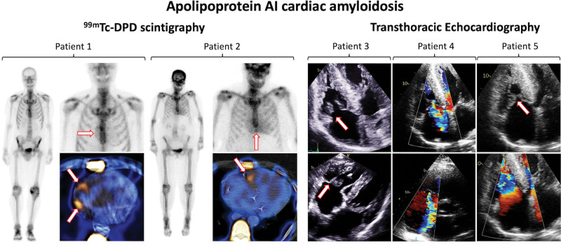
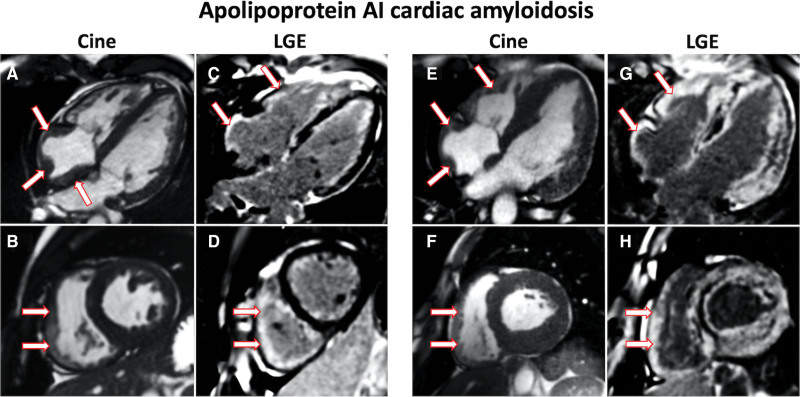
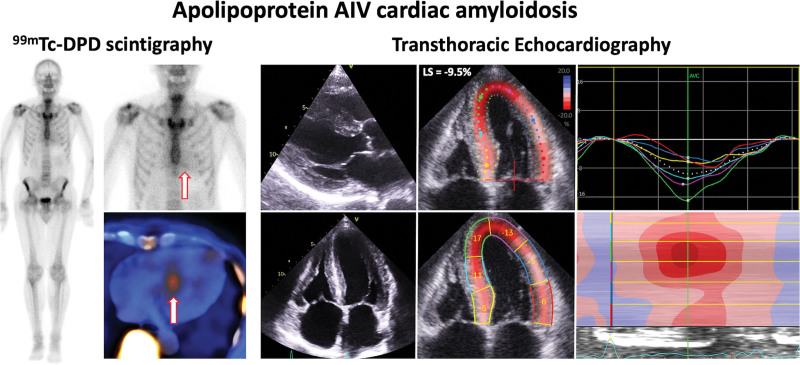
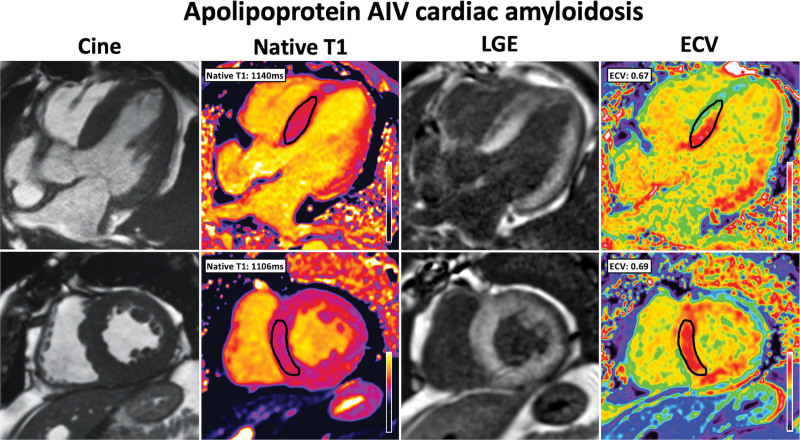
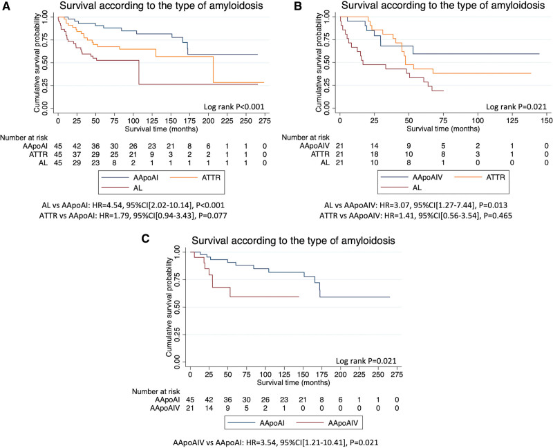
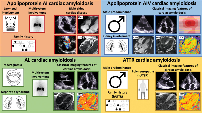
Results: Forty-five patients had AApoAI, 13 (29%) of whom had cardiac involvement, 32 (71%) renal involvement, 28 (62%) splenic involvement, 27 (60%) hepatic involvement, and 7 (16%) laryngeal involvement. AApoAI-CA commonly presented with heart failure (n=8, 62%) or dysphonia (n=7, 54%). The Arg173Pro variant universally caused cardiac and laryngeal involvement (n=7, 100%). AApoAI-CA was associated with right-sided involvement, with a thicker right ventricular free wall (8.6±1.9 versus 6.3±1.3 mm versus 7.7±1.2 mm, P=0.004), greater incidence of tricuspid stenosis (4 [31%] versus 0 [0%] versus 0 [0%], P=0.012) and tricuspid regurgitation (6 [46%] versus 1 [8%] versus 2 [15%], P=0.048) than AL-CA and transthyretin CA. Twenty-one patients had AApoAIV, and cardiac involvement was more common than in AApoAI (15 [71%] versus 13 [29%], P=0.001). AApoAIV-CA most commonly presented with heart failure (n=12, 80%), and a lower median estimated glomerular filtration rate than AL-CA and transthyretin CA (36 mL/[min·1.73 m²] versus 65 mL/[min·1.73 m²] versus 63 mL/[min·1.73 m²], P<0.001). All AApoAIV-CA patients had classical CA features on echocardiography/cardiac magnetic resonance, including an apical-sparing strain pattern, which was less common in AApoAI-CA (15 [100%] versus 7 [54%], P=0.003), whereas cardiac uptake on bone scintigraphy was less common in AApoAIV-CA than AApoAI-CA (all grade 1) (14% versus 82%, P<0.001). Patients with AApoAI and AApoAIV had a good prognosis (median survival >172 and >30 months, respectively), and a lower risk of mortality than matched patients with AL-amyloidosis (AL versus AApoAI: hazard ratio, 4.54 [95% CI, 2.02-10.14]; P<0.001; AL versus AApoAIV: hazard ratio, 3.07 [95% CI, 1.27-7.44]; P=0.013).

Conclusions: Dysphonia, multisystem involvement, or right-sided cardiac disease should raise suspicion of AApoAI-CA. AApoAIV-CA presents most commonly with heart failure and always displays classical CA imaging features, mimicking common forms of CA. Both AApoAI and AApoAIV are associated with a good prognosis and a lower risk of mortality than matched patients with AL-amyloidosis.

Keywords: amyloidosis; apolipoprotein; hereditary; phenotype; prognosis; transthyretin-related.


Conflict of interest statement

Disclosures Dr Fontana has consulting income from Intellia, Novo-Nordisk, Pfizer, Eidos, Prothena, Alnylam, Alexion, Janssen, and Ionis. Dr Gillmore has consulting income from Ionis, Alexion, Eidos, Intellia, Alnylam, and Pfizer. A. Wechelakar has consulting income from Alexia, Astra-Zeneca, Janssen, Attralus, and Prothena. Dr Hawkins has consulting income from Alnylam. The other authors report no conflicts.

Figures

Figure 1.
Figure 1.
99mTechnetium labeled 3,3-diphosphono-1,2-propanodicarboxylic acid (99mTc-DPD) scintigraphy and echocardiographic images of patients with Apo AI cardiac amyloidosis. Patient 1=99mTc-DPD scintigraphy and single-photon emission computed tomography (SPECT) images demonstrating grade 1 cardiac uptake with prominent right atrial uptake (arrows). Patient 2=99mTc-DPD scintigraphy and SPECT images demonstrating grade 1 cardiac uptake with prominent right ventricular uptake (arrows). Patient 3=Transthoracic echocardiography demonstrating marked thickening of the tricuspid valve leaflets (arrows) and subvalvular apparatus and biventricular hypertrophy. Patient 4=Transthoracic echocardiography demonstrating severe tricuspid regurgitation. Patient 5=Transthoracic echocardiography demonstrating tricuspid stenosis (arrows), with turbulent forward flow through the tricuspid valve, biatrial enlargement, and biventricular hypertrophy.
Figure 2.
Figure 2.
Cardiac magnetic resonance images of patients with Apo AI cardiac amyloidosis. A, 4-chamber cine image acquired with steady-state free precession sequence demonstrating prominent right atrial thickening (arrows). B, Short axis cine image demonstrating prominent right ventricular thickening (arrows). C, 4-chamber late gadolinium enhancement (LGE) image acquired using phase-sensitive inversion recovery sequence reconstructions with steady-state free precession read-outs, demonstrating prominent right atrial and right ventricular LGE (arrows). D, Short axis LGE image demonstrating prominent right ventricular LGE (arrows). E, 4-chamber cine image demonstrating prominent right atrial (arrows) and right ventricular thickening (arrows). F, Short axis cine image demonstrating prominent right ventricular thickening (arrows). G, 4-chamber LGE image demonstrating prominent right atrial and right ventricular LGE (arrows). H, Short axis LGE image demonstrating prominent right ventricular LGE (arrows).
Figure 3.
Figure 3.
99mTechnetium labeled 3,3-diphosphono-1,2-propanodicarboxylic acid (99mTc-DPD) scintigraphy and echocardiographic images in Apo AIV cardiac amyloidosis. 99mTc-DPD scintigraphy demonstrating grade 1 cardiac uptake and single-photon emission computed tomography demonstrating uptake in the septum (arrows). Transthoracic echocardiography images demonstrating increased biventricular wall thickness, impaired longitudinal strain, and a typical apical-sparing strain pattern with an increased septal apex-to-base strain ratio measured in the 4-chamber view. Images were acquired using a GE Vivid machine, and the strain assessment was carried out using Q-analysis on EchoPac.
Figure 4.
Figure 4.
Cardiac magnetic resonance images in Apo AIV cardiac amyloidosis demonstrate classical features of cardiac amyloidosis. Cine images were acquired with steady-state free precession (SSFP) sequence, and demonstrate increased wall thickness and left ventricular mass. Native-T1 mapping was acquired using the modified look-locker inversion (MOLLI) recovery sequence and demonstrated an elevated native-T1. Late gadolinium enhancement (LGE) imaging was acquired using phase-sensitive inversion recovery sequence reconstructions with SSFP read-outs and demonstrated diffuse biventricular transmural LGE. T1 mapping was repeated 15 minutes postcontrast using the same slice locations with the MOLLI sequence to produce automated inline extracellular volume (ECV) mapping reconstruction and demonstrated an elevated ECV.
Figure 5.
Figure 5.
Kaplan-Meier curves demonstrating survival in different types of amyloidosis. A, Kaplan-Meier curve comparing survival in patients with Apo AI amyloidosis (AApoAI), to age, sex and cardiac involvement matched patients with transthyretin amyloidosis (ATTR) and immunoglobulin light-chain amyloidosis (AL). B, Kaplan-Meier curve comparing survival in patients with apo AIV amyloidosis (AApoAIV), to age, sex and cardiac involvement matched patients with ATTR and AL-amyloidosis. C, Kaplan-Meier curve comparing survival in patients with AApoAI and AApoAIV.
Figure 6.
Figure 6.
Diagram illustrating key features in the clinical history and on cardiac imaging that should raise the suspicion of the different forms of cardiac amyloidosis. Top left, Apo AI cardiac amyloidosis can present with laryngeal involvement, multiorgan involvement, and a strong family history. Echocardiographic images demonstrate right-sided disease with thickening of the tricuspid valve and tricuspid regurgitation. Cardiac magnetic resonance (CMR) demonstrates right atrial and right ventricular thickening, and right atrial and right ventricular late gadolinium enhancement (LGE). Top right, Apo AIV cardiac amyloidosis has a male predominance and can present with renal involvement. Echocardiographic images demonstrate biventricular wall thickening and a typical apical-sparing strain pattern. CMR demonstrates left ventricular wall thickening, biventricular transmural LGE, and an elevated extracellular volume (ECV). Bottom left, Immunoglobulin light-chain (AL) cardiac amyloidosis can present with macroglossia, multisystem involvement, and nephrotic syndrome. Echocardiographic images demonstrate biventricular wall thickening. CMR demonstrates diffuse biventricular transmural LGE and an elevated ECV. Bottom right, Transthyretin (ATTR) cardiac amyloidosis has a male predominance and can present with polyneuropathy and a strong family history. Echocardiographic images demonstrate biventricular wall thickening. CMR demonstrates diffuse biventricular transmural LGE and an elevated ECV. hATTR indicates hereditary ATTR.

References

    1. Wechalekar AD, Gillmore JD, Hawkins PN. Systemic amyloidosis. Lancet. 2016;387:2641–2654. doi: 10.1016/S0140-6736 (15) 01274-X - PubMed
    1. Martinez-Naharro A, Hawkins PN, Fontana M. Cardiac amyloidosis. Clin Med (Lond). 2018;18:s30–s35. doi: 10.7861/clinmedicine.18-2-s30 - PMC - PubMed
    1. Ioannou A, Patel R, Gillmore JD, Fontana M. Imaging-guided treatment for cardiac amyloidosis. Curr Cardiol Rep. 2022;24:839–850. doi: 10.1007/s11886-022-01703-7 - PMC - PubMed
    1. Ioannou A, Patel RK, Razvi Y, Porcari A, Sinagra G, Venneri L, Bandera F, Masi A, Williams GE, O’Beara S, et al. . Impact of earlier diagnosis in cardiac ATTR amyloidosis over the course of 20 years. Circulation. 2022;146:1657–1670. doi: 10.1161/CIRCULATIONAHA.122.060852 - PMC - PubMed
    1. Martinez-Naharro A, Patel R, Kotecha T, Karia N, Ioannou A, Petrie A, Chacko LA, Razvi Y, Ravichandran S, Brown J, et al. . Cardiovascular magnetic resonance in light-chain amyloidosis to guide treatment. Eur Heart J. 2022;43:4722–4735. doi: 10.1093/eurheartj/ehac363 - PMC - PubMed

Publication types

MeSH terms