. 2023 Sep;32(9):1575-1581.
doi: 10.1111/exd.14885. Epub 2023 Jul 11.

Effects of TP63 mutations on keratinocyte adhesion and migration

Affiliations

Effects of TP63 mutations on keratinocyte adhesion and migration

Maddison N Salois et al. Exp Dermatol. 2023 Sep.

Abstract

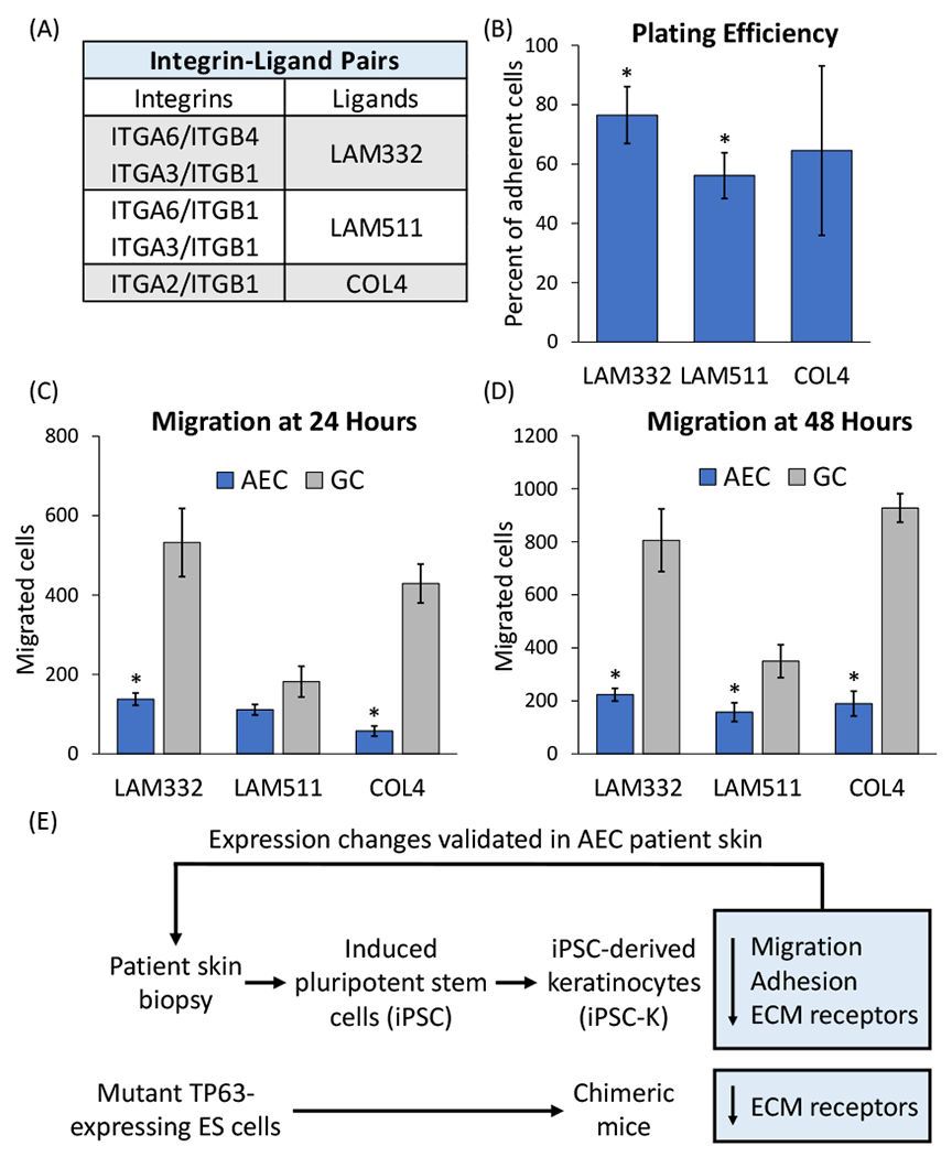
The goal of this study was to investigate the molecular mechanisms responsible for the formation of skin erosions in patients affected by Ankyloblepharon-ectodermal defects-cleft lip/palate syndrome (AEC). This ectodermal dysplasia is caused by mutations in the TP63 gene, which encodes several transcription factors that control epidermal development and homeostasis. We generated induced pluripotent stem cells (iPSC) from AEC patients and corrected the TP63 mutations using genome editing tools. Three pairs of the resulting conisogenic iPSC lines were differentiated into keratinocytes (iPSC-K). We identified a significant downregulation of key components of hemidesmosomes and focal adhesions in AEC iPSC-K compared to their gene-corrected counterparts. Further, we demonstrated reduced AEC iPSC-K migration, suggesting the possibility that a process critical for cutaneous wound healing might be impaired in AEC patients. Next, we generated chimeric mice expressing a TP63-AEC transgene and confirmed a downregulation of these genes in transgene-expressing cells in vivo. Finally, we also observed these abnormalities in AEC patient skin. Our findings suggest that integrin defects in AEC patients might weaken the adhesion of keratinocytes to the basement membrane. We propose that reduced expression of extracellular matrix adhesion receptors, potentially in conjunction with previously identified desmosomal protein defects, contribute to skin erosions in AEC.

Keywords: TP63; ectodermal dysplasia; extracellular matrix adhesion; hemidesmosome; skin erosion.


Conflict of interest statement

Conflict of interest statement:

The authors declare no conflict of interest.

Figures

Figure 1:
Figure 1:. Hemidesmosomal and focal adhesion components are downregulated in AEC iPSC-K.
(A) Images of an AEC patient showing skin erosions. Note the superficial erosions, abnormal and excessive granulation tissue and residual scarring alopecia (left) and the severe scalp erosive dermatitis (with overlying bacterial infection) and patchy alopecia with light wiry hairs (right). The photos are a courtesy of the National Foundation for Ectodermal Dysplasias (NFED) and are shown with parental consent. (B, C) qRT-PCR analysis of three pairs of AEC and GC iPSC-K. (B) All gene expression values were normalized to the same GC iPSC-K sample (GC1). The horizontal line represents the median; the whiskers represent the highest and lowest values in each dataset. (C) Numerical values of qRT-PCR analysis. In contrast to (B), all AEC iPSC-K gene expression values were normalized to their respective GC iPSC-K. Note the downregulation of all genes tested in (B) and (C). (D) Example of Western blot analysis using two pairs of AEC and GC iPSC-K. Numbers indicate ratios of signal intensity of AEC iPSC-K to conisogenic GC iPSC-K. The observed variability in transcript and protein expression between the three GC iPSC-K lines is expected, and reflects the expression variability of cell surface receptors often observed between different individuals ,.
Figure 2:
Figure 2:. Downregulation of cell surface receptors in mutant TP63-expressing mouse tissues and AEC patient skin.
(A) Schematic representation of the lentiviral construct used to generate chimeric mice expressing mutant TP63. The construct is designed to express mutant TP63 and TdTomato in equimolar amounts due to the presence of the T2A self-cleaving peptide. The CAG promoter is active in embryonic stem cells as well as epidermal and dermal skin cells. Transgenic cells can be identified in tissues by TdTomato expression. (B-E) Immunofluorescent staining of mouse tissue with antibodies against TdTomato (red) and ITGA2 (green). (B) Chimeric mouse palate (P) and tongue (T) are shown. Note the downregulation of ITGA2 (green) in areas of the palate that express the transgene (red; arrow). Arrowheads indicate non-transgenic areas with normal ITGA2 expression. (C) Control mouse palate and tongue. (D) Chimeric mouse skin. Note the downregulation of ITGA2 (green) in the transgenic (red) upper permanent portion of the hair follicle (arrow). (E) Control mouse skin. Size bars in (B, C) 50 μm, size bars in (D, E) 20 μm. (F, G) Immunofluorescent staining for ITGA2 on (F) AEC patient skin and (G) control skin. (H, I) Immunofluorescent staining for COL17A1 on (H) AEC patient skin and (I) control skin. As above, arrowheads indicate normal expression; arrows indicate loss of expression. Note the focal loss of ITGA2 and COL17A1 in AEC patient skin. Size bars in (F-I) 50 μm.
Figure 3:
Figure 3:. Reduced plating efficiency and migration of AEC iPSC-K.
(A) Table indicating integrins and their ligands in the epidermis. (B) Plating efficiency was determined by calculating the relative percentage of AEC iPSC-K versus GC iPSC-K that adhered to the indicated ECM substrates. Plating efficiency of GC iPSC-K was set to 100%. (C, D) Assessment of AEC and GC iPSC-K migration on different ECM substrates at (C) 24 and (D) 48 hours. Note that both plating efficiency and migration were reduced in AEC iPSC-K on all tested substrates. *p<0.05. (E) Schematic representation of the outline of our experimental approach and the main findings of this study.

Update of

Similar articles

References

    1. McGrath JA, Duijf PH, Doetsch V, et al. Hay-Wells syndrome is caused by heterozygous missense mutations in the SAM domain of p63. Hum Mol Genet. Feb 1 2001;10(3):221–9. doi:10.1093/hmg/10.3.221 - DOI - PubMed
    1. Koster MI. p63 in skin development and ectodermal dysplasias. J Invest Dermatol. Oct 2010;130(10):2352–8. doi:10.1038/jid.2010.119 - DOI - PMC - PubMed
    1. Rinne T, Brunner HG, van Bokhoven H. p63-associated disorders. Cell Cycle. Feb 1 2007;6(3):262–8. doi:10.4161/cc.6.3.3796 - DOI - PubMed
    1. Clements SE, Techanukul T, Lai-Cheong JE, et al. Mutations in AEC syndrome skin reveal a role for p63 in basement membrane adhesion, skin barrier integrity and hair follicle biology. The British journal of dermatology. Feb 13 2012;167(1):134–144. doi:10.1111/j.1365-2133.2012.10888.x - DOI - PubMed
    1. Koster MI, Marinari B, Payne AS, Kantaputra PN, Costanzo A, Roop DR. DeltaNp63 knockdown mice: A mouse model for AEC syndrome. Am J Med Genet A. Sep 2009;149A(9):1942–7. doi:10.1002/ajmg.a.32794 - DOI - PMC - PubMed

Publication types

Substances

Supplementary concepts