. 2023 Oct 2;220(10):e20221757.
doi: 10.1084/jem.20221757. Epub 2023 Jul 11.

IL-6 selectively suppresses cDC1 specification via C/EBPβ

Affiliations

IL-6 selectively suppresses cDC1 specification via C/EBPβ

Sunkyung Kim et al. J Exp Med. .

Abstract

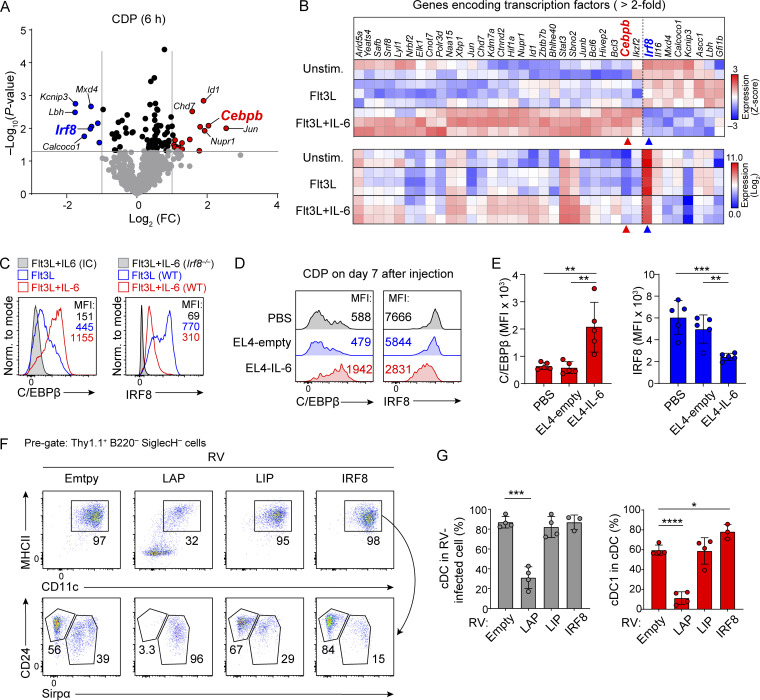
Cytokines produced in association with tumors can impair antitumor immune responses by reducing the abundance of type 1 conventional dendritic cells (cDC1), but the mechanism remains unclear. Here, we show that tumor-derived IL-6 generally reduces cDC development but selectively impairs cDC1 development in both murine and human systems through the induction of C/EBPβ in the common dendritic cell progenitor (CDP). C/EBPβ and NFIL3 compete for binding to sites in the Zeb2 -165 kb enhancer and support or repress Zeb2 expression, respectively. At homeostasis, pre-cDC1 specification occurs upon Nfil3 induction and consequent Zeb2 suppression. However, IL-6 strongly induces C/EBPβ expression in CDPs. Importantly, the ability of IL-6 to impair cDC development is dependent on the presence of C/EBPβ binding sites in the Zeb2 -165 kb enhancer, as this effect is lost in Δ1+2+3 mutant mice in which these binding sites are mutated. These results explain how tumor-associated IL-6 suppresses cDC1 development and suggest therapeutic approaches preventing abnormal C/EBPβ induction in CDPs may help reestablish cDC1 development to enhance antitumor immunity.


Conflict of interest statement

Disclosures: The authors declare no competing interests exist.

Figures

None
Graphical abstract
Figure 1.
Figure 1.
IL-6 suppresses cDC1 development from murine and human progenitors. (A) Sort-purified CDPs cultured with Flt3L and IL-6 (0, 1, 5, and 25 ng/ml) for 4 d and analyzed. Shown is FACS analysis of MerTK B220 SiglecH cells. Data are representative of four independent experiments. (B) The bar-scatter graphs show average cDC frequencies in the single cell gate (left, gray) and cDC1 (right, red) or cDC2 (right, blue) in total cDCs of the indicated conditions (average % ± SD, n = 4). (C) Analysis of human cDC1 differentiated from umbilical cord blood–derived CD34+ progenitors cultured with SCF (20 ng/ml), GM-CSF (20 ng/ml), IL-4 (20 ng/ml), Flt3L (100 ng/ml), and various concentrations of IL-6 (0, 1, and 5 ng/ml) for 14 d. (D) The bar-scatter graphs show average cDC1 frequencies in the single cell gate of the indicated conditions (average % ± SD, n = 3). (E and F) Analysis of cDCs in the (E) spleen and (F) MLNs of Zbtb46egfp/+ mice injected i.p. with PBS, EL4-empty, or EL4-IL-6 tumor (106 cells) on day 14 after injection. The histogram for the splenic cDCs is representative of two independent experiments. (G and H) Sort-purified OT-I and OT-II (2.5 × 10⁵ cells of each) were transferred i.v. into WT or Irf8 +32−/− mice inoculated 14 d earlier with PBS, EL4-empty, or EL4-IL-6 tumors. OVA-loaded splenocytes (5 × 10⁵ cells/mouse) were transferred i.v. after 3 h. (G) In vivo proliferation of OT-I (upper panel) and OT-II (lower panel) on day 3 after transfer. (H) Bar-scatter graphs from G for OT-I (upper) and OT-II (lower) of the indicated conditions (average % ± SD). Individual mice are shown as dots. *P < 0.05, **P < 0.01, ***P < 0.001, ****P < 0.0001 (Student’s t test).
Figure S1.
Figure S1.
Generation and validation of IL-6 expressing EL4 tumor in vitro and in vivo. (A) Sort-purified MDPs were cultured with Flt3L with or without 25 ng/ml IL-6 and analyzed by FACS after 7 d. Shown is FACS analysis of MerTK B220 SiglecH cells. Data is representative of four independent experiments. (B) EL4-empty and EL4-IL-6 cell lines were treated with brefeldin A (Sigma-Aldrich) for 3 h and analyzed by FACS for intracellular staining of IL-6 and EGFP. Rat IgG1 antibody was used for isotype control (ctl) staining. (C) Sort-purified cKithi BM progenitors were cultured for 9 d with Flt3L and the indicated concentration of conditioned media (CM) from EL4-empty or EL4-IL-6 cells or recombinant murine IL-6. (D and F) Related to Fig. 1 E, the bar-scatter graphs show the average (D) frequencies (%) and (F) absolute number of cDC in the spleen ± SD. (E and G) Related to Fig. 1 F, the bar-scatter graphs show the average (E) frequencies (%) and (G) absolute number of cDC in the MLNs ± SD. cDC total (gray and/or white), cDC1 (red), and cDC2 (blue). The numbers of mice for each experimental group were indicated as dots in the bar graphs, PBS (n = 7), EL4-empty (n = 9), EL4-IL-6 (n = 11). *P < 0.05, **P < 0.01, ***P < 0.001, ****P < 0.0001 (Student’s t test).
Figure S2.
Figure S2.
Reduction of DC and monocyte progenitors in BM by EL4-IL-6. (A) Lin BM from Zbtb46egfp/+ mice was analyzed by FACS. cKitint Zbtb46-EGFP+ cells were examined for MHCII expression. Numbers represent the percentage of cells in the indicated gate. (B) Zbtb46egfp/+ mice were inoculated i.p. with PBS, EL4-empty, or EL4-IL-6 (106 cells) as described in Fig. 2. BM cells were analyzed by FACS on day 14 after inoculation. Shown are pre-cDC1 progenitors defined as gate R1 in Fig. 2 A (Grajales-Reyes et al., 2015). (C) Bar-scatter graphs for absolute number of pre-cDC1 from Fig. 2, F and G (average # ± SD). Cells were enumerated from total BM cells collected from two femurs and tibias. Numbers of mice for each experimental group are indicated as dots. (D) Zbtb46egfp/+ mice injected i.p. with PBS, EL4-empty, or EL4-IL-6 (106 cells), and the BM progenitors were analyzed on day 14 after inoculation. Shown are analyses for MDP (cKithi Flt3+ M-CSFR+; red gate), CDP (cKitint Flt3+ M-CSFR+ MHCII CD11c; blue gate) and monocyte progenitors (cKit+ Flt3 M-CSFR+), mostly cMoPs (black gate). Data shown are representative of two independent experiments. (E) Bar-scatter graphs show the frequency of MDP (red), CDP (blue), and monocyte progenitors (cKithi, gray bars; ckitint, white bars) as a fraction of Lin BM cells for the mice in D (average % ± SD). Each dot represents an individual mouse for PBS (n = 7), EL4-empty (n = 9), EL4-IL-6 (n = 11). *P < 0.05, **P < 0.01, ****P < 0.0001 (Student’s t test).
Figure 2.
Figure 2.
IL-6 suppresses development of pre-cDC1. (A) pre-cDC1 (R1) in Zbtb46egfp/+ mice defined by Grajales-Reyes et al. (2015). (B) Upper panel: Pre-cDC1 (R2) in Zbtb46egfp/+ mice defined by Schlitzer et al. (2015). Lower panel: Cells in R2 were analyzed for cKit and Zbtb46-EGFP expression (R3 and R4). A bar-scatter graph shows the percentage (average % of R2 ± SD) of cKitint R2 cells (R3) and cKitlo R2 cells (R4). (C) Sort-purified cells from gate R1 in A and R3 and R4 in B were cultured in Flt3L for 3 d and analyzed by FACS. Arrows indicate the order of gating for successive histograms. Results are representative of three independent experiments. (D) Zbtb46egfp/+ mice were inoculated i.p. with PBS, EL4-empty, or EL4-IL-6 tumor cells (106 cells). BM cells were analyzed by FACS after 7, 10, 12, and 14 d as indicated. Numbers represent the percentage of Lin BM cells in the indicated gate (cKitint Zbtb46-EGFP+). (E) Shown are bar-scatter graphs for mice from D for the frequency of pre-cDC1 in Lin BM (average % ± SD). (F) Pre-cDC1 progenitor as defined in Grajales-Reyes et al. (2015) (R1 in A) from Zbtb46egfp/+ mice at 14 d after inoculation. (G) Upper panel: Pre-cDC1 progenitor defined in Schlitzer et al. (2015) (R2 in B) from Zbtb46egfp/+ mice at 14 d after inoculation. Lower panel: Analysis of R2 in upper panel for cKit and Zbtb46-EGFP+. (H) Bar-scatter graphs for frequency of pre-cDC1 as defined in F and G (average % of Lin BM ± SD). Individual mice are indicated as dots. *P < 0.05, **P < 0.01, ****P < 0.0001 (Student’s t test).
Figure 3.
Figure 3.
IL-6 increases Cebpb expression in CDPs. (A) RNA-seq was performed on CDPs cultured for 6 h with Flt3L alone or with added IL-6. Shown is a volcano plot of differentially expressed transcription factors as the fold change (FC) induced by IL-6. Selected genes with greater than twofold change are highlighted in red (increased) or blue (decreased). (B) Heatmaps of the top 36 differentially expressed transcription factors from A clustered on (upper panel) Z-score or (lower panel) log2 expression value. (C) Intracellular expression of C/EBPβ and IRF8 in sort-purified CDPs treated in vitro with Flt3L (blue) or Flt3L with 25 ng/ml IL-6 (red) for 20 h. IC indicates staining using isotype control antibody. (D) Intracellular expression of C/EBPβ and IRF8 in CDPs from BM of Zbtb46egfp/+ mice analyzed 7 d after tumor inoculation i.p. as indicated. (E) Bar-scatter graphs show the geometric mean fluorescence intensity (MFI) ± SD for experiments shown in E for C/EBPβ (red) and IRF8 (blue). (F) Sort-purified CDPs were transduced in vitro with retroviral vectors (RV) encoding LAP, LIP, or IRF8, cultured in Flt3L for 4 d, and analyzed for FACS. Shown are Thy1.1+ B220 SiglecH cells for expression of MHCII, CD11c, CD24, and Sirpα. Results are representative of four independent experiments. (G) Scatter plots with error bars for cells described in F for total MHCII+ CD11c+ cells (left panel) or for MHCII+ CD11c+CD24+ Sirpα cells (right panel; average % ± SD). *P < 0.05, **P < 0.01, ***P < 0.001, ****P < 0.0001 (Student’s t test).
Figure S3.
Figure S3.
Increases of C/EBPβ expression and bindings at Zeb2 −165 kb in CDP by IL-6. (A) RNA-seq was performed on CDPs cultured for 6 h with Flt3L alone or with added IL-6 (25 ng/ml) as described in Fig. 3. Shown is a volcano plot of differentially expressed genes as fold change (FC) induced by IL-6. Selected genes with greater than threefold changes are highlighted in red (increased) or blue (decreased). (B) Shown are genes from A with greater than threefold decrease (blue) or greater than threefold increase in expression (red) in response to IL-6 in CDPs after 6 h. (C) Heatmap of top 101 differentially expressed genes from A clustered on log2 expression value. (D and E) Characterization of Hoxb8-transduced BM progenitor cell line. (D) FACS analysis of Hoxb8 cells and BM progenitors. (E) Intracellular expression of C/EBPβ in Hoxb8 cells cultured with or without IL-6 (25 ng/ml) for 22 h. Isotype control (IC) is a mouse IgG2a. (F) cDC differentiation from Hoxb8 cell line cultured with or without IL-6 (25 ng/ml) for 7 d. (G and H) CUT&RUN analysis for C/EBPβ bindings to the Zeb2 locus in M-CSFR+ Hoxb8 cells cultured in Flt3L with or without IL-6 (25 ng/ml). (G) A snapshot of UCSC genome browser (chr2:44,912,814-45,315,672). Zeb2 −165 kb enhancer is highlighted in yellow. (H) A scatter plot shows total tag counts normalized to IC experimental group in the Zeb2 −165 kb enhancer. Peak calling was performed with HOMER command findPeaks.
Figure 4.
Figure 4.
IL-6 blocks cDC1 development by activating the Zeb2 −165 kb enhancer. (A) Upper panel: Retroviral reporter construct: Zeb2 −165 kb enhancer (yellow), CMV minimal promoter (red arrow), and EGFP (green). Lower panel: Sort-purified CDPs were transduced with reporter constructs harboring WT or Δ1+2+3 enhancers and cultured for 2 d with 5% Flt3L with or without 25 ng/ml IL-6, and EGFP expression assessed in Thy1.1+ cells. Numbers represent the MFI of cells. LTR, long terminal repeat. (B) A bar-scatter graph of three independent experiments from A for EGFP expression (average MFI ± SD). ***P < 0.001 (Student’s t test). (C) Sort-purified CDPs from WT or Δ1+2+3 mice were cultured in vitro with Flt3L and the indicated concentrations of IL-6 for 4 d. Shown is FACS analysis for MerTK B220 SiglecH cells. Numbers indicate the percentage of cells in the indicated gates. Data shown are representative of two independent similar experiments. (D) Sort-purified CDPs from WT or Δ1+2+3 mice were cultured in vitro with Flt3L alone (blue) or with 25 ng/ml IL-6 (red) for 20 h and analyzed for intracellular C/EBPβ expression. IC indicates staining with isotype control antibody. Number indicates MFI. (E and F) WT and Δ1+2+3 mice were inoculated with EL4-empty or EL4-IL-6 tumors and analyzed after 14 d. Shown is FACS analysis of (E) splenocytes pre-gated as MerTK B220 SiglecH MHCII+ CD11c+ cells and (F) BM DC progenitors pre-gated as Lin SiglecH Flt3+ CD11c+ MHCIIlo-int BM cells for cKit and CD24 expression. (G and H) Sort-purified OT-I and OT-II (2.5 × 10⁵ cells, each) were transferred i.v. into WT or Δ1+2+3 mice inoculated 14 d earlier with EL4 tumors. OVA-loaded splenocytes (5 × 10⁵ cells/mouse, i.v.) were transferred i.v. after 3 h. (G) In vivo proliferation of OT-I (upper panel) and OT-II (lower panel) on day 3 after transfer. (H) Bar-scatter graphs from G for OT-I (upper) and OT-II (lower) of the indicated conditions (average % ± SD). Individual mice are shown as dots. *P < 0.05, **P < 0.01, ****P < 0.0001 (Student’s t test).
Figure S4.
Figure S4.
IL-6 blocks cDC1 development by acting the Zeb2 −165 kb Δ1+2+3 enhancer. (A) CDPs from WT mice or Zeb2ZEB2-EGFP/+ reporter mice were sort-purified and cultured with Flt3L with (red) or without (blue) IL-6 (25 ng/ml) for the indicated time, and analyzed by FACS for EGFP expression. Numbers indicate the MFI for EGFP of cells. The line graph (lower left) shows the ratio of the ZEB2-EGFP MFI and WT MFI at each time point. (B) WT and Δ1+2+3 mice were inoculated i.p. with EL4-empty or EL4-IL-6 (106 cells) and analyzed after 14 d. Shown is FACS analysis of splenocytes gated as MerTK B220 SiglecH cells. (C and D) Bar-scatter graphs for (C) the splenic MerTK B220 SiglecH cells described in B and (D) the splenic MerTK B220 SiglecH MHCII+ CD11c+ cells described in the main Fig. 4 F. (E) WT and Δ1+2+3 mice inoculated in B were analyzed for DC progenitors in the BM. Shown is FACS analysis of Lin BM cells. Numbers indicate the percentage of cells in the indicated gates. (F) Bar-scatter graphs for cells described in E for WT (red) and Δ1+2+3 mice (gray; average % ± SD). *P < 0.05, **P < 0.01, ***P < 0.001 (Student’s t test).

References

    1. Akira, S., Isshiki H., Sugita T., Tanabe O., Kinoshita S., Nishio Y., Nakajima T., Hirano T., and Kishimoto T.. 1990. A nuclear factor for IL-6 expression (NF-IL6) is a member of a C/EBP family. EMBO J. 9:1897–1906. 10.1002/j.1460-2075.1990.tb08316.x - DOI - PMC - PubMed
    1. Anderson, D.A., III, Murphy K.M., and Briseño C.G.. 2018. Development, diversity, and function of dendritic cells in mouse and human. Cold Spring Harb. Perspect. Biol. 10:a028613. 10.1101/cshperspect.a028613 - DOI - PMC - PubMed
    1. Auffray, C., Fogg D.K., Narni-Mancinelli E., Senechal B., Trouillet C., Saederup N., Leemput J., Bigot K., Campisi L., Abitbol M., et al. 2009. CX3CR1+ CD115+ CD135+ common macrophage/DC precursors and the role of CX3CR1 in their response to inflammation. J. Exp. Med. 206:595–606. 10.1084/jem.20081385 - DOI - PMC - PubMed
    1. Bagadia, P., Huang X., Liu T.T., Durai V., Grajales-Reyes G.E., Nitschké M., Modrusan Z., Granja J.M., Satpathy A.T., Briseño C.G., et al. 2019. An Nfil3-Zeb2-Id2 pathway imposes Irf8 enhancer switching during cDC1 development. Nat. Immunol. 20:1174–1185. 10.1038/s41590-019-0449-3 - DOI - PMC - PubMed
    1. Böttcher, J.P., and Reis e Sousa C.. 2018. The role of type 1 conventional dendritic cells in cancer immunity. Trends Cancer. 4:784–792. 10.1016/j.trecan.2018.09.001 - DOI - PMC - PubMed

Publication types