. 2023 Jul 26;145(29):16166-16175.
doi: 10.1021/jacs.3c04810. Epub 2023 Jul 11.

Stacking Interactions and Flexibility of Human Telomeric Multimers

Affiliations

Stacking Interactions and Flexibility of Human Telomeric Multimers

Benedetta Petra Rosi et al. J Am Chem Soc. .

Abstract

G-quadruplexes (G4s) are helical four-stranded structures forming from guanine-rich nucleic acid sequences, which are thought to play a role in cancer development and malignant transformation. Most current studies focus on G4 monomers, yet under suitable and biologically relevant conditions, G4s undergo multimerization. Here, we investigate the stacking interactions and structural features of telomeric G4 multimers by means of a novel low-resolution structural approach that combines small-angle X-ray scattering (SAXS) with extremely coarse-grained (ECG) simulations. The degree of multimerization and the strength of the stacking interaction are quantitatively determined in G4 self-assembled multimers. We show that self-assembly induces a significant polydispersity of the G4 multimers with an exponential distribution of contour lengths, consistent with a step-growth polymerization. On increasing DNA concentration, the strength of the stacking interaction between G4 monomers increases, as well as the average number of units in the aggregates. We utilized the same approach to explore the conformational flexibility of a model single-stranded long telomeric sequence. Our findings indicate that its G4 units frequently adopt a beads-on-a-string configuration. We also observe that the interaction between G4 units can be significantly affected by complexation with benchmark ligands. The proposed methodology, which identifies the determinants that govern the formation and structural flexibility of G4 multimers, may be an affordable tool aiding in the selection and design of drugs that target G4s under physiological conditions.


Conflict of interest statement

The authors declare no competing financial interest.

Figures

Figure 1
Figure 1
The log–log plot of the SAXS intensities of G4 multimeric samples at different DNA concentrations. Data are reported in absolute scale and normalized to the molar concentration C of DNA. For comparison, the SAXS intensities of Tel22 monomeric solutions at C = 0.5 mM, prepared in both K+ and Na+ environments, are shown. In the inset, the same SAXS profiles are shown on a linear–log scale.
Figure 2
Figure 2
Simulation model for the G4 unit. (a) Each G4 is modeled as a HC characterized by a diameter D and a length L. (b) Each cylinder is decorated with two attractive sites at the basis. Sites belonging to different cylinders interact through the SW potential u(r).
Figure 3
Figure 3
Experimental (symbols) and simulated (lines) scattering intensities for monomeric (panels a and b) and multimeric (panels c and d) G4 samples. Best accordance between experimental and simulated data has been evaluated through the residual sum of squares (RSS), calculated at different values of T* and K (panels a and c). At the best value of K, the best-temperature simulated curve is reproduced as a solid line.
Figure 4
Figure 4
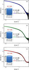
Best accordance between experimental and simulated scattering intensities, respectively, for the G4 solution at C = 0.6 mM (a), 1.2 mM (b), and 4.5 mM (c).
Figure 5
Figure 5
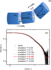
High-order G4 sequences. (a) Each sequence is modeled by three HCs whose shape and stacking interaction (yellow patch) are identical to those used in the previous sections. An additional covalent interaction (light purple patch) has been employed, as described in the Methods section. (b) Experimental (symbols) and simulated (lines) scattering intensities for Tel72. At the best value of K, the best-temperature simulated curve is reported as a solid red line.
Figure 6
Figure 6
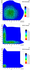
Distributions of angles θ1 and θ2 formed by two adjacent cylinders within the trimer, as obtained from the best-fit simulation. The distributions corresponding to (a) 0 bonds, (b) 1 bond, and (c) 2 bonds between the HCs are reported.
Figure 7
Figure 7
Experimental and simulated scattering intensities for Tel22+TMPyP4 (a) and Tel22+BRACO19 (b) solutions. Insets show in the semilogarithmic logscale the same curves together with the monomer profiles (blue lines).

References

    1. Neidle S. Quadruplex nucleic acids as targets for anticancer therapeutics. Nat. Rev. Chem. 2017, 1, 0041.10.1038/s41570-017-0041. - DOI
    1. Webba da Silva M. Geometric Formalism for DNA Quadruplex Folding. Chem. Eur. J. 2007, 13, 9738–45. 10.1002/chem.200701255. - DOI - PubMed
    1. Neidle S.; Parkinson G. N. The structure of telomeric DNA. Curr. Opin. Struct. Biol. 2003, 13, 275–283. 10.1016/S0959-440X(03)00072-1. - DOI - PubMed
    1. Karsisiotis A. I.; Hessari N. M.; Novellino E.; Spada G. P.; Randazzo A.; Webba da Silva M. Topological Characterization of Nucleic Acid G-Quadruplexes by UV Absorption and Circular Dichroism. Angew. Chem., Int. Ed. 2011, 50, 10645–10648. 10.1002/anie.201105193. - DOI - PubMed
    1. Huppert J. L. Structure, location and interactions of G-quadruplexes. The FEBS Journal 2010, 277, 3452–3458. 10.1111/j.1742-4658.2010.07758.x. - DOI - PubMed

Publication types