. 2023 Aug;243(2):319-333.
doi: 10.1111/joa.13872. Epub 2023 Apr 7.

A workflow for the creation of photorealistic 3D cadaveric models using photogrammetry

Affiliations

A workflow for the creation of photorealistic 3D cadaveric models using photogrammetry

Morgan Titmus et al. J Anat. 2023 Aug.

Abstract

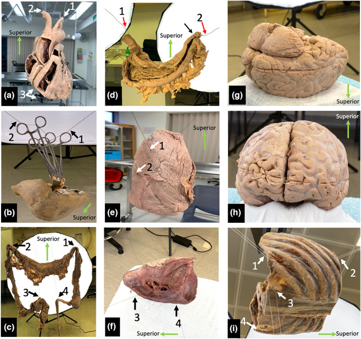
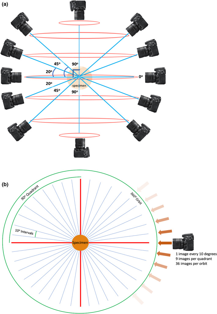
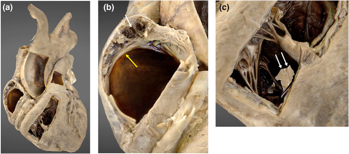
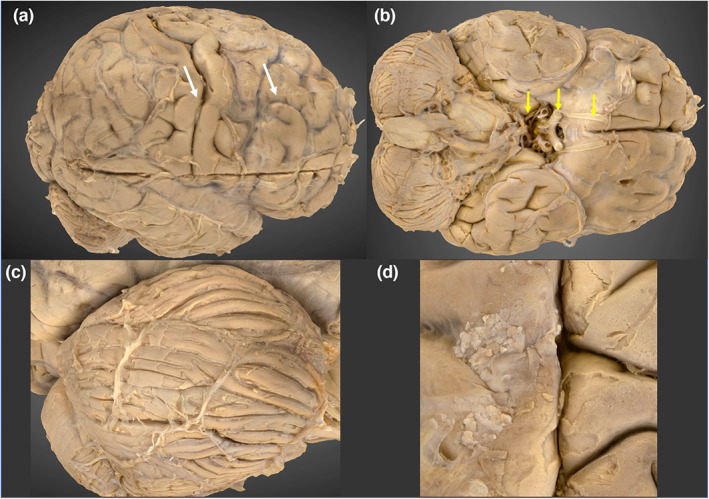
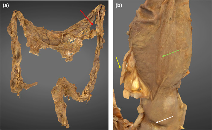
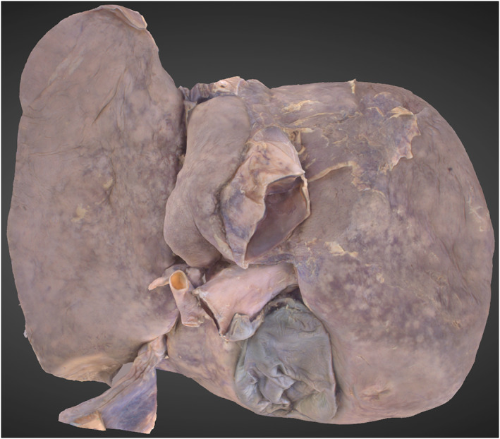
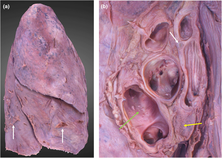
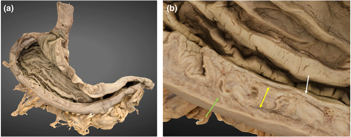
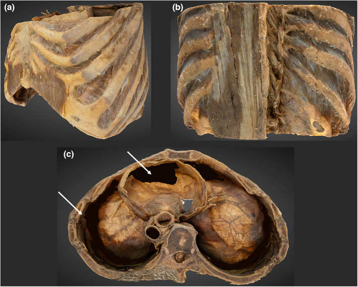
Three-dimensional (3D) representations of anatomical specimens are increasingly used as learning resources. Photogrammetry is a well-established technique that can be used to generate 3D models and has only been recently applied to produce visualisations of cadaveric specimens. This study has developed a semi-standardised photogrammetry workflow to produce photorealistic models of human specimens. Eight specimens, each with unique anatomical characteristics, were successfully digitised into interactive 3D models using the described workflow and the strengths and limitations of the technique are described. Various tissue types were reconstructed with apparent preservation of geometry and texture which visually resembled the original specimen. Using this workflow, an institution could digitise their existing cadaveric resources, facilitating the delivery of novel educational experiences.

Keywords: 3D visualisation; anatomy visualisation; cadaver; photogrammetry.


Figures

FIGURE 1
FIGURE 1
Specimen staging. (a) Plastinated heart suspended via the cut lumina of the aortic arch branches with additional inferior stabilisation. (b) Plastinated liver suspended with haemostats, wood and thread. The suspension line is indicated with black arrows. (c) Posterior view of the suspended specimen with full lighting. The small bowel has been dissected, but not suspended, and was not included in the image capture. (d) Posterior view of a fixed, wet stomach suspended using steel wire which is hidden in the rugal folds (red arrows). Sutures have been placed to secure the wire within the superior part of the duodenum (black arrow). (e) demonstrates vertical suspension of the right lung, white arrowheads indicate placement of suspension lines. (f) demonstrates horizontal suspension of the left lung with suspension lines (black arrows), which was found to be a more stable configuration. (g) brain position during capture of inferior structures. (h) brain position during capture of superior structures. (i) Suspended plastinated thorax (left posterior oblique view). White arrows indicate suspension points.
FIGURE 2
FIGURE 2
Image acquisition planning. (a) Horizontal orbits (red) and camera angles (blue) used for image acquisition planning, relative to the anatomical specimen in the coronal plane. (b) Image acquisition plan in the transverse plane, that is image capture in one orbit of rotation. Straight red lines represent 90 degree angles, relative to the specimen. Angled blue lines represent camera perspectives.
FIGURE 3
FIGURE 3
3D photogrammetric model of the heart. (a) (left) demonstrates the entire heart model. (b) (middle) demonstrates internal structures displayed in the lateral wall of the right atrium including the pectinate muscle (white arrowhead) and crista terminalis (yellow arrowhead). (c) (right) demonstrates the internal structures within the right ventricle and incomplete model development in the region of the interventricular septum (white arrows).
FIGURE 4
FIGURE 4
3D photogrammetric model of the brain. (a) (top left) Lateral view of the brain, demonstrating the transverse seam at the model merge site (white arrowheads). (b) (top right) Inferior view, demonstrating the cerebroarterial circle and cranial nerves I, II and III (yellow arrows). (c) (bottom left) Right cerebellar folia. (d) (bottom right) Arachnoid granulations.
FIGURE 5
FIGURE 5
3D photogrammetric model of the colon. (a) (left) demonstrates the entire colon model with reconstruction defect in greater omentum (red arrow). (b) (right) demonstrates a haustral fold (white arrow), epiploic appendage (yellow arrow) and taenia coli (green arrow).
FIGURE 6
FIGURE 6
3D Photogrammetric model of the liver. A posterior view shows the reconstruction of the porta hepatis, hepatic lobes and gallbladder.
FIGURE 7
FIGURE 7
3D photogrammetric model of the right lung. (a) (left) demonstrates the entire model (lateral view), dissection artefacts are indicated by white arrows. (b) (right) demonstrates the hilar structures including hyaline cartilage of the bronchial wall (white arrow), hilar lymph node (yellow arrow) and lumina of the pulmonary veins (green arrow).
FIGURE 8
FIGURE 8
3D photogrammetric model of the stomach. (a) (left) demonstrates the entire model (posterior view). (b) (right) demonstrates a cross section of the gastric wall layers including mucosa (white arrow), submucosa (yellow arrow) and muscularis externa (green arrow).
FIGURE 9
FIGURE 9
3D photogrammetric model of the thorax. (a) (top left) demonstrates the entire thorax model from a left anterolateral perspective. (B) (top right) demonstrates the reconstruction of the erector spinae and transversospinales. (c) (bottom, middle) superior view of the thorax. Shows reconstruction errors of the inferior pericardium and costophrenic recesses that have been digitally filled (white arrowheads).

References

    1. Apollonio, F.I. , Fantini, F. , Garagnani, S. & Gaiani, M. (2021) A photogrammetry‐based workflow for the accurate 3D construction and visualization of museums assets. Remote Sensing, 13, 486.
    1. Aziz, M.A. , Mckenzie, J.C. , Wilson, J.S. , Cowie, R.J. , Ayeni, S.A. & Dunn, B.K. (2002) The human cadaver in the age of biomedical informatics. The Anatomical Record: An Official Publication of the American Association of Anatomists, 269, 20–32. - PubMed
    1. Benjamin, A. , O'brien, D. , Barnes, G. , Wilkinson, B. & Volkmann, W. (2017) Assessment of structure from motion (SfM) processing parameters on processing time, spatial accuracy, and geometric quality of unmanned aerial system derived mapping products. Journal of Unmanned Aerial Systems, 3, 27.
    1. Bergman, E. , van der Vleuten, C.P. & Scherpbier, A.J. (2011) Why don't they know enough about anatomy? A narrative review. Medical Teacher, 33, 403–409. - PubMed
    1. Bhat, D. , Chittoor, H. , Murugesh, P. , Basavanna, P.N. & Doddaiah, S. (2019) Estimation of occupational formaldehyde exposure in cadaver dissection laboratory and its implications. Anatomy & Cell Biology, 52, 419–425. - PMC - PubMed

Publication types