. 2023 Sep 15;4(3):102431.
doi: 10.1016/j.xpro.2023.102431. Epub 2023 Jul 9.

Protocol for inducing branching morphogenesis in human cholangiocyte and cholangiocarcinoma organoids

Affiliations

Protocol for inducing branching morphogenesis in human cholangiocyte and cholangiocarcinoma organoids

Kimberley Ober et al. STAR Protoc. .

Abstract

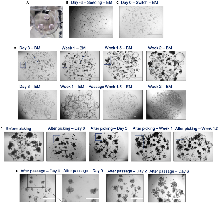
Bile ducts are essential for bile transport and consist of complex branching tubular networks. Human patient-derived cholangiocyte develops a cystic rather than branching duct morphology. Here, we present a protocol to establish branching morphogenesis in cholangiocyte and cholangiocarcinoma organoids. We describe steps for the initiation, maintenance, and expansion of intrahepatic cholangiocyte organoids branching morphology. This protocol enables the study of organ-specific and mesenchymal-independent branching morphogenesis and provides an improved model to study biliary function and diseases. For complete details on the use and execution of this protocol, please refer to Roos et al. (2022).1.

Keywords: Cancer; Cell Biology; Developmental biology; Organoids.


Conflict of interest statement

Declaration of interests The authors declare no competing interests.

Figures

None
Graphical abstract
Figure 1
Figure 1
Bright field microscopic images of ICO culture maintenance after tissue or cryopreserved organoid culture initiation (A) Immediately after tissue-derived ICO initiation and SEM addition, showing residual cellular debris and only small potential ICO structures (Ø 5–10 μm; zoom in black squared box). (B) While residual cellular debris is still present, the SEM media is switched for EM after day 3 of initiation, showing ICO structures arising (Ø 10–100 μm; zoom in black squared box). (C) The maximum density of the residual cellular debris level upon tissue-derived ICO initiation, which will still result in viable ICO culture (zoom in black squared box). (D) ICO initiation from previously established cryopreserved ICO with an overall 50% debris density upon thawing. Only few small potential ICO structures are visible (Ø 5–10 μm; zoom in black squared box). (E) Upon multiple small ICO structures (Ø 10–100 μm; zoom in black squared box) the SEM media is switched for EM, after approximately 5 days. (F) 95% ICO density within the BME dome with a collapsed darkened ICO within the zoom in black squared box, indicating that passaging the culture is desired. (G) Examples of ICO cultures with densities <80% that only require media refreshment, no passaging yet, showing entrapped debris within an ICO in the zoom in black squared box. (H) Examples of ICO cultures with densities >80% that require passaging for culture expansion, in which the zoom in black squared box indicates the formation of thickened ICO borders within a dense ICO culture. (All images: 2× magnification; scale bar indicates 2,000 μm).
Figure 2
Figure 2
Bright field microscopic images of the branching initiation process in ICO cultures (A) The lay-out of the BME dome seeding in a well of a 12-wells plate upon branching initiation. (B) The desired (20%–50%) ICO seeding size and density to promote successful BRCO initiation. (C) After 3 days of EM and the presence of small ICOs (Ø 10–100 μm), the media is switched to BM. (D) BM incubation of 3 days will result in darkened ICO structures with a thickened border (blue arrow). The blue squared box tracks the branching formation of one of the BRCO over 2 weeks’ time. This specific ICO line already showed clear small tubular branching structures (Ø 100–500 μm) after 2 weeks of BM refreshment, while the EM control maintained exponential growth and required passaging after 1 week. (E) Tracking the outgrowth of branching structures until 1.5 weeks after manual BRCO clone selection, showing the formation of tubular structures after careful structure breaking during picking (blue squared box). (F) The 6 day outgrowth of BRCO after normal passaging procedures for the expansion of BRCO up to 1,000 μm (zoomed scale bar indicated 1,000 μm at 4× magnification), these results have been published as supplementary data in Roos et al., 2022; Supplementary figure S1 (All images, except for A and zoom in: 2× magnification; scale bar indicates 2,000 μm).
Figure 3
Figure 3
The manual clone selection of BRCO to enable the outgrowth of larger BRCO structures and the purification of the BRCO culture from cystic ICO structures (A) The BRCO density (<70%) without overlapping structures that could be picked by applying an inverted microscope, as indicated by the example in the picture. (B) The manual BRCO selection of high density BRCO cultures (>70%) with overlapping structures utilizing an automated hybrid microscope cell imaging system (EVOS Cell Imaging System, Thermo Fisher Scientific). The blue arrows indicate the BRCO structures that should be manually picked. (All bright field images; 2× magnification; scale bar indicates 2,000 μm).
Figure 4
Figure 4
Bright field microscopic images of the expected outcomes of a successful BRCO initiation procedure (A) The formation of an elaborate tubular network of BRCO structures within one healthy ICO line (zoomed scale bar indicates 1,000 μm at 4× magnification). (B) Upon BM media switch, the percentage of BRCO outgrow differs between different healthy ICO lines, ranging from 70%‒25% of total organoid structures within the BME dome including different size ranges of (100–500 μm), these results have been published as supplementary data in Roos et al., 2022; Supplementary figure S1. (C) Successful branching of BRCCAO, showing the distinct dense branching structures without large elaborate networks or tubular formations reaching outwards (zoomed scale bar indicates 1,000 μm at 4× magnification). (D) The successful branching of PCLDO showing large elaborate tubular networks exceeding the 3,000 μm (zoomed scale bar indicates 1,000 μm at 4× magnification). (All images, except zoom in; 2× magnification; scale bar indicates 2,000 μm).
Figure 5
Figure 5
An overview of bright field microscopic images presenting potential problems that could arise during the BRCO initiation procedure (A) The presence of large cystic ICO structures (Ø 1,000 μm) only, even after 4 weeks of BM culture. The blue arrows indicate the somewhat thickened border and darkened coloring of the cystic organoids after 4 weeks of BM culture. (B) Exceeding the advised ICO seeding density >70% for BRCO initiation will result in the formation of smaller, dense and seemingly less viable BRCO structure, creating an overall less successful BRCO culture. (C) While decreasing the ICO seeding to <20% could result in the outgrowth of faster growing cystic ICO cultures, with only 1 or 2 BRCO that would be eligible for manual clone selection (increasing overall procedure times drastically). (D) When BRCO are not broken or passaged on time, the BRCO structure will start to expire over time showing very dark dense thickened tubular structures. The zoomed in black square indicates the release of dying cells into the BME dome. (E) Mycoplasma contamination of an ICO culture showing the distinct bearded ICO structures and the small dots (Ø 1–5 μm) that are both clear indicators for mycoplasma contamination (20× magnification; scale bar indicates 200 μm) (All images, except zoom in; 2× magnification; scale bar indicates 2,000 μm).

Similar articles

Cited by

References

    1. Roos F.J.M., van Tienderen G.S., Wu H., Bordeu I., Vinke D., Albarinos L.M., Monfils K., Niesten S., Smits R., Willemse J., et al. Human branching cholangiocyte organoids recapitulate functional bile duct formation. Cell Stem Cell. 2022;29:776–794.e13. - PubMed
    1. Huch M., Gehart H., van Boxtel R., Hamer K., Blokzijl F., Verstegen M.M.A., Ellis E., van Wenum M., Fuchs S.A., de Ligt J., et al. Long-term culture of genome-stable bipotent stem cells from adult human liver. Cell. 2015;160:299–312. - PMC - PubMed
    1. Broutier L., Mastrogiovanni G., Verstegen M.M., Francies H.E., Gavarró L.M., Bradshaw C.R., Allen G.E., Arnes-Benito R., Sidorova O., Gaspersz M.P., et al. Human primary liver cancer–derived organoid cultures for disease modeling and drug screening. Nat. Med. 2017;23:1424–1435. doi: 10.1038/nm.4438. - DOI - PMC - PubMed
    1. Broutier L., Andersson-Rolf A., Hindley C.J., Boj S.F., Clevers H., Koo B.K., Huch M. Culture and establishment of self-renewing human and mouse adult liver and pancreas 3D organoids and their genetic manipulation. Nat. Protoc. 2016;11:1724–1743. - PubMed
    1. Barker N., Huch M., Kujala P., van de Wetering M., Snippert H.J., van Es J.H., Sato T., Stange D.E., Begthel H., van den Born M., et al. Lgr5+ve Stem Cells Drive Self-Renewal in the Stomach and Build Long-Lived Gastric Units In Vitro. Cell Stem Cell. 2010;6:25–36. - PubMed

Publication types