. 2024 Feb;13(2):53-69.
doi: 10.1089/wound.2023.0036. Epub 2023 Aug 14.

Targeting Anaerobic Respiration in Pseudomonas aeruginosa with Chlorate Improves Healing of Chronic Wounds

Affiliations

Targeting Anaerobic Respiration in Pseudomonas aeruginosa with Chlorate Improves Healing of Chronic Wounds

Jane H Kim et al. Adv Wound Care (New Rochelle). 2024 Feb.

Abstract

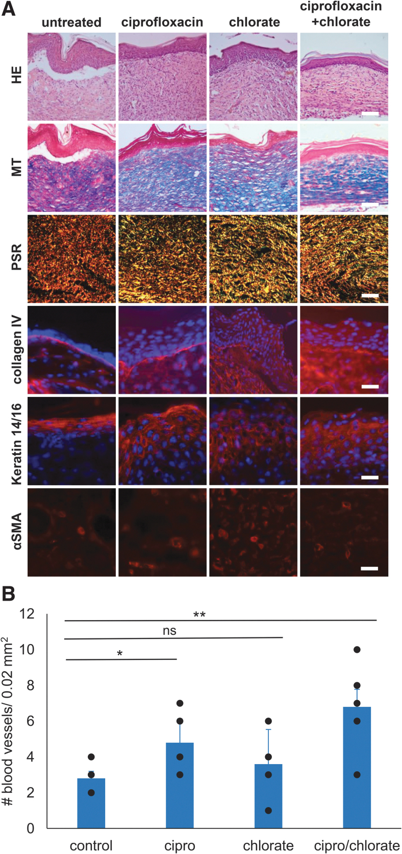
Objective: Pseudomonas aeruginosa is an opportunistic pathogen that can establish chronic infections and form biofilm in wounds. Because the wound environment is largely devoid of oxygen, P. aeruginosa may rely on anaerobic metabolism, such as nitrate respiration, to survive in wounds. While nitrate reductase (Nar) typically reduces nitrate to nitrite, it can also reduce chlorate to chlorite, which is a toxic oxidizing agent. Therefore, chlorate can act as a prodrug to specifically eradicate hypoxic/anoxic, nitrate-respiring P. aeruginosa populations, which are often tolerant to conventional antibiotic treatments. Approach: Using a diabetic mouse model for chronic wounds, we tested the role that anaerobic nitrate respiration plays in supporting chronic P. aeruginosa infections. Results: P. aeruginosa forms biofilm deep within the wound where the environment is anoxic. Daily treatment of P. aeruginosa-infected wounds with chlorate supported wound healing. Chlorate treatment was as effective as a treatment with ciprofloxacin (a conventional antibiotic that targets both oxic and hypoxic/anoxic P. aeruginosa populations). Chlorate-treated wounds showed markers of good-quality wound healing, including well-formed granulation tissue, reepithelialization and microvessel development. Loss- and gain-of-function experiments showed that P. aeruginosa requires nitrate respiration to establish a chronic wound infection and form biofilms. Innovation: We show that the small molecule chlorate, kills the opportunistic pathogen, P. aeruginosa, by targeting a form of anaerobic metabolism called nitrate respiration. Conclusion: Chlorate holds promise as a treatment to combat diverse bacterial infections where oxygen is limiting and/or where pathogens grow as biofilms because many other pathogens possess Nar and survive using anaerobic metabolism.

Keywords: MiPACT-HCR; Pseudomonas aeruginosa; anaerobic respiration; antibiotics; chlorate; chronic wounds.


Conflict of interest statement

The authors state no competing financial interests. The content of this article was expressly written by the authors listed. No ghostwriters were used to write this article.

Figures

None
Manuela Martins-Green, PhD
None
Dianne K. Newman, PhD
Figure 1.
Figure 1.
RPA requires Nar for chlorate susceptibility. (A) Nar is a hypoxically induced enzyme that reduces chlorate to toxic chlorite. (B) Survival of WT and Δnar RPA after treatment with ciprofloxacin, chlorate, or both drugs under oxic and anoxic planktonic conditions and grown as biofilms. For each treatment, n ≥ 3 replicates and bars show mean ± standard error of the mean. RPA, Riverside Pseudomonas aeruginosa; Nar, nitrate respiration.
Figure 2.
Figure 2.
Ciprofloxacin and chlorate treatment support healing of RPA-infected wounds. Wounds were infected with RPA (106 CFU) 24 h after injury and daily treatment began 10 days after infection. Wounds were treated daily with either vehicle (n = 5), ciprofloxacin (cip, n = 7), chlorate (chlor, n = 8), or both ciprofloxacin and chlorate (cip/chlor, n = 9). (A) Visualization of RPA biofilms using MiPACT-HCR on a section of chronic wound tissue collected at 10 days postwounding and before treatment (wound surface is on the top). Yellow fluorescence shows RPA, as detected by 16S rRNA amplification, and DAPI (blue) was used to visualize the nuclei of the cells in the mouse tissue. (B) Representative images of untreated and treated RPA-infected wounds over 40 days. Untreated RPA-infected wounds did not undergo wound closure and have robust biofilm formation at day 40, whereas treated wounds had decreased amount of biofilm and were smaller in size. (C) Quantification of wound areas over time for untreated and treated RPA-infected wounds. The student t-test was used to determine significant differences between treatment groups compared with the untreated control. *p-value <0.05 is between RPA+cip/chlor and RPA. (D) Individual data points for control and treatments groups are shown for RPA-infected wounds at day 40. Student's t-test was used to determine significant differences between treatment groups compared with the untreated control. *p-value <0.05. CFU, colony-forming unit; MiPACT-HCR, Microbial identification after Passive Clarity Technique–Hybridization Chain Reaction; ns, non significant.
Figure 3.
Figure 3.
Ciprofloxacin and chlorate treatment support wound healing. (A) Wound tissue was collected at day 40 for treated wounds that had healed and also for untreated wounds to perform histology and immunofluorescence staining. Cryosections of the skin were taken from wound tissue for HE, MT, and PSR. Scale bar = 100 μm. Collagen III is shown in green, collagen I is shown in red, and the colocalization of collagen III and collagen I is shown in yellow. Wound tissues were immunolabeled with collagen IV (red), αSMA (red), keratin 14/16 (red), and all samples were labeled with DAPI to stain the DNA in the nuclei of the mouse cells (blue). Scale bar = 20 μm. (B) The number of blood vessels were counted in five frames (area = 0.02 mm2) of the granulation tissue. Blood vessel of RPA-infected wounds were compared using one-way ANOVA: ***p-value <0.001. H&E, Hematoxylin & Eosin; MT, Masson's Trichrome; ns, non significant; PSR, Picrosirius Red.
Figure 4.
Figure 4.
RPA requires Nar to establish a chronic wound infection. Wounds were infected with Δnar RPA (106 CFU) 24 h after injury and daily treatment began 10 days after infection. Wounds were treated daily with either vehicle (n = 6), ciprofloxacin (cip, n = 6), chlorate (chlor, n = 6), or both ciprofloxacin and chlorate (cip/chlor, n = 6). (A) Representative images of untreated and treated Δnar RPA-infected wounds over 40 days. (B) Quantification of wound areas over time. All untreated and treated Δnar RPA-infected wounds underwent wound closure. The Student's t-test was used to determine significant differences between treatment groups compared with the untreated control group. No significant differences in wound areas were observed. (C) Individual data points for control and treatment groups are shown for Δnar RPA-infected wounds at day 40. The Student's t-test was used to determine significant differences between treatment groups compared with the untreated control group. No significant (ns) differences in wound areas were observed. Nar, nitrate respiration; ns, non significant.
Figure 5.
Figure 5.
Ciprofloxacin and chlorate treatment enhance healing of Δnar RPA-infected wounds. Wound tissue was collected at day 40 for untreated and treated Δnar RPA-infected wounds to perform histology and immunofluorescence staining. (A) Cryosections of the skin were taken from wound tissue for HE, MT, and PSR. Collagen III is shown in green, collagen I is shown in red, and the colocalization of collagen III and collagen I is shown in yellow. Scale bar = 100 μm. Wounds tissues were immunolabeled with collagen IV (red), αSMA (red), keratin 14/16 (red), and all samples were labeled with DAPI to stain the DNA in the nuclei of the mouse cells (blue) (scale bar = 20 μm). (B) The number of blood vessels were counted in six frames (area = 0.02 mm2) of the granulation tissue. Blood vessel of Δnar RPA-infected wounds were compared using one-way ANOVA: *p-value <0.05, **p-value <0.01; ns, non significant.
Figure 6.
Figure 6.
Mice infected with the Δnar-complement strain (Δnar+att::mTnnar) develop chronic wounds. (A) Representative images of wounds infected with either WT (106 CFU), Δnar (106 CFU), or Δnar+att::mTnnar (104 CFU) RPA 24 h after injury and left undisturbed for 40 days. Wounds infected with either WT or Δnar+att::mTnnar RPA displayed biofilm formation throughout the 40-day experiment, whereas Δnar RPA-infected wounds did not display biofilm at day 40. (B) Quantification of wound areas. Both WT and Δnar+att::mTnnar RPA-infected wounds were open 40 days after injury, whereas Δnar RPA-infected wounds had closed. Note: the data shown for WT and Δnar RPA-infected wounds are the same as the data shown in Figs. 2C and 4B, respectively, and have been added for ease of comparison to the Δnar+att::mTnnar RPA strain. (C) Individual data points for control and treatment groups are shown for wounds infected with either RPA, Δnar RPA, or Δnar+att::mTnnar at day 30 and 40. The Student's t-test was used to determine significant differences in wound areas between mutant strains compared with RPA. **p-value <0.01, ***p-value <0.001. ns, non significant.

Similar articles

Cited by

References

    1. Galaviz KI, Narayan KMV, Lobelo F, et al. . Lifestyle and the prevention of type 2 diabetes: A status report. Am J Lifestyle Med 2018;12:4–20; doi: 10.1177/1559827615619159 - DOI - PMC - PubMed
    1. Chen L, Magliano DJ, Zimmet PZ. The worldwide epidemiology of type 2 diabetes mellitus—present and future perspectives. Nat Rev Endocrinol 2011;8:228–236; doi: 10.1038/nrendo.2011.183 - DOI - PubMed
    1. Edmonds M, Manu C, Vas P. The current burden of diabetic foot disease. J Clin Orthop Trauma 2021;17:88–93; doi: 10.1016/j.jcot.2021.01.017 - DOI - PMC - PubMed
    1. Gardiner M, Vicaretti M, Sparks J, et al. . A longitudinal study of the diabetic skin and wound microbiome. PeerJ 2017;5:e3543; doi: 10.7717/peerj.3543 - DOI - PMC - PubMed
    1. Smith K, Collier A, Townsend EM, et al. . One step closer to understanding the role of bacteria in diabetic foot ulcers: Characterising the microbiome of ulcers. BMC Microbiol 2016;16:54; doi: 10.1186/s12866-016-0665-z - DOI - PMC - PubMed