. 2023 Jul 11;18(7):e0285806.
doi: 10.1371/journal.pone.0285806. eCollection 2023.

Dermokine mutations contribute to epithelial-mesenchymal transition and advanced melanoma through ERK/MAPK pathways

Affiliations

Dermokine mutations contribute to epithelial-mesenchymal transition and advanced melanoma through ERK/MAPK pathways

Wenqiong Ma et al. PLoS One. .

Retraction in

Abstract

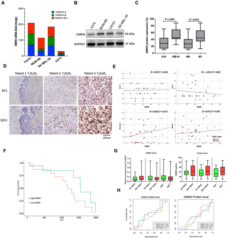
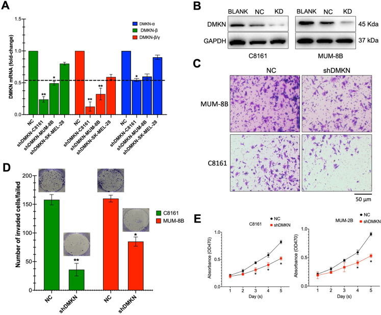
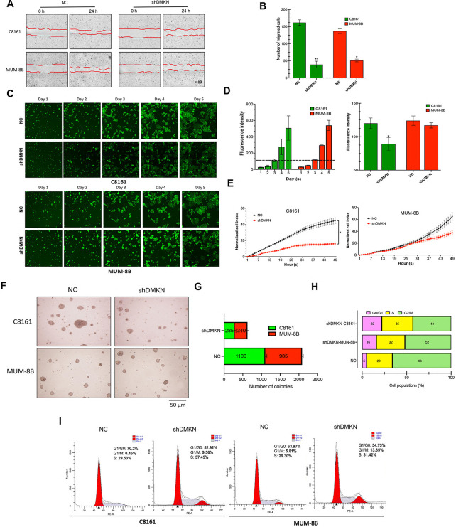
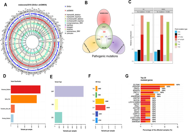
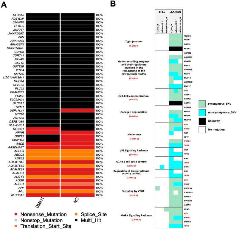
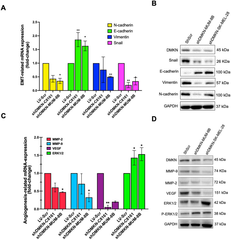
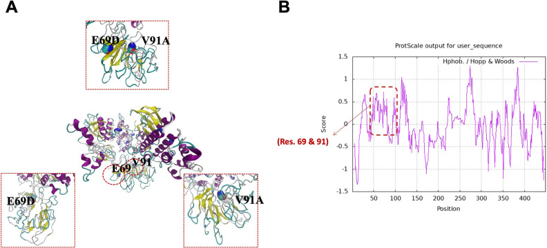
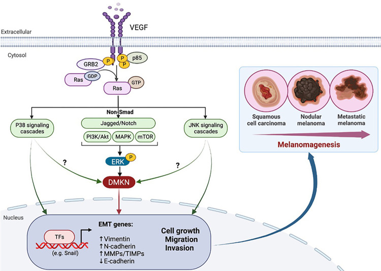
To discover vulnerabilities associated with dermokine (DMKN) as a new trigger of the epithelial-mesenchymal transition (EMT) -driven melanoma, we undertook a genome-wide genetic screening using transgenic. Here, we showed that DMKN expression could be constitutively increased in human malignant melanoma (MM) and that this correlates with poor overall survival in melanoma patients, especially in BRAF-mutated MM samples. Furthermore, in vitro, knockdown of DMKN inhibited the cell proliferation, migration, invasion, and apoptosis of MM cancer cells by the activation of ERK/MAPK signaling pathways and regulator of STAT3 in downstream molecular. By interrogating the in vitro melanoma dataset and characterization of advanced melanoma samples, we found that DMKN downregulated the EMT-like transcriptional program by disrupting EMT cortical actin, increasing the expression of epithelial markers, and decreasing the expression of mesenchymal markers. In addition, whole exome sequencing was presented with p.E69D and p.V91A DMKN mutations as a novel somatic loss of function mutations in those patients. Moreover, our purposeful proof-of-principle modeled the interaction of ERK with p.E69D and p.V91A DMKN mutations in the ERK-MAPK kinas signaling that may be naturally associated with triggering the EMT during melanomagenesis. Altogether, these findings provide preclinical evidence for the role of DMKN in shaping the EMT-like melanoma phenotype and introduced DMKN as a new exceptional responder for personalized MM therapy.


Conflict of interest statement

The authors have declared that no competing interests exist.

Figures

Fig 1
Fig 1. The cancerous role of DMKN in advanced melanoma.
(A) Relative levels of DMKN-α, DMKN-β, and DMKN-β/γ mRNA expression in advanced melanoma cell lines. (B) DMKN protein level in advanced melanoma cell lines. (C-D) Quantification and representative immunobiological staining imaging of DMKN expression in tissue samples from different stages. (E) Linear logistic regression analysis between DMKN and routine diagnostic biomarkers (Ki67, p53, Melanin A, and S-100) in stage II/III melanoma (blue cycle) and stage III/IV melanoma (red cycle). (F) Disease-free survival (DFS) rate based on DMKN expression in 15 patients with low DMKN and 16 patients with high DMKN. (G) DMKN expression in advanced melanoma with different mutation stages (BRAF, NRAS) and EMT features. (H) Receiver operating characteristic curve for DMKN mRNA and protein expression in EMT+ MM group (blue line), NRAS mutant MM group (green line), and BRAF mutation group (red line). The diagnostic values were calculated based on DMKN mRNA expression and protein levels. Patients were categorized as low-DMKN when FC < 0.18 for DMKN mRNA expression and < 42.78 for protein DMKN levels. The grouping of western blot gel images was cropped from different parts of the same gel. The full-length gels are included in S1 Raw images. High DMKN is defined as FC ≥ 0.18 for DMKN mRNA expression and ≥ 42.78 for protein DMKN levels.
Fig 2
Fig 2. DMKN knockdown inhibits proliferation and invasion in advanced melanoma cell lines.
The dmkn gene (A) and protein (B) levels in DMKN-shRNA-transduced cells (shDMKN) compare to that in lentivirus-scrambled control (NC). Analysis of cell invasion by using a modified Boyden Chamber assay. in lentivirus control and DMKN-shRNA-transfected in the C8161 and MUM-2B cell lines. Microscopic picture of invaded cells (C) represents Bar graph (D) and the number of cells invaded the matrigel. (E) Relative cell viability of shDMKN (red line) or NC (black line) cells. Cell proliferation values are presented as the mean SEM of three independent tests. Values are as mean ± SEM, n = 3, **p < 0.001 for NC. The grouping of western blot gel cropped from different parts of the same gel. During the invasion assay, the leftover supernatants on the upper chamber of the transwell insert were collected from all the treatments of both cells.
Fig 3
Fig 3. DMKN knockdown reduced cell migration in advanced melanoma cell lines.
(A) Scratch wound-healing assay of C8161 and MUM-8B cells transfected with GFP lentivirus (NC) or DMKN-shRNA (shDMKN). The movement of cells into the wounds was shown at 0 and 24 hrs after scratching (magnification: 10x). (B) The bar graph represents the number of cells that migrated into the scratched area. (C) Representative fluorescence microscopy photos in C8161 and MUM-8B cells transfected with NC or shDMKN within 5 days of treatment. (D) Bar graphs showing means fluorescence of migrating in C8161 and MUM-8B cells transfected with NC or shDMKN within 5 days of treatment. (E) Real-time detection cell index of C8161 and MUM-8B with NC or shDMKN in the xCELLigence real-time cell analysis. (F) Representative microscopic photos of the colony formation assays to examine the effect of DMKN on the proliferation of advanced melanoma cells. (G) The bar graph represents the number of cells that migrated into the scratched area. The cells were recovered in regular media for 10 days and colonies were counted. (H) Bar graphs showed the percentage of each cell cycle phase in C8161 and MUM-8B cells transfected with shDMKN or NT siRNAs for 3 days. (I) Representative percentage of each cell cycle phase in C8161 and MUM-8B cells transfected with shDMKN or NT siRNAs for 3 days. Expression of DMKN triggers cell cycle arrest and inhibits cell invasion in advanced melanoma cell lines. Values are as mean ± SEM, n = 3, **p < 0.001, ***p < 0.0001 for NC.
Fig 4
Fig 4. The whole genome and targeted exome sequencing results after the knockdown of DMKN in advanced melanoma cell lines.
(A) Visualization of chromosomal breakpoint junctions between the shDMKN- C8161 and GFP lentivirus (NC) control. (B) The differentially expressed pathogenic genes with the largest fold changes between shDMKN-KD and NC in the different melanoma cell lines. (C) The ratio of somatically point mutation variants between shDMKN-KD and GFP lentivirus (NC) control in C8161 cells. The down panels above describe the variant classification (D), variant type (E), SNV class (F), and variant per sample in all shDMKN cell lines. (G) Top 20 frequently implicated genes out of the 221 genes mutated in the shDMKN-C8161 melanoma cell lines genome.
Fig 5
Fig 5. DMKN interactome is reprogrammed in MM cells.
(A) The gene set enrichment heatmap indicates the mutation patterns of the top 20 most frequently implicated cancer driver genes with a high number of mutations in the shDMKN-C8161 melanoma cell lines genome. Each column represents an affected individual, and each row represents a gene. (B) Gene set enrichment analysis of most deregulated cellular processes among 984 genes carrying mutations/aberrations in the shDMKN (KD) group. The genes are halighlited in red color involved in melnomagesis. The p-value for each process represents the significance level for the enrichment of mutations/aberrations that affect this pathway in the DMKN-shRNA lentivirus (KD) group compared to the GFP lentivirus (NC) control and was calculated by two-sided Fisher’s exact test.
Fig 6
Fig 6. Analysis of the oncogenic pathways regulated by DMKN.
(A-B) the DMK disruption of MET/EMT cortical actin, increased expression of epithelial markers (E-cadherin), and decreased expression of mesenchymal markers (N-cadherin, Vimentin, and Snail). Expression of EMT-associated and oncogenic pathway proteins in DMKN-shRNA (KD), and GFP lentivirus (NC) were assayed in both gene (A) and protein levels (B). (C-D) ERK/MAPK signaling pathway was active with shDMKN downregulation as compared to NC in C8161 and MUM-2B MM cell lines. Expression of ERK/MAPK oncogenic pathway proteins in advanced melanoma cells transfected with either DMKN-shRNA (KD) or GFP lentivirus (NC) were assayed in both gene (C) and protein levels (D). The grouping of western blot gel is cropped from different parts of the gel. Whole-cell lysates (50–70 μg) were subjected to Western blotting analysis. GAPDH as loading control is shown with a representative blot. Values are as mean ± SEM, n = 3, **p < 0.001, ***p < 0.0001 for NC.
Fig 7
Fig 7. Functional mimicry effects of DMKN pathogenic mutation with pERK.
(A) The DMKN protein [Genbank: Q6EOU4] and human ERK1 [Genbank: P27361] and human ERK2 [Genbank: P28482] protein sequences were aligned using the MUSCLE method with default settings in Geneious [26]. (B) Structural analyses of the novel mutation identified in the dmkn gene. Predicted crystal structures of wild type and both novel mutants identified in the dmkn gene. Red dashes show seven hydrogen bonds between mutated amino acid and 6Å relative to its around amino acids. H-bonds include the following distances 1.8Å, 2.2Å, 2.3Å, 2.5Å, and 2.6Å. C-D). Predicted 3-D structure assessment of p-ERK the magnified image of binding site with DMKN. nsSNP E69D and V91A made the pathogenic deletion in the C-terminal of the DMKN protein. The results point to the probability that nsSNP E69D ‎frameshift mutation is most likely larger structurally abnormal, unstable, and certainly functional DMKN ‎in the interaction with the p-ERK.
Fig 8
Fig 8. The possible molecular mechanism of DMKN during melanomagenesis.
DMKN triggers the EMT via DMKN/ERK/MAPK-initiated signaling pathways. This schematic carton shows the major triggering of EMT-inducing signaling pathways.

Similar articles

Cited by

References

    1. Erdmann F, Lortet-Tieulent J, Schuz J, Zeeb H, Greinert R, Breitbart EW, et al.. International trends in the incidence of malignant melanoma 1953-2008—are recent generations at higher or lower risk? Int J Cancer. 2013;132(2):385–400. Epub 2012/04/26. doi: 10.1002/ijc.27616 . - DOI - PubMed
    1. Wu Y, Wang Y, Wang L, Yin P, Lin Y, Zhou M. Burden of melanoma in China, 1990–2017: Findings from the 2017 global burden of disease study. Int J Cancer. 2020;147(3):692–701. Epub 2019/11/02. doi: 10.1002/ijc.32764 . - DOI - PubMed
    1. Davar D, Lin Y, Kirkwood JM. Unfolding the mutational landscape of human melanoma. J Invest Dermatol. 2015;135(3):659–62. Epub 2015/02/11. doi: 10.1038/jid.2014.467 ; PubMed Central PMCID: PMC4466551. - DOI - PMC - PubMed
    1. Timar J, Ladanyi A. Molecular Pathology of Skin Melanoma: Epidemiology, Differential Diagnostics, Prognosis and Therapy Prediction. Int J Mol Sci. 2022;23(10). Epub 2022/05/29. doi: 10.3390/ijms23105384 ; PubMed Central PMCID: PMC9140388. - DOI - PMC - PubMed
    1. Ernst M, Giubellino A. The Current State of Treatment and Future Directions in Cutaneous Malignant Melanoma. Biomedicines. 2022;10(4). Epub 2022/04/24. doi: 10.3390/biomedicines10040822 ; PubMed Central PMCID: PMC9029866. - DOI - PMC - PubMed

Publication types

Substances