. 2023 Jul 25;120(30):e2221413120.
doi: 10.1073/pnas.2221413120. Epub 2023 Jul 11.

The human milk component myo-inositol promotes neuronal connectivity

Affiliations

The human milk component myo-inositol promotes neuronal connectivity

Andrew F Paquette et al. Proc Natl Acad Sci U S A. .

Abstract

Effects of micronutrients on brain connectivity are incompletely understood. Analyzing human milk samples across global populations, we identified the carbocyclic sugar myo-inositol as a component that promotes brain development. We determined that it is most abundant in human milk during early lactation when neuronal connections rapidly form in the infant brain. Myo-inositol promoted synapse abundance in human excitatory neurons as well as cultured rat neurons and acted in a dose-dependent manner. Mechanistically, myo-inositol enhanced the ability of neurons to respond to transsynaptic interactions that induce synapses. Effects of myo-inositol in the developing brain were tested in mice, and its dietary supplementation enlarged excitatory postsynaptic sites in the maturing cortex. Utilizing an organotypic slice culture system, we additionally determined that myo-inositol is bioactive in mature brain tissue, and treatment of organotypic slices with this carbocyclic sugar increased the number and size of postsynaptic specializations and excitatory synapse density. This study advances our understanding of the impact of human milk on the infant brain and identifies myo-inositol as a breast milk component that promotes the formation of neuronal connections.

Keywords: DHA; brain development; myo-inositol; nutritional neuroscience; synapse formation.


Conflict of interest statement

S.D.M., B.L., S.C.P., and N.P. are present and C.K. and D.H. past employees of Reckitt/Mead Johnson Nutrition. This funder performed and analyzed the human milk study. The funder had no other role in research design, data collection, and analysis and had no role in study conceptualization and result interpretation.

Figures

Fig. 1.
Fig. 1.
Myo-inositol peaks in human milk during early infant brain development and this carbocyclic sugar promotes the abundance and size of excitatory postsynaptic sites in human glutamatergic neurons. (A) Free myo-inositol content in human milk is highest in the first wk of lactation. Samples were longitudinally collected from ten mothers per site in Shanghai, Mexico City, and Cincinnati over 52 wk. Circles, myo-inositol concentration per mother. Lines, mean concentration per geography. (B) Temporal profiles of processes underlying cortical developmental. (C, Top), representative confocal images of human glutamatergic–enriched cortical neurons 24 d after plating, cultured under control conditions (C, Left) or with myo-inositol at 2 mM (C, Right). Immunostainings were performed for presynaptic Bassoon (red), excitatory postsynaptic Homer (green), and dendritic MAP2 (blue). (C, Bottom) enlarged dendritic segments. (D and E) Quantification of images as in C determined that myo-inositol increases in human glutamatergic neurons the abundance of postsynaptic Homer measured as immunostaining intensity per dendritic area (D) and the size of Homer-positive specializations (E). Violin plots show data distribution. Solid lines mark the median and dotted lines the quartiles. Asterisks show statistical differences of mean values, which are not plotted. (Student’s t test, two-tailed unpaired; dendritic segments from N = 86 control/71 myo-inositol-treated neurons) ***P < 0.001. (F and G) Myo-inositol treatment did not significantly increase presynaptic Bassoon abundance (F) and size of Bassoon-positive sites (G). Violin plots show data as in D and E. n.s., not significant.
Fig. 2.
Fig. 2.
Synapse size and number are increased by myo-inositol in a dose-dependent manner, and it promotes neuronal responsiveness to synaptogenic Neuroligin 1. (A) Representative confocal images of dendritic segments of control primary rat neurons at 14 div (A, Top), or of neurons treated with myo-inositol at 1 mM (A, Center) or 13 mM (A, Bottom). Immunostainings show presynaptic Bassoon (green), postsynaptic Homer (red), and dendritic MAP2 (blue). (BF) Quantification of images as in A determined that myo-inositol increases in a dose-dependent manner the density of Bassoon (B) and Homer (C) puncta and of the synaptic sites where they colocalize (D). Myo-inositol also increased the size of Bassoon and Homer puncta (E and F), with data distribution shown in Violin plots as in Fig. 1 (one-way ANOVA with Tukey’s multiple comparison test; N = 16 to 18 dendritic segments per condition). (G) Confocal images of rat hippocampal neurons cultured in the presence of vehicle control or myo-inositol at 1 mM as indicated. Neurons were cocultured with HEK293-expressing CFP as transfection marker (cyan) that served as negative control, or with HEK293 cells coexpressing CFP and Neuroligin 1. At 14 div., immunostaining was performed for the presynaptic active zone marker Bassoon (green) and the dendritic marker MAP2 (blue). Insets show enlarged cocultured HEK293 cells. (H) Quantification of images as in G showed that contact with HEK293 cells expressing Neuroligin 1 induced neurons to assemble Bassoon-positive specializations, as expected. Myo-inositol treatment increases this synaptogenic response by a further 50 ± 13 %. Violin plots show data distribution (one-way ANOVA with Tukey’s multiple comparison test; N = 57 to 72 HEK293 cells per condition from three independent experiments).
Fig. 3.
Fig. 3.
Cortical synapse size increases when myo-inositol is supplemented during development. (A) For analysis of the visual cortex area V1, the diet of mouse pups was supplemented from birth with myo-inositol until postnatal day 35. (B, Top), single optical confocal sections of synaptic Homer immunostaining in binocular primary visual cortex area V1b, layer II/III in control (B, Left), and myo-inositol-supplemented mice (B, Right) at postnatal day 35. Mice received 50 mg myo-inositol/kg body weight/day from birth. (B, Bottom), puncta masks after image processing. (C and D) Quantification in layer II/III of V1 from images as in B of synaptic Homer puncta density (C) and size (D) of control and myo-inositol-supplemented animals. Animal averages are shown, and means were compared (Student’s t test, two-tailed unpaired; control, N = 5 mice, data averaged from 3/2/2/2/2 sections per mouse; myo-inositol, N = 4, data averaged from 2/2/2/2 sections per mouse). (E and F) Quantification in layer V of V1b of synaptic Homer puncta density (E) and size (F) of mice treated as in A and imaged as in B (Student’s t test, two-tailed unpaired; control, N = 5 mice; myo-inositol, N = 4).
Fig. 4.
Fig. 4.
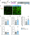
Myo-inositol is synaptogenic in mature hippocampal organotypic slice cultures. (A) Organotypic slice cultures were prepared from the hippocampus of mouse pups and cultured for 90 d. A subset of cultured slices was treated from 60 to 90 div with myo-inositol at 1 mM. (B) Representative confocal images of CA3 stratum lucidum of organotypic hippocampal slice cultures for 90 div under control conditions (B, Left) or after treatment with myo-inositol at 1 mM from 60 to 90 div (B, Right). Slice cultures were stained for presynaptic Bassoon (red) or the excitatory postsynaptic marker PSD-95 (green). (CE) Quantification of immunostainings from the hippocampal CA3 area as in B showed that myo-inositol increases the density of presynaptic Bassoon (C) and postsynaptic PSD-95-positive specializations (D) and of sites where these pre- and post-synaptic markers colocalize (E). (F and G) Quantification of images as in B determined that myo-inositol does not change the size of Bassoon-positive sites (F) but enlarges PSD-95-positive specializations (G) in the hippocampal CA3 area (CG, one-way ANOVA with Tukey’s multiple comparison test; N = 25 control and N = 10 myo-inositol slices from two independent experiments).

Comment in

  • Bridging a mechanistic gap from diet to synapses.
    Benson DL, Layé S. Benson DL, et al. Proc Natl Acad Sci U S A. 2023 Aug 15;120(33):e2309992120. doi: 10.1073/pnas.2309992120. Epub 2023 Aug 2. Proc Natl Acad Sci U S A. 2023. PMID: 37531376 Free PMC article. No abstract available.

Similar articles

Cited by

References

    1. Huttenlocher P. R., Synaptic density in human frontal cortex - developmental changes and effects of aging. Brain Res. 163, 195–205 (1979). - PubMed
    1. Rakić P., Bourgeois J. P., Eckenhoff M. F., Zecevic N., Goldman-Rakic P. S., Concurrent overproduction of synapses in diverse regions of the primate cerebral cortex. Science 232, 232–235 (1986). - PubMed
    1. Eidelman A. I., Schanler R. J., Breastfeeding and the use of human milk. Pediatrics 129, e827 (2012). - PubMed
    1. Horta B. L., Loret de Mola C., Victora C. G., Breastfeeding and intelligence: A systematic review and meta-analysis. Acta Paediatr. 104, 14–19 (2015). - PubMed
    1. Bode L., Raman A. S., Murch S. H., Rollins N. C., Gordon J. I., Understanding the mother-breastmilk-infant “triad”. Science 367, 1070–1072 (2020). - PubMed

Publication types