. 2023 Aug 11;9(8):1499-1507.
doi: 10.1021/acsinfecdis.3c00049. Epub 2023 Jul 11.

Nanoscaled Discovery of a Shunt Rifamycin from Salinispora arenicola Using a Three-Color GFP-Tagged Staphylococcus aureus Macrophage Infection Assay

Affiliations

Nanoscaled Discovery of a Shunt Rifamycin from Salinispora arenicola Using a Three-Color GFP-Tagged Staphylococcus aureus Macrophage Infection Assay

Nhan T Pham et al. ACS Infect Dis. .

Abstract

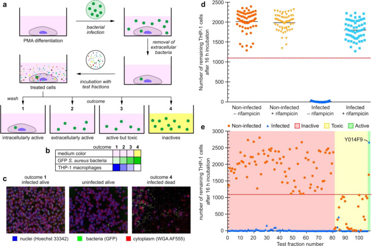
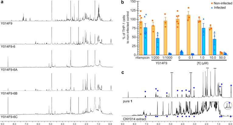
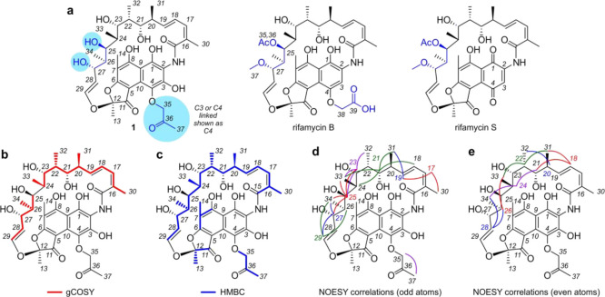
Antimicrobial resistance has emerged as a global public health threat, and development of novel therapeutics for treating infections caused by multi-drug resistant bacteria is urgent. Staphylococcus aureus is a major human and animal pathogen, responsible for high levels of morbidity and mortality worldwide. The intracellular survival of S. aureus in macrophages contributes to immune evasion, dissemination, and resilience to antibiotic treatment. Here, we present a confocal fluorescence imaging assay for monitoring macrophage infection by green fluorescent protein (GFP)-tagged S. aureus as a front-line tool to identify antibiotic leads. The assay was employed in combination with nanoscaled chemical analyses to facilitate the discovery of a new, active rifamycin analogue. Our findings indicate a promising new approach for the identification of antimicrobial compounds with macrophage intracellular activity. The antibiotic identified here may represent a useful addition to our armory in tackling the silent pandemic of antimicrobial resistance.

Keywords: Salinispora arenicola; Staphylococcus aureus; fluorescence imaging assay; macrophage; rifamycin.


Conflict of interest statement

The authors declare no competing financial interest.

Figures

Figure 1
Figure 1
Fluorescent macrophage infection assay. (a) Schematic representation of the three-color confocal fluorescence macrophage infection assay run in 96-well plate format on a PerkinElmer Opera instrument using a 20× objective. The assay process begins with infecting PMA differentiated THP-1-macrophage cells with GFP-USA300 S. aureus. Co-culturing of these cells results in infected mammalian cells after washing and removal of the media. The resulting bacterial infected cells are then subjected to test samples, which can result in four different outcomes. The first, arising from intracellular active compounds, is identified by a significant number of infected THP-1 cells remaining after the overnight incubation (indicated as dark blue in Figure 1b), and the cell media is red/pink (indicated as rose in panel b). The number of GFP-USA300 S. aureus bacteria is low (indicated as light green in panel b). The next two, extracellular active and active but toxic compounds, are marked by a low number of remaining THP-1 cells (indicated as light blue in panel b), some extracellular bacteria (indicated as lighter green in Figure 1b, and red/pink cell media (indicated as rose in panel b). The only difference between these two outcomes is that in the non-infected control, only a few to no THP-1 cells are found for toxic compounds. The final outcome, arising from non-active compounds, contains an absence of THP-1 cells as well as yellow cell media (panel b, lane 4), caused by saturated growth of bacteria (indicated by a dark green colored field in panel b, lane 4), which turns the cell media acidic, causing a color change to yellow. The cell membranes and the nuclei of the THP-1 macrophages were stained with wheat germ agglutinin Alexa Fluor 555 (WGA AF555) and Hoechst 33342 to enable blue/green/orange detection scheme. (b) Heatmap showing the outcome for each of the four different possibilities. The color in the table illustrates whether there is a color change in the cell medium, and the strength of the color illustrates the number of bacteria or THP-1 cells found in the sample. Medium color: color change from pink (red) to yellow only for outcome 4. GFP S. aureus bacteria (green) are most abundant for outcome 4. THP-1 macrophages (blue) are most abundant for outcome 1 and absent for outcome 4. (c) Typical Opera images for the various outcomes of the assay, THP-1 nuclei and plasma membrane are blue and red, respectively, and S. aureus bacteria are in green. Left image visualizes the outcome for an intracellularly active compound, the middle for an extracellularly active compound, and right image for an inactive compound. (d) Assay sensitivity. The separation between positive control containing infected THP-1 cells in 50 nM rifampicin (light blue square) and negative control (dark blue upright triangle) containing infected THP-1 cells in media only. For comparison, non-infected THP-1 cells with (yellow upside-down triangle) and without (orange round dots) 50 nM rifampicin are also shown. The red dashed line is the hit threshold which is the lower 3σ (standard deviation) or limit of the average of infected THP-1 cells in 50 nM rifampicin (parameters providing in the Supporting Information). (e) Results of the screen of 108 marine microbial test fractions. More than 80 fractions were inactive (red shaded area), and the rest of the fractions were toxic to THP-1 cells (yellow shaded area) except fractionY014F9, which was identified to be active, with a high number of infected and non-infected THP-1 cells remaining after overnight incubation.
Figure 2
Figure 2
Nanomole-scaled isolation of 1 from the test fraction Y014F9. (a) 1H NMR spectra collected on 50 μL sample of the test fraction Y014F9. A 100 μL sample of the test fraction in DMSO was then further fractionated into eight bands by pTLC to afford fractions Y014F9-1 to Y014F9-8. The second purification of Y014F9-6 afforded three cuts Y014F9-6A to Y014F9-6C. (b) Testing of compound 1 obtained from Y014F9 at 0.1, 1.0, 10, or 50 μM in the THP-1 macrophage infection assay. For comparison, 50 nM rifampicin and the parent test fraction Y014F9 at 1/200 and 1/1000 dilution are shown. (c) 1H NMR spectra comparing purified 1 against the CYN014 extract.
Figure 3
Figure 3
Structure elucidation. (a) Structures of 4-(propan-2-one)-25-O-deacetyl-27-O-desmethylrifamycin (1), rifamycin B, and rifamycin S. Reference NMR data sets were collected from rifamycin B (Supporting Table S3) and rifamycin S (Supporting Table S4) to aid in the structure elucidation process. (b) Summary of the 1H-1H gCOSY data obtained from 1 in CD3OD (Supporting Table S2). Key gCOSY (red) correlations are shown on 1. (c) Summary of the 1H-13C HMBC data obtained from 1 in CD3OD (Supporting Table S2). Key HMBC (blue) correlations are shown mapped on 1. (d, e) Stereochemical assignments in 1 were confirmed by tabulating the 1H-1H NOESY data from 1 (Supporting Table S2) and comparing them to that expected from X-ray crystal structural data from rifampicin (Supporting Figure 3). Key NOE interactions are displayed in (d) for the odd numbered protons from H17-H29 and (e) even numbered protons from H18–H28. Search engines including Scifinder, MarinLit, and Coconut indicate that 1 is a new member of the rifamycin class. While 25-O-deacetyl-27-O-desmethylrifamycin is a known motif, all evidence from these searches suggest that this is the first member of this large family of natural products with a propan-2-one modification.

Similar articles

References

    1. Tommasi R.; Brown D. G.; Walkup G. K.; Manchester J. I.; Miller A. A. ESKAPEing the labyrinth of antibacterial discovery. Nat. Rev. Drug Discov. 2015, 14, 529–542. 10.1038/nrd4572. - DOI - PubMed
    1. Silver L. L. Challenges of antibacterial discovery. Clin. Microbiol. Rev. 2011, 24, 71–109. 10.1128/CMR.00030-10. - DOI - PMC - PubMed
    1. Payne D. J.; Gwynn M. N.; Holmes D. J.; Pompliano D. L. Drugs for bad bugs: confronting the challenges of antibacterial discovery. Nat. Rev. Drug Discov. 2007, 6, 29–40. 10.1038/nrd2201. - DOI - PubMed
    1. Lewis K. Platforms for antibiotic discovery. Nat. Rev. Drug Discov. 2013, 12, 371–387. 10.1038/nrd3975. - DOI - PubMed
    1. Brodin P.; Christophe T. High-content screening in infectious diseases. Curr. Opin. Chem. Biol. 2011, 15, 534–539. 10.1016/j.cbpa.2011.05.023. - DOI - PubMed

Publication types

Supplementary concepts