. 2023 Jul;10(1):e001148.
doi: 10.1136/bmjgast-2023-001148.

Toll-like receptor 5 tunes hepatic and pancreatic stellate cells activation

Affiliations

Toll-like receptor 5 tunes hepatic and pancreatic stellate cells activation

Pietro Di Fazio et al. BMJ Open Gastroenterol. 2023 Jul.

Abstract

Objective: Stellate cells are responsible for liver and pancreas fibrosis and strictly correlate with tumourigenesis. Although their activation is reversible, an exacerbated signalling triggers chronic fibrosis. Toll-like receptors (TLRs) modulate stellate cells transition. TLR5 transduces the signal deriving by the binding to bacterial flagellin from invading mobile bacteria.

Design: Human hepatic and pancreatic stellate cells were activated by the administration of transforming growth factor-beta (TGF-β). TLR5 was transiently knocked down by short-interference RNA transfection. Reverse Transcription-quantitativePCR and western blot were performed to analyse the transcript and protein level of TLR5 and the transition players. Fluorescence microscopy was performed to identify these targets in spheroids and in the sections of murine fibrotic liver.

Results: TGF-β-activated human hepatic and pancreatic stellate cells showed an increase of TLR5 expression. TLR5 knockdown blocked the activation of those stellate cells. Furthermore, TLR5 busted during murine liver fibrosis and co-localised with the inducible Collagen I. Flagellin suppressed TLR5, COL1A1 and ACTA2 expression after the administration of TGF-β. Instead, the antagonist of TLR5 did not block the effect of TGF-β. Wortmannin, a specific AKT inhibitor, induced TLR5 but not COL1A1 and ACTA2 transcript and protein level.

Conclusion: TGF-β-mediated activation of hepatic and pancreatic stellate cells requires the over-expression of TLR5. Instead, its autonomous signalling inhibits the activation of the stellate cells, thus prompting a signalling through different regulatory pathways.

Keywords: CELL SIGNALLING; GASTROINTESTINAL PATHOLOGY; HEPATIC FIBROSIS; HEPATIC STELLATE CELL; PANCREATIC FIBROSIS.


Conflict of interest statement

Competing interests: None declared.

Figures

Figure 1
Figure 1
Expression of trans-activation markers after TGF-β and TLR5 knockdown. Transcript level of the transactivation markers COL1A1, ACTA2, VIM, SNAI1, SNAI2 and TLR5 in LX-2 (A) and HPSC2.2 (B) cells after 48 hours of treatment with 2.5 ng/mL of rhTGF-β. (C) Transcript level of MYD88, TICAM TLR(1-6) in LX-2 and HPSC2.2 cells treated for 48 hours with 2.5 ng/mL of rhTGF-β. Transcript level of the transactivation markers COL1A1, ACTA2, VIM, SNAI1, SNAI2 and TLR5 in LX-2 (D) and HPSC2.2 (E) cells after 48 hours of treatment with 2.5 ng/mL of rhTGF-β and TLR5 knockdown with specific siRNA. The expression was normalised to untreated cells. GAPDH was detected as a housekeeping gene. Shown are mean triplicate values±SEM. *p<0.05 regarded as significant for untreated/NTC versus rhTGF-β/TLR5 kd cells. NTC, negative transfection control, kd, knockdown; rh, recombinant human; siRNA, short-interference RNA; TGF-β, transforming growth factor-beta; TLR, Toll-like receptor.
Figure 2
Figure 2
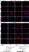
Protein level of the transactivation markers in monolayer cells. Immunofluorescence detection of Collagen I, TLR5, Vimentin and SNAIL in LX-2 (A) and HPSC2.2 (B) cells treated for 48 hours with 2.5 ng/mL of rhTGF-β and siRNA for TLR5. Western blot detection of the protein level of the transactivation markers smooth muscle actin, Collagen I, TLR5 and SNAIL in LC-2 (C) and HPSC2.2 (D) cells treated for 48 hours with 2.5 ng/mL of rhTGF-βand siRNA for TLR5. Β-actin was detected as equal loading control. rh, recombinant human; siRNA, short-interference RNA; TGF-β, transforming growth factor-beta; TLR, Toll-like receptor.
Figure 3
Figure 3
Detection of the transactivation markers in LX-2 and HPSC2.2 spheroids. Immunofluorescence signal (A) and transcript level (B) of the transactivation markers in LX-2 cells treated for 48 hours with 2.5 ng/mL of rhTGF-β and siRNA for TLR5. Immunofluorescence signal (C) and transcript level (D) of the transactivation markers in HPSC2.2 cells treated for 48 hours with 2.5 ng/mL of rhTGF-β. The expression of the transcripts was normalised to untreated cells. GAPDH transcript was detected as a housekeeping gene. Shown are mean triplicate values (B, D) ± SEM. *p<0.05 regarded as significant for untreated/NTC versus rhTGF-β/TLR5 kd cells. AllStar/NTC, negative transfection control, kd, knockdown; rh, recombinant human; siRNA, short-interference RNA; TGF-β, transforming growth factor-beta; TLR, Toll-like receptor
Figure 4
Figure 4
Implication of flagellin in the modulation of TLR5, COL1A1 and ACTA2 in monolayer and spheroids. Detection of the transcript level of TLR5, COL1A1 and ACTA2 in LX-2 and HPSC2.2 monolayer (A, C) and spheroids (B, D) treated for 48 hours with solo and combined administration of 1000 ng/mL of flagellin, 2.5 ng/mL of rhTGF-β, 1 ng/ml of TLR5 antagonist and 1 µmol/L of wortmannin. The expression of the transcripts was normalised to untreated cells. GAPDH transcript was detected as a housekeeping gene. Shown are mean triplicate values±SEM. *p<0.05 regarded as significant for untreated versus treated LX-2/HPSC2.2 cells. TGF-β, transforming growth factor-beta; TLR, Toll-like receptor
Figure 5
Figure 5
Implication of Akt in the transactivation of the hepatic and pancreatic stellate cells. Detection of the protein level of Collagen I, smooth muscle actin, TLR5, Akt and P-Akt in LX-2 and HPSC2.2 cells treated for 48 hours with single and combined 1000 ng/mL of flagellin, 2.5 ng/mL of rhTGF-β and 1 µmol/L of wortmannin. Β-actin was detected as equal loading control. Graphs represent the densitometry analysis of the protein bands. Shown are mean triplicate values±SEM. *p<0.05 regarded as significant for untreated versus treated LX-2/HPSC2.2 cells. rh, recombinant human; TGF-β, transforming growth factor-beta; TLR, Toll-like receptor
Figure 6
Figure 6
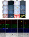
Detection of fibrosis and transactivation markers in murine fibrotic liver tissue. Detection of the three different collagen fibres in thioacetamide/ethanol induced liver fibrosis (A). Picrosirius stained collagen fibres type I are evidenced by the red colour (normal light) and yellow-orange (polarised light). The yellow (normal light) green colour (polarised light) evidence the deposit of muscle/vascular fibers of collagen type III. (B) Immunofluorescence detection of the transactivation marker collagen I (red) and tlr5 (green) in mice treated for 6 weeks with thioacetamide/ethanol. Nuclei were stained with Hoechst 33342 (blue).
Figure 7
Figure 7
Detection of collagen I and tlr5 in murine fibrotic liver tissue after 12 weeks of treatment. Immunofluorescence detection of the transactivation marker collagen I (red) and tlr5 (green) in mice treated for 12 weeks with thioacetamide/ethanol. Nuclei were stained with Hoechst 33342 (blue).
Figure 8
Figure 8
Detection of collagen I and tlr5 in murine fibrotic liver tissue after 18 weeks of treatment. Immunofluorescence detection (left panels) of the transactivation marker collagen I (red) and tlr5 (green) in mice treated for 12 weeks with thioacetamide/ethanol. Nuclei were stained with Hoechst 33342 (blue). Box and whiskers plots of the transcript expression of col1a1, tlr5 and acta2. Shown are means of six mice values±SEM.

Similar articles

Cited by

References

    1. Bonnans C, Chou J, Werb Z. Remodelling the extracellular matrix in development and disease. Nat Rev Mol Cell Biol 2014;15:786–801. 10.1038/nrm3904 - DOI - PMC - PubMed
    1. Eming SA, Wynn TA, Martin P. Inflammation and metabolism in tissue repair and regeneration. Science 2017;356:1026–30. 10.1126/science.aam7928 - DOI - PubMed
    1. Hyun J, Han J, Lee C, et al. . Pathophysiological aspects of alcohol metabolism in the liver. Int J Mol Sci 2021;22:5717. 10.3390/ijms22115717 - DOI - PMC - PubMed
    1. Watari N, Hotta Y, Mabuchi Y. Morphological studies on a vitamin A-storing cell and its complex with macrophage observed in mouse pancreatic tissues following excess vitamin a administration. Okajimas Folia Anat Jpn 1982;58:837–58. 10.2535/ofaj1936.58.4-6_837 - DOI - PubMed
    1. Hernandez-Gea V, Friedman SL. Pathogenesis of liver fibrosis. Annu Rev Pathol 2011;6:425–56. 10.1146/annurev-pathol-011110-130246 - DOI - PubMed

LinkOut - more resources