Review
. 2023 Jul 12;8(1):267.
doi: 10.1038/s41392-023-01486-5.

Role of neuroinflammation in neurodegeneration development

Affiliations
Review

Role of neuroinflammation in neurodegeneration development

Weifeng Zhang et al. Signal Transduct Target Ther. .

Abstract

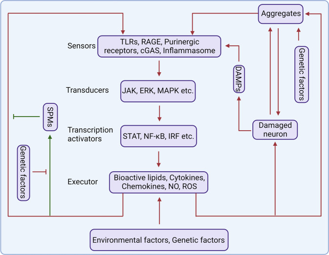
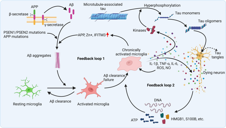
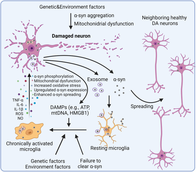
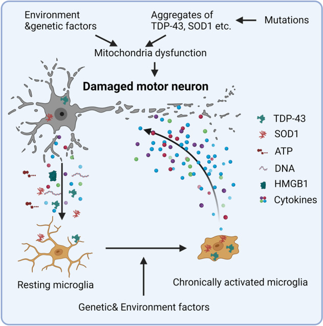
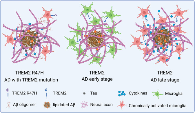
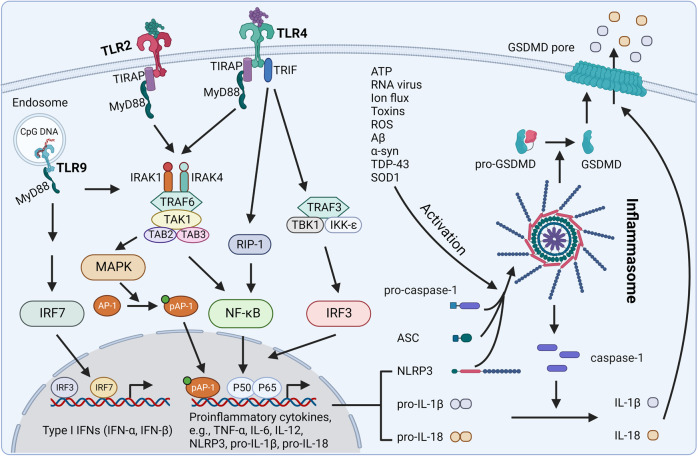
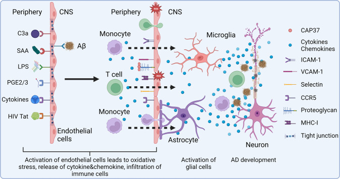
Studies in neurodegenerative diseases, including Alzheimer's disease, Parkinson's disease and Amyotrophic lateral sclerosis, Huntington's disease, and so on, have suggested that inflammation is not only a result of neurodegeneration but also a crucial player in this process. Protein aggregates which are very common pathological phenomenon in neurodegeneration can induce neuroinflammation which further aggravates protein aggregation and neurodegeneration. Actually, inflammation even happens earlier than protein aggregation. Neuroinflammation induced by genetic variations in CNS cells or by peripheral immune cells may induce protein deposition in some susceptible population. Numerous signaling pathways and a range of CNS cells have been suggested to be involved in the pathogenesis of neurodegeneration, although they are still far from being completely understood. Due to the limited success of traditional treatment methods, blocking or enhancing inflammatory signaling pathways involved in neurodegeneration are considered to be promising strategies for the therapy of neurodegenerative diseases, and many of them have got exciting results in animal models or clinical trials. Some of them, although very few, have been approved by FDA for clinical usage. Here we comprehensively review the factors affecting neuroinflammation and the major inflammatory signaling pathways involved in the pathogenicity of neurodegenerative diseases, including Alzheimer's disease, Parkinson's disease, and Amyotrophic lateral sclerosis. We also summarize the current strategies, both in animal models and in the clinic, for the treatment of neurodegenerative diseases.


Conflict of interest statement

The authors declare no competing interests.

Figures

Fig. 1
Fig. 1
The role of inflammation in neurodegeneration. Inflammatory receptors on the surface of immune cells, especially glial cells, act as sensors to detect abnormality in the human body. Stimulation of the sensors by DAMPs or PAMPs, such as protein aggregates, virus, bacteria, leads to activation of signal transducers which then activate transcription activators. Subsequently, activated transcription factors induce the secretion of inflammatory mediators which further amplify inflammation. Generally, activated glial cells should kill the dangers and then induce an inflammation resolution process to clear the DAMPs or PAMPs and stop inflammatory response. However, owing to some reasons, activated immune cells fail to resolve inflammation and generate chronic inflammation which cause neuronal toxicity and enhance protein aggregation. Protein aggregates and DAMPs released from damaged neurons further amplify neuroinflammation and aggravate disease. Some protein aggregates, such as TDP-43 and α-synuclein, may even invade mitochondria which can induce death of neuron directly
Fig. 2
Fig. 2
Inflammation in AD. In the earliest stages of AD, the formation of Aβ occurs due to abnormal cleavage of APP by β- and γ-secretases. Aβ monomers are intrinsically disordered and have a propensity to oligomerize and aggregate into Aβ plaques, which can be promoted by genetic mutations in APP or PSEN1/PSEN2 genes. The Aβ aggregates activates microglia, causing them to clear Aβ via phagocytosis and proteolysis. Yet when that clearance fails, microglia become chronically activated, which further enhances the aggregation of Aβ by improving the expression of APP and IFITM3 and increasing the release of irons, such as Zn+. This process forms a positive feedback loop, which leads to persistent, chronic inflammation. Chronically activated microglia also release proinflammatory cytokines and toxic products, including ROS and nitric oxide (NO), which amplify the immune response and lead to neurotoxicity. DAMPs released from dying neurons, including ATP, HMGB1, S100B, DNA, etc., also amplify inflammation and lead to the second positive feedback loop. Inflammatory cytokines activate kinases, leading to hyperphosphorylation of tau, the dissociation of tau monomers from microtubules, and the subsequent formation of tau tangles in the cytosol of neurons. Thus, inflammation acts as a link between the aggregation of Aβ and the accumulation of tau tangles
Fig. 3
Fig. 3
Inflammation in PD. Lewy bodies, mainly composed of α-synuclein aggregates, are one of the pathological features of PD. It is generally accepted that some genetic and environment factors lead to the aggregation of α-synuclein. Excessive α-synuclein in neuron is transported into the mitochondria, leading to the mitochondrial dysfunction that is central to the progression of PD. Mutations in mitochondrial-associated proteins like LRRK2, PINK1, PARK7, and PRKN, which are found in familial cases of PD, also induce mitochondrial dysfunction and lead to neurotoxicity. Excessive aggregation of α-synuclein or failure to clear it from the cell will result in its release either directly from the neuron or through the exosome, which activates microglia and amplifies neurotoxicity by spreading α-synuclein to the neighboring healthy DA neurons. DAMPs released from dying neurons further enhance the activation of microglia. In addition, genetic and environment factors also promote the activation of microglia and other immune cells. Activated microglia further exacerbate disease by enhancing α-synuclein pathogenicity, increasing oxidative stress, and promoting mitochondrial dysfunction
Fig. 4
Fig. 4
Inflammation in ALS. TDP-43 or SOD1 forms aggregates in the cytoplasm due to genetic and/or environmental factors, causing deleterious effects to neuron. SOD1 aggregation induces oxidative stress, while TDP-43 aggregates invade mitochondria and release mtDNA into the cytoplasm, inducing inflammation through the cGAS-STING pathway. Some mitochondria related genetic variants directly lead to dysfunction of mitochondria and oxidative stress in motor neurons. Proinflammatory cytokines and DAMPs released from damaged motor neurons activate microglia and other immune cells, leading to a persistent inflammatory attack on the motor neurons. Genetic and environment factors also promote the activation of microglia and other immune cells directly, benefiting the development of ALS
Fig. 5
Fig. 5
Function of TREM2 in AD. TREM2 expression enhances the proliferation and migration of microglia, and improves the phagocytosis ability. At the early stage of AD development, TREM2 expression microglia surround the Aβ plaque, interact with lipidated Aβ to efficiently clear them. The surrounding microglia also prevent the spread of Aβ. But at the late stage of AD, TREM2 expression microglia fail to clear the aggregates, leading to chronic inflammation, which then attenuates the phagocytosis ability of microglia, induces tau phosphorylation and aggregation. For individuals with TREM2 mutation, the affinity of TREM2 was weakened, which inhibits the proliferation and migration of microglia, leading to the failure of microglia surrounding Aβ aggregates. TREM2 mutation also enhance the secretion of cytokines from microglia. As a result, TREM2 mutation in microglia benefits the spread of Aβ, and the phosphorylation and aggregation of tau, exacerbating AD pathology
Fig. 6
Fig. 6
Crosstalk between TLRs and the inflammasome pathway. TLR2, TLR4, and TLR9 are the most involved TLR receptors in neurodegenerative diseases. The binding of aggregates (e.g., Aβ, α-synuclein) or other PAMPs or DAMPs activates TLRs. Activation of TLR2 induces the MyD88-dependent pathway, which activates MAPK and NF-κB, leading to the release of proinflammatory cytokines. In addition to the MyD88-dependent pathway, TLR4 activation also transduces signal through an MyD88-independent pathway, which leads to the activation of the IRF3 transcription factor and the subsequent release of type I IFNs. TLR9 is located on internal vesicles and binds to bacterial and viral nucleic acids or endogenous CpG DNA. Activation of TLR9 induces MyD88-dependent signaling, translocation of IRF7 into the nucleus, and the release of type I IFNs. TLR activation also induces the expression of NLRP3, pro-IL-1β, and pro-IL-18, acting as the first signal (or priming signal) for the NLRP3 inflammasome pathway. A variety of stimuli, including ATP, RNA virus, and aggregates, act as the second signal (or activation signal) activating NLRP3 and inducing the assembly of NLRP3, ASC, and pro-caspase-1 into an inflammasome. This process activates caspase-1, which in turn cleaves pro-IL-1β and pro-IL-18 into IL-1β and IL-18, respectively. NLRP3 inflammasome activation also induces the maturation of GSDMD, which translocates to the plasma membrane and forms a pore through which the cleaved IL-1β and IL-18 molecules can be released into the extracellular space. In addition, GSDMD can induce an inflammatory form of cell death termed pyroptosis. The cleaved cytokines have both autocrine and paracrine effects that further amplify the inflammatory response
Fig. 7
Fig. 7
Involvement of endothelial cells in AD. Endogenous stimuli (e.g., Aβ aggregates) or exogenous stimuli (e.g., C3a, SAA, LPS, prostaglandins, cytokines, virus proteins) can bind to its receptors in endothelial cells and induce the activation of endothelial cells. Activated endotheliocyte secrets a low level of inflammatory cytokines and chemokines, increases the expression of adhesion molecules and reduces the expression of tight junction proteins. Activation of endothelial cells also increases the expression of iNOS and NOX, which lead to oxidative stress, amplifying inflammation and damaging BBB. Secretion of cytokines and chemokine recruits peripheral immune cells, activates microglia and astrocytes in the CNS. The impaired permeability of endothelial cells and increased expression of adhesion molecules facilitate the infiltration of peripheral immune cells into the CNS, further augmenting inflammation in the CNS. As a result, stimulation on endothelial cells leads to strong inflammation in the CNS, ultimately induces Aβ and tau aggregation, and neurodegeneration

Similar articles

Cited by

References

    1. Mayne K, White JA, McMurran CE, Rivera FJ, de la Fuente AG. Aging and neurodegenerative disease: is the adaptive immune system a friend or foe? Front. Aging Neurosci. 2020;12:572090. doi: 10.3389/fnagi.2020.572090. - DOI - PMC - PubMed
    1. Parkinson J. An essay on the shaking palsy. 1817. J. Neuropsychiatry Clin. Neurosci. 2002;14:223–236. doi: 10.1176/jnp.14.2.223. - DOI - PubMed
    1. Goetz CG. The history of Parkinson’s disease: early clinical descriptions and neurological therapies. Cold Spring Harb. Perspect. Med. 2011;1:a008862. doi: 10.1101/cshperspect.a008862. - DOI - PMC - PubMed
    1. Rogers J, Luber-Narod J, Styren SD, Civin WH. Expression of immune system-associated antigens by cells of the human central nervous system: relationship to the pathology of Alzheimer’s disease. Neurobiol. Aging. 1988;9:339–349. doi: 10.1016/S0197-4580(88)80079-4. - DOI - PubMed
    1. Duffy PE, Rapport M, Graf L. Glial fibrillary acidic protein and Alzheimer-type senile dementia. Neurology. 1980;30:778–782. doi: 10.1212/WNL.30.7.778. - DOI - PubMed

Publication types

Substances