. 2023 Oct 20;136(20):2484-2495.
doi: 10.1097/CM9.0000000000002763. Epub 2023 Jul 12.

Effect of down-regulation of let-7c/g on triggering a double-negative feedback loop and promoting restenosis

Affiliations

Effect of down-regulation of let-7c/g on triggering a double-negative feedback loop and promoting restenosis

Qian Zhang et al. Chin Med J (Engl). .

Abstract

Background: Excessive proliferation and migration of vascular smooth muscle cells (VSMCs) are the main causes of restenosis (RS) in diabetic lower extremity arterial disease (LEAD). However, the relevant pathogenic mechanisms are poorly understood.

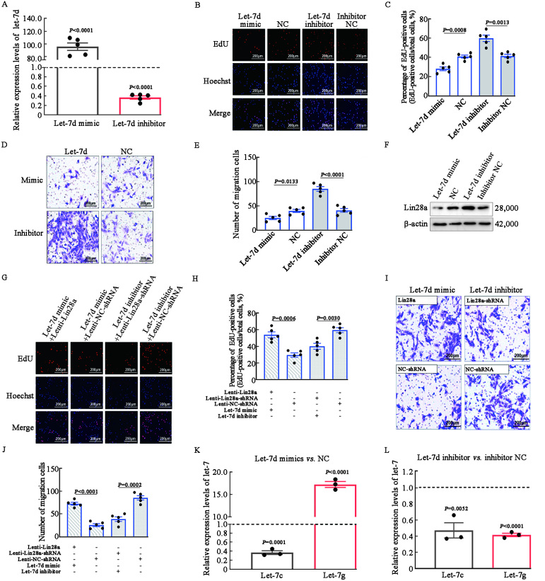
Methods: In this study, we introduced a "two-step injury protocol" rat RS model, which started with the induction of atherosclerosis (AS) and was followed by percutaneous transluminal angioplasty (PTA). Hematoxylin-eosin (HE) staining and immunohistochemistry staining were used to verify the form of RS. Two-step transfection was performed, with the first transfection of Lin28a followed by a second transfection of let-7c and let-7g, to explore the possible mechanism by which Lin28a exerted effects. 5-ethynyl-2΄-deoxyuridine (EdU) and Transwell assay were performed to evaluate the ability of proliferation and migration of VSMCs. Western blotting and quantitative real-time polymerase chain reaction (qRT-PCR) were performed to detect the expression of Lin28a protein and let-7 family members.

Results: Using a combination of in vitro and in vivo experiments, we discovered that let-7c, let-7g, and microRNA98 (miR98) were downstream targets of Lin28a. More importantly, decreased expression of let-7c/let-7g increased Lin28a, leading to further inhibition of let-7c/let-7g. We also found an increased level of let-7d in the RS pathological condition, suggesting that it may function as a protective regulator of the Lin28a/let-7 loop by inhibiting the proliferation and migration of VSMCs.

Conclusion: These findings indicated the presence of a double-negative feedback loop consisting of Lin28a and let-7c/let-7g, which may be responsible for the vicious behavior of VSMCs in RS.


Conflict of interest statement

None.

Figures

Figure 1
Figure 1
Expression of Lin28a and the let-7 family in VSMCs in RS. (A) Expression and colocalization of Lin28a (red) and α-SMA (green) in human post-PTA RS and AS samples. α-SMA is the specific marker of VSMCs. The immunofluorescence image was obtained at magnification ×40 and the scale bar represents 500 µm; a magnification ×400 image is shown in the inset on the bottom right corner of the merged image; the scale bar represents 50 µm. (B) Quantification of Lin28a+ α-SMA+ cells in the neointima area. (C) The expression levels of let-7 miRNAs in human AS and RS (n = 5). P <0.05 vs. AS. (D) The immunohistochemical staining showed the expression of α-SMA in AS and RS plaques in rat models. The image was obtained at magnification ×100 and the scale bar represents 200 µm. (E) Quantification of positive staining for α-SMA in the AS and RS groups. (F) Expression of let-7 miRNAs in rats with AS and PTA-related RS. P <0.05 vs. AS. (G) Western blotting was performed to examine Lin28a expression after transfection with up- or down-regulating Lenti-virus in cultured primary VSMCs. (H) Statistical analysis of Western blotting results displayed by the bar graph. P <0.05 vs. Lenti-NC-shRNA group. qRT-PCR was performed to examine let-7 miRNAs after transfection with up-regulation (I) or down-regulating (J) Lin28a Lenti-virus in VSMCs. The results showed that let-7c, let-7g, and miR98 were negatively regulated by Lin28a. P <0.05 vs. Lenti-NC-shRNA group. α-SMA: α-smooth muscle actin; AS: Atherosclerosis; DAPI: 4′,6-diamidino-2-phenylindole; Lenti-NC-shRNA: Normal control short-hairpin RNA lentivirus; miRNA: microRNA; miR98: microRNA98; PTA: Percutaneous transluminal angioplasty; qRT-PCR: Quantitative real-time polymerase chain reaction; RS: Restenosis; shRNA: Short-hairpin RNA; VSMCs: Vascular smooth muscle cells.
Figure 2
Figure 2
Lin28a interacted with let-7c, let-7g, and miR98, and further regulates the proliferation and migration of VSMCs. (A–E) Let-7c, let-7g, miR98, NC and inhibitor NC oligos were transfected into VSMCs. (A) qRT-PCR validation of successful let-7c, let-7g or miR98 transfections. P <0.05 (let-7c, let-7g, miR98 mimic) vs. NC; P <0.05 (let-7c, let-7g, miR98 inhibitor) vs. inhibitor NC. (B) The EdU assay illustrated the proliferation of VSMCs. VSMCs were labeled with nucleoside analog EdU (red) and nuclei were labeled with Hoechst 33342 dye (blue). The colocalization of EdU (red) and Hoechst 33342 (blue) was shown as pink color, indicative of the proliferative VSMCs. (C) The quantification of the EdU-positive cells. (D) The Transwell assay illustrates the migration of VSMCs. The migrated VSMCs are presented in purple. (E) The quantification of the migration of VSMCs. (F–I) Let-7c, let-7g, miR98, NC and inhibitor NC oligo were transfected into the Lin28a Lenti-virus transfected VSMCs. (F,G) The EdU assay illustrated the proliferation of VSMCs. (H,I) The Transwell assay illustrated the migration of VSMCs. The images of EdU and Transwell assays were taken at magnification ×200 and the scale bar was 75 µm (n = 5). EdU: 5-ethynyl-2΄-deoxyuridine; miR98: microRNA98; NC: Normal control; qRT-PCR: Quantitative real-time polymerase chain reaction; shRNA: Short-hairpin RNA; VSMCs: Vascular smooth muscle cells.
Figure 3
Figure 3
Lin28a and let-7c and let-7g interacted in a double-negative feedback loop. (A–C) Let-7c, let-7d, let-7g, miR98, and NC mimic and inhibitor oligos were transfected into VSMCs. (A) qRT-PCR analysis to validate Lin28a expression in mRNA level. P <0.05 (let-7c, let-7d, let-7g, miR98 mimic) vs. NC; P <0.05 (let-7c, let-7d, let-7g, miR98 inhibitor) vs. inhibitor NC. (B) Western blotting validation of Lin28a expression in protein level. (C) Quantification of the Lin28a protein levels by Western blotting. P <0.05 (let-7c, let-7d and let-7g mimic) vs. NC; P <0.05 (let-7c, let-7d, and let-7g inhibitor) vs. inhibitor NC. (D–G) Let-7c, let-7g individually or a mixture of let-7c and let-7g mimic or inhibitor oligos were transfected into the Lenti-Lin28a or Lenti-Lin28a-shRNA VSMCs. (D) The EdU assay illustrated the proliferation of VSMCs. (E) Quantification of the EdU-positive cells. (F) The Transwell assay illustrated the migration of VSMCs. (G) Quantification of the migration of VSMCs. Images the of EdU and Transwell assays were obtained at magnification ×200 and the scale bar was 75 µm (n = 5). EdU: 5-ethynyl-2΄-deoxyuridine; miR98: microRNA98; mRNA: Messenger RNA; NC: Normal control; qRT-PCR: Quantitative real-time polymerase chain reaction; shRNA: Short-hairpin RNA; VSMCs: Vascular smooth muscle cells.
Figure 4
Figure 4
Let-7d restricted the proliferation and migration of VSMCs by downregulating Lin28a and upregulating let-7g. (A) Let-7d mimic and inhibitor oligo were transfected into VSMCs and their expression levels were validated by qRT-PCR. P <0.05 (let-7d mimic) vs. NC; P <0.05 (let-7d inhibitor) vs. inhibitor NC. (B) The EdU assay illustrated the proliferation of VSMCs. (C) Quantification of EdU-positive cells. (D) The Transwell assay illustrated the migration of VSMCs. (E) Quantification of the migration of VSMCs. (F) Western blotting validation of Lin28a expression in protein level. (G–J) Let-7d, NC, and NC inhibitor oligos were transfected into the Lin28a lentivirus-transfected VSMCs. (G,H) The EdU assay illustrated the proliferation of VSMCs. (I,J) The Transwell assay illustrated the migration of VSMCs. (K,L) qRT-PCR was performed to examine let-7c, let-7g levels with up- or down-regulated let-7d in VSMCs. Results showed that let-7g expression was promoted by let-7d. P <0.05 (let-7d mimic) vs. NC; P <0.05 (let-7d inhibitor) vs. inhibitor NC. The images of EdU and Transwell assays were taken at magnification ×200 and the scale bar was 75 µm (n = 5). EdU: 5-ethynyl-2΄-deoxyuridine; mRNA: Messenger RNA; NC: Normal control; qRT-PCR: Quantitative real-time polymerase chain reaction; shRNA: Short-hairpin RNA; VSMCs: Vascular smooth muscle cells.
Figure 5
Figure 5
Let-7c and let-7g attenuated RS development by downregulating Lin28a in vivo. (A,B) qRT-PCR was performed to examine let-7c and let-7g expression after transfection with upregulation of AAV9. P <0.05 (AAV9-let-7c/g RS) vs. AAV9-EV RS. (C) qRT-PCR was performed to examine Lin28a expression after transfection with upregulating let-7c or let-7g AAV9 in RS rats. The results showed that Lin28a was negatively regulated by let-7c and let-7g. Data are presented as fold changes relative to the AAV9-EV RS group. P <0.05 (AAV9-let-7c/g RS) vs. AAV9-EV RS. (D) Representative HE staining of iliac arteries. The image of hematoxylin-eosin (HE) staining was taken at magnification ×100 and the scale bar represents 200 µm. (E) Quantitative analysis of lesion areas in arteries. (F) Quantitative analysis of neointima/media ratio. (G) The immumohistochemical staining was performed to examine the expression of α-SMA in different RS plaques. The image was taken at magnification ×100 and the scale bar represents 200 µm, and taken at magnification ×40 and the scale bar represents 50 µm. α-SMA: α-smooth muscle actin; AAV: Adeno-associated virus; AS: Atherosclerosis; EV: Empty vector; qRT-PCR: Quantitative real-time polymerase chain reaction; RS: Restenosis.

Similar articles

References

    1. National Clinical Guideline Centre (UK) . Lower limb peripheral arterial disease: Diagnosis and management. London: National Institute for Health and Clinical Excellence: Guidance, 2012.
    1. Low Wang CC Blomster JI Heizer G Berger JS Baumgartner I Fowkes FGR, et al. . Cardiovascular and limb outcomes in patients with diabetes and peripheral artery disease: The EUCLID trial. J Am Coll Cardiol 2018;72: 3274–3284. doi: 10.1016/j.jacc.2018.09.078. - PubMed
    1. Matsuzaki K, Hayashi R, Okabe K, Aramaki-Hattori N, Kishi K. Prognosis of critical limb ischemia: Major vs. minor amputation comparison. Wound Repair Regen 2015;23: 759–764. doi: 10.1111/wrr.12329. - PubMed
    1. Cardaioli P Rigatelli G Dell'avvocata F Giordan M Lisato G Mollo F, et al. . Endovascular treatment of diabetic foot syndrome: Results from a single center prospective registry using mixed coronary and peripheral techniques and equipment. J Interv Cardiol 2011; 24: 562–568. doi: 10.1111/j.1540-8183.2011.00676.x. - PubMed
    1. Tepe G Zeller T Albrecht T Heller S Schwarzwälder U Beregi JP, et al. . Local delivery of paclitaxel to inhibit restenosis during angioplasty of the leg. N Engl J Med 2008;358: 689–699. doi: 10.1056/NEJMoa0706356. - PubMed