. 2023 Aug;28(8):3397-3413.
doi: 10.1038/s41380-023-02130-y. Epub 2023 Jul 11.

Fetal cannabidiol (CBD) exposure alters thermal pain sensitivity, problem-solving, and prefrontal cortex excitability

Affiliations

Fetal cannabidiol (CBD) exposure alters thermal pain sensitivity, problem-solving, and prefrontal cortex excitability

Karli S Swenson et al. Mol Psychiatry. 2023 Aug.

Abstract

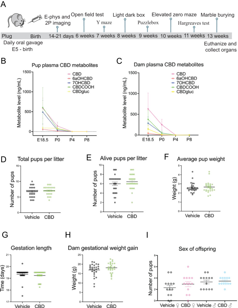
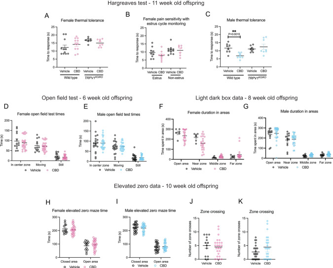
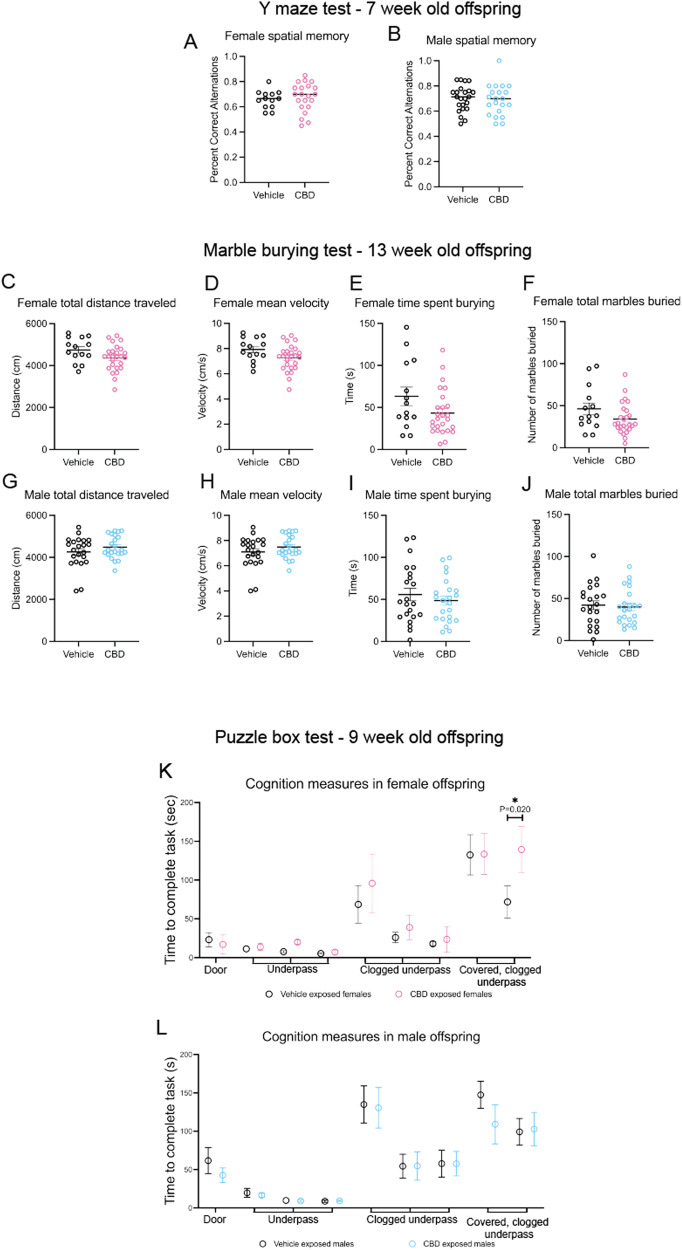
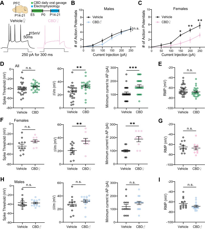
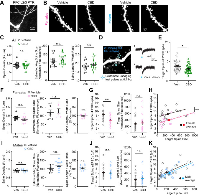
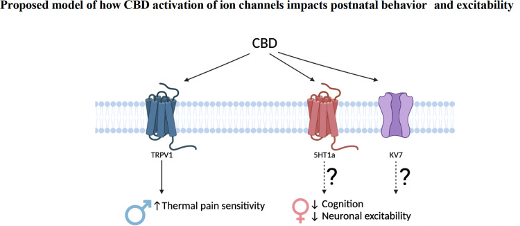
Thousands of people suffer from nausea with pregnancy each year. Nausea can be alleviated with cannabidiol (CBD), a primary component of cannabis that is widely available. However, it is unknown how fetal CBD exposure affects embryonic development and postnatal outcomes. CBD binds and activates receptors that are expressed in the fetal brain and are important for brain development, including serotonin receptors (5HT1A), voltage-gated potassium (Kv)7 receptors, and the transient potential vanilloid 1 receptor (TRPV1). Excessive activation of each of these receptors can disrupt neurodevelopment. Here, we test the hypothesis that fetal CBD exposure in mice alters offspring neurodevelopment and postnatal behavior. We administered 50 mg/kg CBD in sunflower oil or sunflower oil alone to pregnant mice from embryonic day 5 through birth. We show that fetal CBD exposure sensitizes adult male offspring to thermal pain through TRPV1. We show that fetal CBD exposure decreases problem-solving behaviors in female CBD-exposed offspring. We demonstrate that fetal CBD exposure increases the minimum current required to elicit action potentials and decreases the number of action potentials in female offspring layer 2/3 prefrontal cortex (PFC) pyramidal neurons. Fetal CBD exposure reduces the amplitude of glutamate uncaging-evoked excitatory post-synaptic currents, consistent with CBD-exposed female problem-solving behavior deficits. Combined, these data show that fetal CBD exposure disrupts neurodevelopment and postnatal behavior in a sex specific manner.


Conflict of interest statement

The authors declare no competing interests.

Figures

Fig. 1
Fig. 1. Dosing schematic, validation of CBD metabolites and litter factors.
A timeline shows CBD administration and age of offspring when tests were performed (A). A graph shows CBD and CBD metabolites in the dam blood plasma from E18.5, P0, P4, and P8 (B) and pooled pup litter plasma from each group (C) from E18.5, P0, P4, and P8. Graphs show gestational CBD consumption does not alter total pups per litter (D), alive pups per litter (E), average pup weight (F), gestation length (G), gestation weight gain (H), or sex of offspring (I) from 27 vehicle administered dams and 26 CBD administered dams. Error bars represent S.E.M. No measures were significantly different based on treatment (p > 0.1 by t-test). Mean values, SEM, and p values by t-test are reported in Tables 1 and 2.
Fig. 2
Fig. 2. Fetal CBD exposure increases thermal sensitivity in male mice, but not female mice.
Fetal CBD exposure does not affect latency to response to thermal stimulus in the Hargreaves test in wildtype or TRPV1KO/KO female mice (A), and over the estrus cycle (B). Fetal CBD exposure decreases latency to response in wild-type CBD-exposed male mice (11.58 ± 0.64 s for vehicle-exposed vs 6.87 ± 3.27 s, P = 4.993E−8, t-test), but does not affect latency response in TRPV1KO/KO mice (11.089 ± 0.649 s vehicle-exposed, vs. 12.429 ± 1.610 s CBD-exposed P = 0.453, t-test) (C). Graphs show fetal CBD exposure does not affect time in the center zone, time moving, or time still in the Open field test in 6-week-old female (D), or male offspring (E). Graphs show fetal CBD exposure does not affect time in open area, near zone or far zone in the Light Dark Box in 8-week-old female (F) or male (G) offspring. The elevated zero maze shows that fetal CBD exposure does not affect time in closed or open areas in 10-week-old female (H), and male (I) offspring. Fetal CBD exposure did not affect zone crossings in female (J), nor male offspring (K) in the elevated zero maze. Error bars represent the S.E.M. All mean values of measurements, S.E.M., and p values from t-tests are reported in Table 2.
Fig. 3
Fig. 3. Fetal CBD exposure decreases female offspring problem-solving behaviors.
Fetal CBD exposure does not affect female spatial memory (A) or male spatial memory (B) via the Y maze test. Graphs show fetal CBD exposure does not affect offspring compulsivity, including female total distance traveled (C), mean velocity (D), time spent burying (E) or marbles buried (F), nor male distance traveled (G), mean velocity (H), time spent burying (I) or total marbles buried (J), via the marble burying test. Graphs show fetal CBD exposure decreases female problem-solving at trial 9, (71.75 ± 20.71 s vehicle-exposed females, 139.42 ± 26.91 s CBD-exposed females, N = 12 each, P = 0.201, Wilcoxon rank sum test) (K), but not male problem-solving (L) via the puzzle box test.
Fig. 4
Fig. 4. Fetal CBD exposure decreases excitability of PFC layer 2/3 pyramidal neurons in a sex specific manner.
A Experimental timeline and representative traces of a stimulus train elicited by 250pA current injection for 300 ms. B, C Fetal CBD exposure decreased the intrinsic excitability of layer 2/3 pyramidal neurons in P14 females but not males (vehicle females: n = 6 cells, 1 mouse; vehicle males: n = 12 cells, 2 mice; CBD females: n = 5 cells, 1 mouse; CBD males: n = 14 cells, 2 mice; female treatment effect: P < 0.0001; Sidak’s multiple comparison, *<0.05, **<0.01). D Fetal CBD exposure did not alter spike thresholds of P14-21 mice (left, vehicle: −39.84 ± 2.613 mV, n = 25 cells, 4 mice; CBD: −35.92 ± 1.523 mV, n = 24 cells, 4 mice; Welch’s t-test, P = 0.2018). Fetal CBD exposure significantly increased membrane potential change (middle, vehicle: 24.23 ± 2.251 mV; CBD: 33.21 ± 1.963 mV; two-tailed t-test, P = 0.0043) and minimum currents (right, vehicle: 110 ± 9.574 pA; CBD: 162.5 ± 11.36; Mann–Whitney, P = 0.0007) required to evoke action potentials. E Resting membrane potential of P14-21 mice remained unchanged following fetal CBD exposure (vehicle: −64.07 ± 2.008 mV, n = 25 cells, 4 mice; CBD: −68.06 ± 1.773 mV, n = 25 cells, 4 mice; Mann–Whitney test, P = 0.1261). F The effect of fetal CBD exposure on changes of membrane potential (vehicle: 20.76 ± 2.826 mV, n = 12 cells, 2 mice; CBD: 35.19 ± 3.963, n = 10 cells, 2 mice; two-tailed t-test, P = 0.0066) and minimum current for action potential firing stemmed from females (vehicle: 100 ± 8.704 pA, n = 12 cells, 2 mice; CBD: 188.9 ± 20.03 pA, n = 10 cells, 2 mice; Welch’s t-test, P = 0.0018). G Resting membrane potential was unchanged in females following fetal CBD exposure (vehicle: −65.97 ± 2.966 mV, n = 12 cells, 2 mice; CBD: −67.16 ± 3.506 mV, n = 10 cells, 2 mice; two-tailed t-test, P = 0.6370). H, I Male mice showed no significant differences in spike threshold (vehicle: −35.81 ± 3.321 mV, n = 13 cells, 2 mice; CBD: −36.65 ± 1.725 pA, n = 15 cells, 2 mice; Mann–Whitney test, 0.7856), change in membrane potential (vehicle: 27.43 ± 3.309 mV; CBD: 32.02 ± 2.115 mV; Mann–Whitney test, P = 0.0648),and minimum currents for action potential spikes (vehicle: 119.2 ± 16.54 pA; CBD: 146.7 ± 12.41 pA; two-tailed t-test, P = 0.1893), or resting membrane potential (vehicle: −63.24 ± 2.820 mV; CBD: −68.67 ± 1.910, Mann–Whitney test, P = 0.0977). *P < 0.05, **P < 0.01, ***P < 0.001; error bars represent SEM. n.s. not significant.
Fig. 5
Fig. 5. Fetal CBD exposure decreases synaptic strength of layer 2/3 pyramidal neurons in PFC of female mice.
A, B Two-photon images of a whole-cell PFC layer 2/3 pyramidal neuron and dendritic segments from CBD and vehicle treated female and male mice at P14-22. C Fetal CBD exposure has no effect on spine structure for combined male and female mice (vehicle: n = 70 dendrites, 16 cells, 4 mice; CBD n = 84 dendrites, 19 cells, 4 mice). D A two-photon image from a dendritic segment of PFC layer 2/3 pyramidal neuron and two-photon glutamate uncaging evoked EPSC (uEPSC) traces (average of 5–8 test pulses) recorded by whole-cell voltage-clamp recording (blue crosses indicate glutamate uncaging timepoint). E uEPSC amplitudes are significantly decreased in CBD-exposed offspring (vehicle: 7.49 ± 0.42 pA; CBD: 6.09 ± 0.33 pA; P = 0.0116, two-tailed t-test) (vehicle: n = 41 spines, 14 cells, 3 mice; CBD n = 39 spines, 13 cells, 3 mice). F In female offspring, fetal CBD exposure has no effect on spine density, average spine size, or spine length/width ratio. (vehicle: n = 35 dendrites, 8 cells, 2 mice; CBD n = 36 dendrites, 8 cells, 2 mice). G uEPSCs recorded from similar sizes of target spines are significantly smaller in fetal CBD-exposed female offspring (vehicle: 8.66 ± 0.55 pA; CBD: 5.89 ± 0.51; P = 0.0009, two-tailed t-test) (vehicle: n = 19 spines, 7 cells, 1 mouse; CBD n = 17 spines, 6 cells, 1 mouse). H Scatter plots showing significantly smaller uEPSCs in fetal CBD-exposed mice. I In male offspring, fetal CBD exposure had no effect on spine density, average spine size, or spine length/width ratio. (vehicle: n = 35 dendrites, 8 cells, 2 mice; CBD n = 48 dendrites, 11 cells, 2 mice). J In male offspring, CBD has no effect on uEPSCs (vehicle: n = 22 spines, 7 cells, 2 mice; CBD n = 22 spines, 7 cells, 2 mice). K Scatter plots showing comparable uEPSCs between fetal CBD-exposed and control mice. *P < 0.05, **P < 0.01; ***P < 0.001; error bars represent SEM. n.s. not significant.
Fig. 6
Fig. 6. Model of effects of fetal CBD exposure.
Fetal CBD exposure increases male thermal pain sensitivity, decreases female problem-solving behaviors, and alters female prefrontal cortex pyramidal neurons. CBD activates TRPV1, 5HT1A, and Kv7.2/3 receptors. TRPV1 regulates thermal pain sensitivity. 5HT1a and Kv7.2/3 have documented roles in neuronal excitability and cognitive function.

References

    1. Wibowo N, Purwosunu Y, Sekizawa A, Farina A, Tambunan V, Bardosono S. Vitamin B6 supplementation in pregnant women with nausea and vomiting. Int J Gynaecol Obstet. 2012;116:206–10. doi: 10.1016/j.ijgo.2011.09.030. - DOI - PubMed
    1. Volkow ND, Han B, Compton WM, McCance-Katz EF. Self-reported medical and nonmedical cannabis use among pregnant women in the United States. JAMA. 2019;322:167. doi: 10.1001/jama.2019.7982. - DOI - PMC - PubMed
    1. Metz TD, Silver RM, McMillin GA, Allshouse AA, Jensen TL, Mansfield C, et al. Prenatal marijuana use by self-report and umbilical cord sampling in a state with marijuana legalization. Obstet Gynecol. 2019;133:98–104. doi: 10.1097/AOG.0000000000003028. - DOI - PMC - PubMed
    1. Bidwell CL, YorkWilliams SL, Mueller RL, Bryan AD, Hutchison KE. Exploring cannabis concentrates on the legal market: user profiles, product strength, and health-related outcomes. Addict Behav Rep. 2018;8:102–6. doi: 10.1016/j.abrep.2018.08.004. - DOI - PMC - PubMed
    1. Parker LA, Rock EM, Limebeer CL. Regulation of nausea and vomiting by cannabinoids. Br J Pharmacol. 2011;163:1411–22. doi: 10.1111/j.1476-5381.2010.01176.x. - DOI - PMC - PubMed