

Apoptotic bodies: bioactive treasure left behind by the dying cells with robust diagnostic and therapeutic application potentials
- PMID: 37434199
- PMCID: PMC10337089
- DOI: 10.1186/s12951-023-01969-1
Apoptotic bodies: bioactive treasure left behind by the dying cells with robust diagnostic and therapeutic application potentials
Abstract
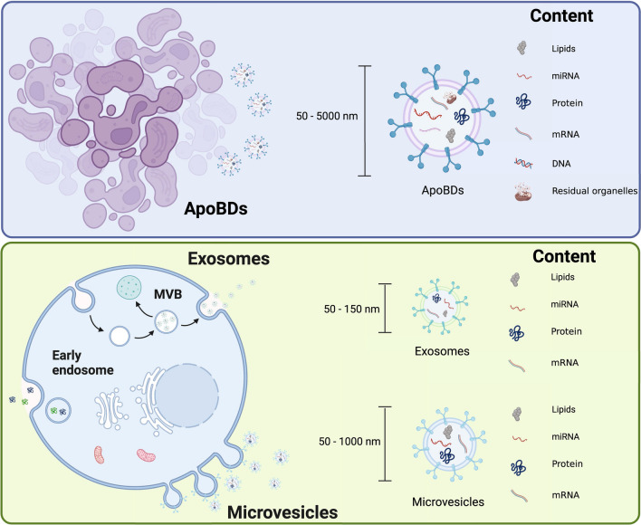
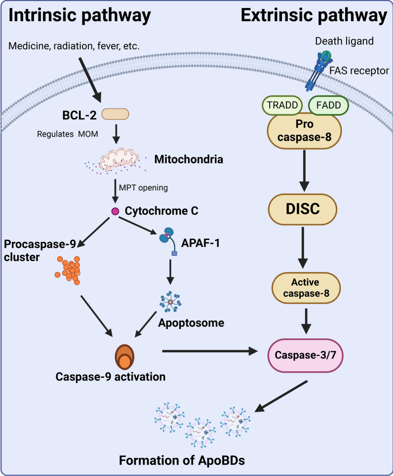
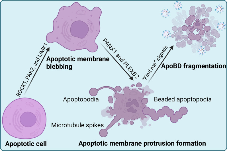
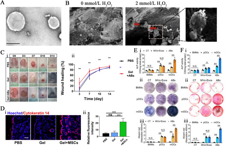
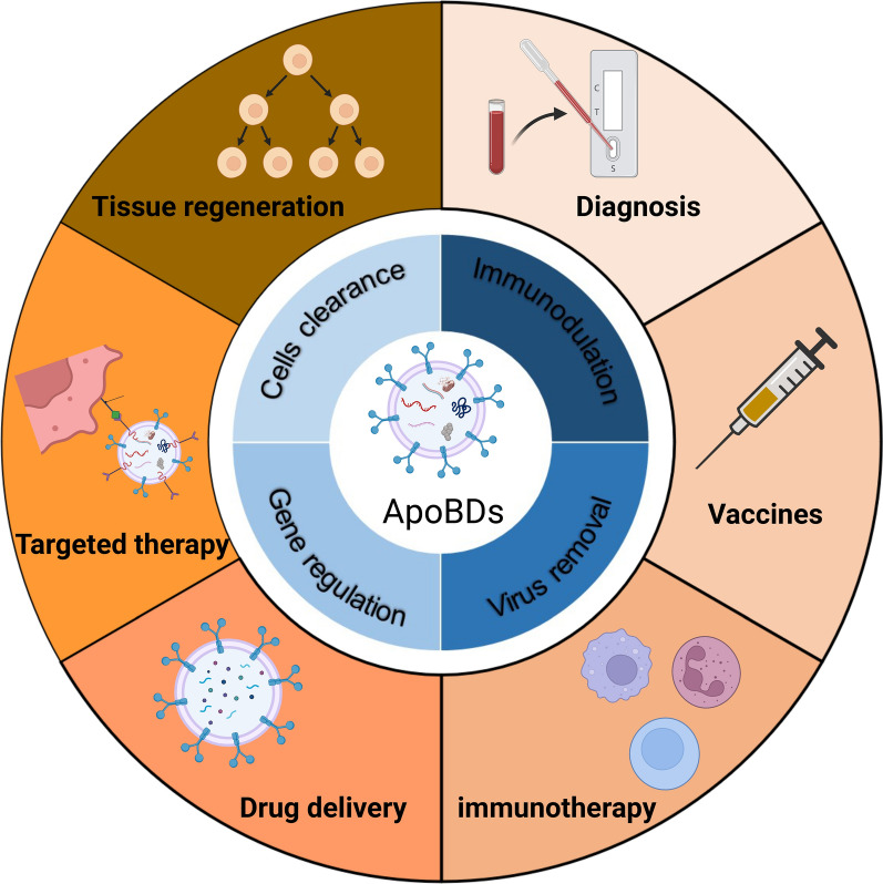
Apoptosis, a form of programmed cell death, is essential for growth and tissue homeostasis. Apoptotic bodies (ApoBDs) are a form of extracellular vesicles (EVs) released by dying cells in the last stage of apoptosis and were previously regarded as debris of dead cells. Recent studies unraveled that ApoBDs are not cell debris but the bioactive treasure left behind by the dying cells with an important role in intercellular communications related to human health and various diseases. Defective clearance of ApoBDs and infected-cells-derived ApoBDs are possible etiology of some diseases. Therefore, it is necessary to explore the function and mechanism of the action of ApoBDs in different physiological and pathological conditions. Recent advances in ApoBDs have elucidated the immunomodulatory, virus removal, vascular protection, tissue regenerative, and disease diagnostic potential of ApoBDs. Moreover, ApoBDs can be used as drug carriers enhancing drug stability, cellular uptake, and targeted therapy efficacy. These reports from the literature indicate that ApoBDs hold promising potential for diagnosis, prognosis, and treatment of various diseases, including cancer, systemic inflammatory diseases, cardiovascular diseases, and tissue regeneration. This review summarizes the recent advances in ApoBDs-related research and discusses the role of ApoBDs in health and diseases as well as the challenges and prospects of ApoBDs-based diagnostic and therapeutic applications.
Keywords: Apoptotic bodies; Diseases; Immunomodulation; Mesenchymal stem cells; Tissue regeneration.
© 2023. The Author(s).
Conflict of interest statement
No potential conflict of interest was reported by the authors.
Figures


Similar articles
-
Advances in the Therapeutic Effects of Apoptotic Bodies on Systemic Diseases.Int J Mol Sci. 2022 Jul 26;23(15):8202. doi: 10.3390/ijms23158202. Int J Mol Sci. 2022. PMID: 35897778 Free PMC article. Review.
-
Comparison of in-vitro immunomodulatory capacity between large and small apoptotic bodies from human bone marrow mesenchymal stromal cells.Int Immunopharmacol. 2025 Apr 24;153:114480. doi: 10.1016/j.intimp.2025.114480. Epub 2025 Mar 18. Int Immunopharmacol. 2025. PMID: 40101418
-
ApoBDs: a paradigm shift from cellular debris to therapeutic vehicles.Front Endocrinol (Lausanne). 2025 Jul 17;16:1626796. doi: 10.3389/fendo.2025.1626796. eCollection 2025. Front Endocrinol (Lausanne). 2025. PMID: 40747304 Free PMC article. Review.
-
Apoptotic Bodies: Mechanism of Formation, Isolation and Functional Relevance.Subcell Biochem. 2021;97:61-88. doi: 10.1007/978-3-030-67171-6_4. Subcell Biochem. 2021. PMID: 33779914
-
Determining the contents and cell origins of apoptotic bodies by flow cytometry.Sci Rep. 2017 Oct 31;7(1):14444. doi: 10.1038/s41598-017-14305-z. Sci Rep. 2017. PMID: 29089562 Free PMC article.
Cited by
-
Extracellular Vesicles: Advanced Tools for Disease Diagnosis, Monitoring, and Therapies.Int J Mol Sci. 2024 Dec 29;26(1):189. doi: 10.3390/ijms26010189. Int J Mol Sci. 2024. PMID: 39796048 Free PMC article. Review.
-
Extracellular Vesicles in the Mesenchymal Stem Cell/Macrophage Axis: Potential Targets for Inflammatory Treatment.Int J Mol Sci. 2025 Mar 20;26(6):2827. doi: 10.3390/ijms26062827. Int J Mol Sci. 2025. PMID: 40141469 Free PMC article. Review.
-
Biogenesis and delivery of extracellular vesicles: harnessing the power of EVs for diagnostics and therapeutics.Front Mol Biosci. 2024 Jan 3;10:1330400. doi: 10.3389/fmolb.2023.1330400. eCollection 2023. Front Mol Biosci. 2024. PMID: 38234582 Free PMC article. Review.
-
Circulating extracellular vesicles in Systemic Lupus Erythematosus: physicochemical properties and phenotype.Lupus Sci Med. 2024 Aug 17;11(2):e001243. doi: 10.1136/lupus-2024-001243. Lupus Sci Med. 2024. PMID: 39153822 Free PMC article.
-
Extracellular particles: emerging insights into central nervous system diseases.J Nanobiotechnology. 2025 Apr 1;23(1):263. doi: 10.1186/s12951-025-03354-6. J Nanobiotechnology. 2025. PMID: 40170148 Free PMC article. Review.
References
-
- Liu D, Kou X, Chen C, Liu S, Liu Y, Yu W, Yu T, Yang R, Wang R, Zhou Y, Shi S. Circulating apoptotic bodies maintain mesenchymal stem cell homeostasis and ameliorate osteopenia via transferring multiple cellular factors. Cell Res. 2018;28(9):918–933. doi: 10.1038/s41422-018-0070-2. - DOI - PMC - PubMed
Publication types
MeSH terms
Grants and funding
LinkOut - more resources
Full Text Sources
