Review
. 2023 Jul 11;18(1):46.
doi: 10.1186/s13024-023-00640-5.

Pathophysiology and probable etiology of cerebral small vessel disease in vascular dementia and Alzheimer's disease

Affiliations
Review

Pathophysiology and probable etiology of cerebral small vessel disease in vascular dementia and Alzheimer's disease

Yasuteru Inoue et al. Mol Neurodegener. .

Abstract

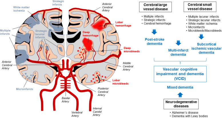
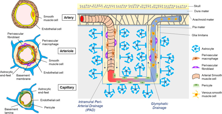
Vascular cognitive impairment and dementia (VCID) is commonly caused by vascular injuries in cerebral large and small vessels and is a key driver of age-related cognitive decline. Severe VCID includes post-stroke dementia, subcortical ischemic vascular dementia, multi-infarct dementia, and mixed dementia. While VCID is acknowledged as the second most common form of dementia after Alzheimer's disease (AD) accounting for 20% of dementia cases, VCID and AD frequently coexist. In VCID, cerebral small vessel disease (cSVD) often affects arterioles, capillaries, and venules, where arteriolosclerosis and cerebral amyloid angiopathy (CAA) are major pathologies. White matter hyperintensities, recent small subcortical infarcts, lacunes of presumed vascular origin, enlarged perivascular space, microbleeds, and brain atrophy are neuroimaging hallmarks of cSVD. The current primary approach to cSVD treatment is to control vascular risk factors such as hypertension, dyslipidemia, diabetes, and smoking. However, causal therapeutic strategies have not been established partly due to the heterogeneous pathogenesis of cSVD. In this review, we summarize the pathophysiology of cSVD and discuss the probable etiological pathways by focusing on hypoperfusion/hypoxia, blood-brain barriers (BBB) dysregulation, brain fluid drainage disturbances, and vascular inflammation to define potential diagnostic and therapeutic targets for cSVD.

Keywords: Arteriolosclerosis; Blood–brain barriers (BBB); Cerebral amyloid angiopathy (CAA); Cerebral small vessel disease (cSVD); Glymphatic drainage; Hypoperfusion/Hypoxia; Intramural periarterial drainage (IPAD); Vascular cognitive impairment and dementia (VCID); Vascular inflammation.


Conflict of interest statement

G.B. is an employee of SciNeuro Pharmaceuticals. Other authors declare no competing interests.

Figures

Fig. 1
Fig. 1
Cerebral vascular lesions and vascular cognitive impairment and dementia. Vascular cognitive impairment and dementia (VCID) is a major cause of age-related cognitive decline related to cerebrovascular damages in cerebral large and small vessels. The internal carotid arteries and the vertebral arteries mediate the arterial blood entry into the brain. The blood supply to the cerebrum is mediated by anterior cerebral arteries (ACA) and middle cerebral arteries (MCA) branched from internal carotid arteries. Posterior cerebral arteries (PCA) arising from vertebral arteries are responsible for the blood supply to the brainstem, cerebellum, and occipital cortex. Leptomeningeal arteries from the cerebral arteries form a network of vessels on the pial surface, which branch into the parenchyma. Based on vascular lesions, severe VCID is generally subtyped as post-stroke dementia, subcortical ischemic vascular dementia, multi-infarct dementia, and mixed dementia. Alzheimer’s disease often coexists with cerebrovascular lesions resulting in mixed dementia
Fig. 2
Fig. 2
Structural and cellular compositions of the cerebral small vessels. Leptomeningeal arteries penetrate the pia mater and glia limitans from the subarachnoid space into the parenchyma, ramifying into arterioles and capillaries. In leptomeningeal arteries, endothelial cells make up a single luminal layer which are covered by multiple smooth muscle cell layers. In penetrating arterioles, endothelial cells and smooth muscle cells are single layers. In capillaries, endothelial cells form the blood–brain barrier (BBB) with pericytes and basement membrane, surrounded by astrocytic end-feet. The capillaries connect to venules flowing back into veins. In contrast to arterial smooth muscle cells, venous smooth muscle cells have flattened cell bodies and multiple processes, not fully sheathing the venules and veins. Perivascular fibroblasts and macrophages are mainly localized on arterioles and venules. Subarachnoid cerebrospinal fluid (CSF) distributes into the brain parenchyma through para-arterial spaces referred as glymphatic periarterial CSF influx. Interstitial fluid (ISF) as well as CSF diffuse into the perivenous space by bulk flow, and finally efflux into the CSF-dural sinus or cervical lymph nodes. In addition, the ISF and CSF in brain parenchyma can also enter the periarterial space from the capillary level and flow countercurrent to blood flow along arterial vessels, referred as intramural periarterial drainage (IPAD) pathway
Fig. 3
Fig. 3
Risk factors and pathogenic mechanisms of cerebral small vessel disease. The cerebral small vessel disease (cSVD) can be classified into six groups including arteriolosclerosis, sporadic and hereditary cerebral amyloid angiopathy (CAA), inherited or genetic cSVD distinct from CAA, inflammatory and immunologically mediated cSVD, venous collagenosis, and others. Neuroimaging hallmarks of cSVD include white matter hyperintensities (WMHs), microbleeds, subcortical infarcts, lacunes, and enlarged perivascular space. While various molecular mechanisms are involved in cSVD pathogenesis, hypoperfusion/hypoxia, blood–brain barrier (BBB) dysregulation, interstitial fluid (ISF)/cerebrospinal fluid (CSF) drainage disturbances, and vascular inflammation are likely the major etiological pathways. Hypertension, smoking, diabetes, and sleep apnea are strongly associated with the risk of cSVD, where aging, lifestyle, and genetic factors also contribute to the pathogenic pathways as modifiers

References

    1. O’Brien JT, Erkinjuntti T, Reisberg B, Roman G, Sawada T, Pantoni L, Bowler JV, Ballard C, DeCarli C, Gorelick PB, et al. Vascular cognitive impairment. Lancet Neurol. 2003;2:89–98. - PubMed
    1. Iadecola C. The pathobiology of vascular dementia. Neuron. 2013;80:844–866. - PMC - PubMed
    1. Gorelick PB, Scuteri A, Black SE, Decarli C, Greenberg SM, Iadecola C, Launer LJ, Laurent S, Lopez OL, Nyenhuis D, et al. Vascular contributions to cognitive impairment and dementia: a statement for healthcare professionals from the american heart association/american stroke association. Stroke. 2011;42:2672–2713. - PMC - PubMed
    1. Skrobot OA, O’Brien J, Black S, Chen C, DeCarli C, Erkinjuntti T, Ford GA, Kalaria RN, Pantoni L, Pasquier F, et al. The Vascular Impairment of Cognition Classification Consensus Study. Alzheimers Dement. 2017;13:624–33. - PubMed
    1. Pendlebury ST, Rothwell PM. Prevalence, incidence, and factors associated with pre-stroke and post-stroke dementia: a systematic review and meta-analysis. Lancet Neurol. 2009;8:1006–1018. - PubMed

Publication types