

rWTC-MBTA: autologous vaccine prevents metastases via antitumor immune responses
- PMID: 37434263
- PMCID: PMC10337177
- DOI: 10.1186/s13046-023-02744-8
rWTC-MBTA: autologous vaccine prevents metastases via antitumor immune responses
Abstract
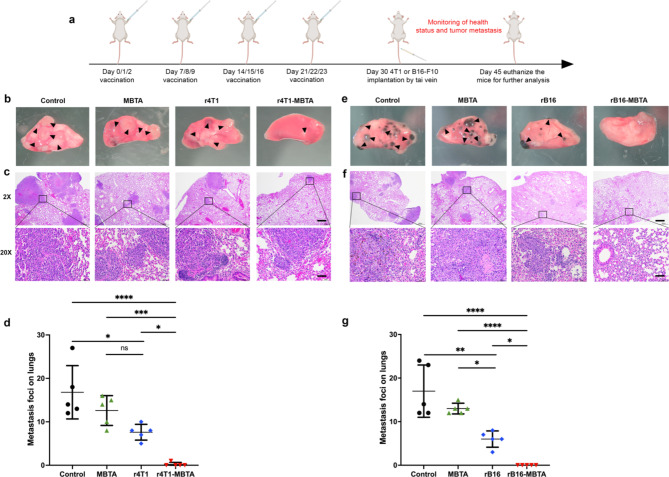
Background: Autologous tumor cell-based vaccines (ATVs) aim to prevent and treat tumor metastasis by activating patient-specific tumor antigens to induce immune memory. However, their clinical efficacy is limited. Mannan-BAM (MB), a pathogen-associated molecular pattern (PAMP), can coordinate an innate immune response that recognizes and eliminates mannan-BAM-labeled tumor cells. TLR agonists and anti-CD40 antibodies (TA) can enhance the immune response by activating antigen-presenting cells (APCs) to present tumor antigens to the adaptive immune system. In this study, we investigated the efficacy and mechanism of action of rWTC-MBTA, an autologous whole tumor cell vaccine consisting of irradiated tumor cells (rWTC) pulsed with mannan-BAM, TLR agonists, and anti-CD40 antibody (MBTA), in preventing tumor metastasis in multiple animal models.
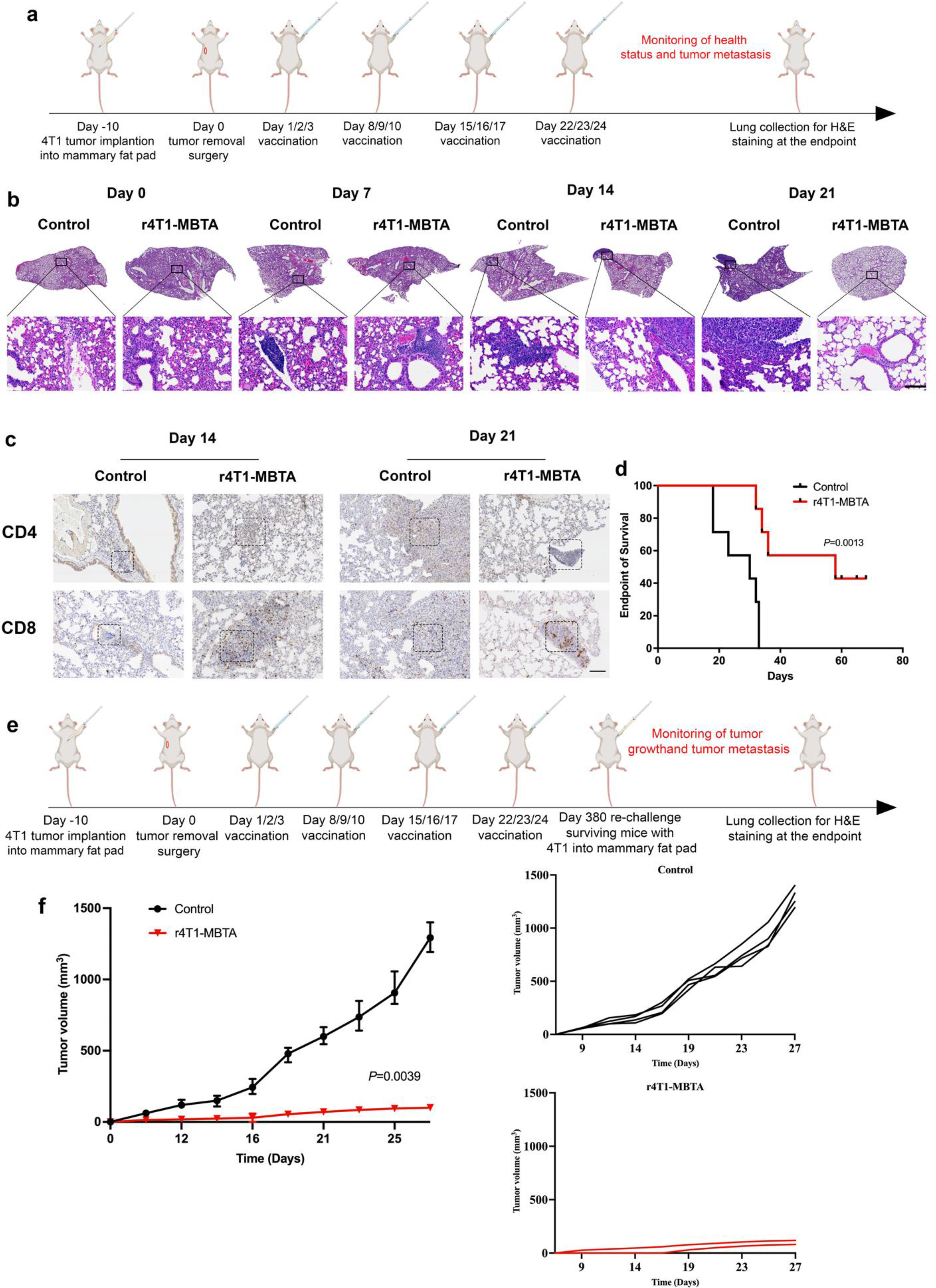
Methods: The efficacy of the rWTC-MBTA vaccine was evaluated in mice using breast (4T1) and melanoma (B16-F10) tumor models via subcutaneous and intravenous injection of tumor cells to induce metastasis. The vaccine's effect was also assessed in a postoperative breast tumor model (4T1) and tested in autologous and allogeneic syngeneic breast tumor models (4T1 and EMT6). Mechanistic investigations included immunohistochemistry, immunophenotyping analysis, ELISA, tumor-specific cytotoxicity testing, and T-cell depletion experiments. Biochemistry testing and histopathology of major tissues in vaccinated mice were also evaluated for potential systemic toxicity of the vaccine.
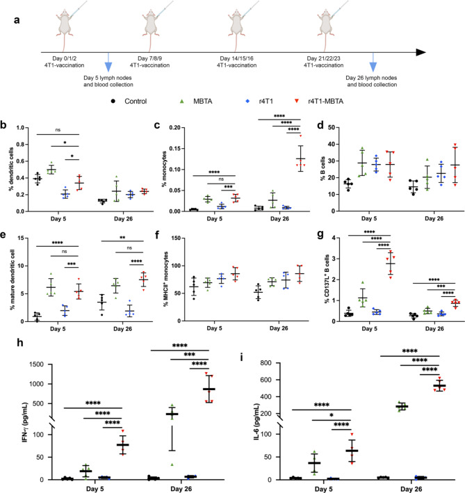
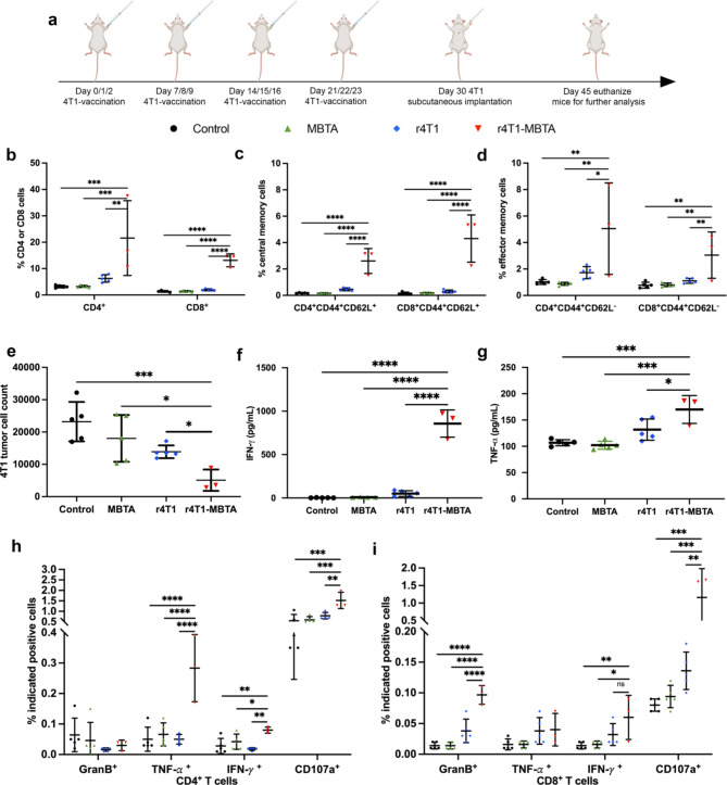
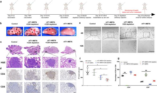
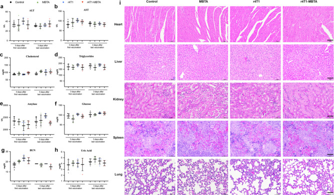
Results: The rWTC-MBTA vaccine effectively prevented metastasis and inhibited tumor growth in breast tumor and melanoma metastatic animal models. It also prevented tumor metastasis and prolonged survival in the postoperative breast tumor animal model. Cross-vaccination experiments revealed that the rWTC-MBTA vaccine prevented autologous tumor growth, but not allogeneic tumor growth. Mechanistic data demonstrated that the vaccine increased the percentage of antigen-presenting cells, induced effector and central memory cells, and enhanced CD4+ and CD8+ T-cell responses. T-cells obtained from mice that were vaccinated displayed tumor-specific cytotoxicity, as shown by enhanced tumor cell killing in co-culture experiments, accompanied by increased levels of Granzyme B, TNF-α, IFN-γ, and CD107a in T-cells. T-cell depletion experiments showed that the vaccine's antitumor efficacy depended on T-cells, especially CD4+ T-cells. Biochemistry testing and histopathology of major tissues in vaccinated mice revealed negligible systemic toxicity of the vaccine.
Conclusion: The rWTC-MBTA vaccine demonstrated efficacy in multiple animal models through T-cell mediated cytotoxicity and has potential as a therapeutic option for preventing and treating tumor metastasis with minimal systemic toxicity.
Keywords: Mannan-BAM; Metastasis; T-cell cytotoxicity; TLR agonists; rWTC-MBTA vaccine.
© 2023. This is a U.S. Government work and not under copyright protection in the US; foreign copyright protection may apply.
Conflict of interest statement
The authors declare no potential conflict of interest. Author SC is employed by NE1 Inc. The remaining authors declare that the research was conducted in the absence of any commercial or financial relationships that could be construed as a potential conflict of interest.
Figures


Similar articles
-
Application of toll-like receptors (TLRs) and their agonists in cancer vaccines and immunotherapy.Front Immunol. 2023 Oct 23;14:1227833. doi: 10.3389/fimmu.2023.1227833. eCollection 2023. Front Immunol. 2023. PMID: 37936697 Free PMC article. Review.
-
rWTC-MBTA Vaccine Induces Potent Adaptive Immune Responses Against Glioblastomas via Dynamic Activation of Dendritic Cells.Adv Sci (Weinh). 2024 Apr;11(14):e2308280. doi: 10.1002/advs.202308280. Epub 2024 Jan 31. Adv Sci (Weinh). 2024. PMID: 38298111 Free PMC article.
-
Optimizing rWTC-MBTA Vaccine Formulations, Dosing Regimens, and Cryopreservation Techniques to Enhance Anti-Metastatic Immunotherapy.Int J Mol Sci. 2025 Feb 5;26(3):1340. doi: 10.3390/ijms26031340. Int J Mol Sci. 2025. PMID: 39941108 Free PMC article.
-
Mannan-BAM, TLR Ligands, Anti-CD40 Antibody (MBTA) Vaccine Immunotherapy: A Review of Current Evidence and Applications in Glioblastoma.Int J Mol Sci. 2021 Mar 26;22(7):3455. doi: 10.3390/ijms22073455. Int J Mol Sci. 2021. PMID: 33810617 Free PMC article. Review.
-
Neoadjuvant intratumoral MBT(A) immunotherapy prevents distant metastases and recurrence in murine models.Cancer Lett. 2025 Mar 1;612:217464. doi: 10.1016/j.canlet.2025.217464. Epub 2025 Jan 12. Cancer Lett. 2025. PMID: 39809356
Cited by
-
Application of toll-like receptors (TLRs) and their agonists in cancer vaccines and immunotherapy.Front Immunol. 2023 Oct 23;14:1227833. doi: 10.3389/fimmu.2023.1227833. eCollection 2023. Front Immunol. 2023. PMID: 37936697 Free PMC article. Review.
-
Current state of cancer immunity cycle: new strategies and challenges of using precision hydrogels to treat breast cancer.Front Immunol. 2025 Mar 7;16:1535464. doi: 10.3389/fimmu.2025.1535464. eCollection 2025. Front Immunol. 2025. PMID: 40124373 Free PMC article. Review.
-
The Immune Landscape of Pheochromocytoma and Paraganglioma: Current Advances and Perspectives.Endocr Rev. 2024 Jul 12;45(4):521-552. doi: 10.1210/endrev/bnae005. Endocr Rev. 2024. PMID: 38377172 Free PMC article. Review.
-
rWTC-MBTA Vaccine Induces Potent Adaptive Immune Responses Against Glioblastomas via Dynamic Activation of Dendritic Cells.Adv Sci (Weinh). 2024 Apr;11(14):e2308280. doi: 10.1002/advs.202308280. Epub 2024 Jan 31. Adv Sci (Weinh). 2024. PMID: 38298111 Free PMC article.
-
Nanomaterial Delivery Vehicles for the Development of Neoantigen Tumor Vaccines for Personalized Treatment.Molecules. 2024 Mar 25;29(7):1462. doi: 10.3390/molecules29071462. Molecules. 2024. PMID: 38611742 Free PMC article. Review.
References
MeSH terms
Substances
LinkOut - more resources
Full Text Sources
Other Literature Sources
Medical
Research Materials
Miscellaneous
