. 2024 Jan 8;5(1):100659.
doi: 10.1016/j.xplc.2023.100659. Epub 2023 Jul 10.

Activation of MAPK-mediated immunity by phosphatidic acid in response to positive-strand RNA viruses

Affiliations

Activation of MAPK-mediated immunity by phosphatidic acid in response to positive-strand RNA viruses

Jiayu Lin et al. Plant Commun. .

Abstract

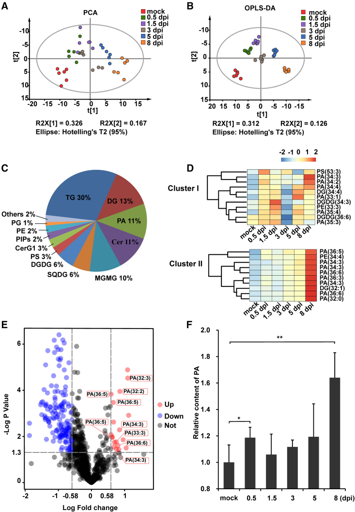
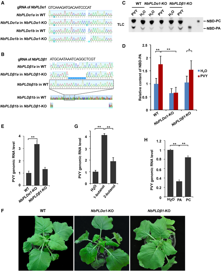
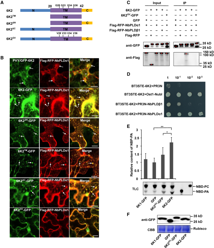
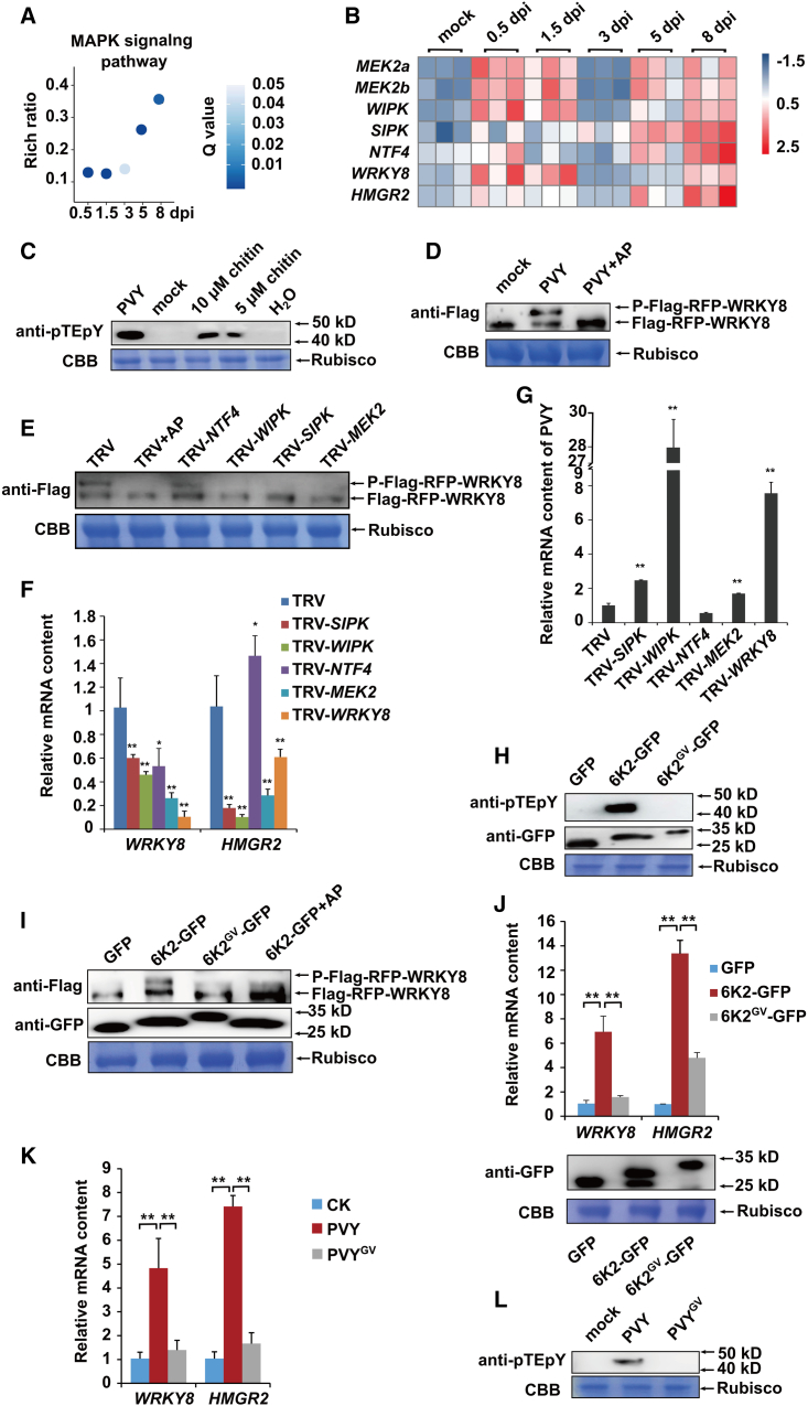
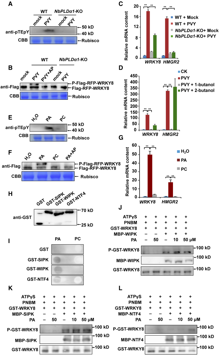
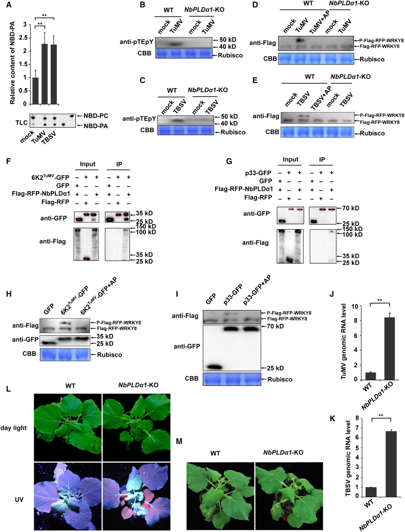
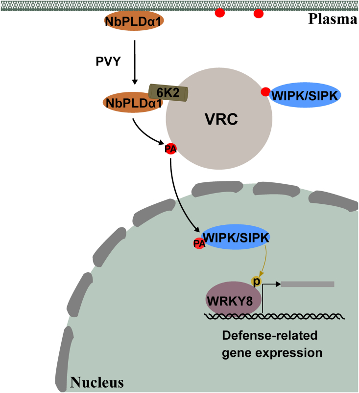
Increasing evidence suggests that mitogen-activated protein kinase (MAPK) cascades play a crucial role in plant defense against viruses. However, the mechanisms that underlie the activation of MAPK cascades in response to viral infection remain unclear. In this study, we discovered that phosphatidic acid (PA) represents a major class of lipids that respond to Potato virus Y (PVY) at an early stage of infection. We identified NbPLDα1 (Nicotiana benthamiana phospholipase Dα1) as the key enzyme responsible for increased PA levels during PVY infection and found that it plays an antiviral role. 6K2 of PVY interacts with NbPLDα1, leading to elevated PA levels. In addition, NbPLDα1 and PA are recruited by 6K2 to membrane-bound viral replication complexes. On the other hand, 6K2 also induces activation of the MAPK pathway, dependent on its interaction with NbPLDα1 and the derived PA. PA binds to WIPK/SIPK/NTF4, prompting their phosphorylation of WRKY8. Notably, spraying with exogenous PA is sufficient to activate the MAPK pathway. Knockdown of the MEK2-WIPK/SIPK-WRKY8 cascade resulted in enhanced accumulation of PVY genomic RNA. 6K2 of Turnip mosaic virus and p33 of Tomato bushy stunt virus also interacted with NbPLDα1 and induced the activation of MAPK-mediated immunity. Loss of function of NbPLDα1 inhibited virus-induced activation of MAPK cascades and promoted viral RNA accumulation. Thus, activation of MAPK-mediated immunity by NbPLDα1-derived PA is a common strategy employed by hosts to counteract positive-strand RNA virus infection.

Keywords: +RNA virus; MAPK; NbPLDα1; PA; PVY; Potato virus Y; WIPK/SIPK; phosphatidic acid; positive-strand RNA virus.


Figures

Figure 1
Figure 1
Lipidomics analysis reveals increased phosphatidic acid level in Potato virus Y–infected leaves of Nicotiana benthamiana. (A and B) Principal component analysis (PCA) and orthogonal partial least-squares discriminant analysis (OPLS-DA) showing overall differences in lipidomes of samples collected at 0.5, 1.5, 3, 5, and 8 dpi (days post inoculation) with Potato virus Y (PVY) or water (collected at 0.5 dpi and designated as mock). (C) Classification of the 178 differentially abundant lipid species (VIP value > 1) identified by OPLS-DA. (D) Clusters I and II excerpted from the hierarchical clustering analysis of 178 differentially abundant lipid species. (E) Significance test of differences in 869 lipid species between mock and 0.5 dpi samples. (F) Relative content of the total phosphatidic acid (PA) class in different treatment groups. Asterisks indicate significant differences (∗P < 0.05, ∗∗P < 0.01) based on two-sided Student’s t-test. Data were collected from six biological replicates, each consisting of a pool of three individual plants.
Figure 2
Figure 2
NbPLDα1 contributes to the increased PA level induced by PVY and plays an antiviral role. (A) Nucleic acid sequences of NbPLDα1 in NbPLDα1-KO (knockout) and wild-type (WT) N. benthamiana plants. (B) Nucleic acid sequences of NbPLDβ1 in NbPLDβ1-KO and WT N. benthamiana plants. (C and D) PLD-derived PA contents in mutant and WT plants infected with PVY or water (as a mock control). Inoculated leaves were analyzed at 0.5 dpi. (C) Lipids were separated by thin-layer chromatography (TLC), and NBD-PC and NBD-PA were used as markers. (D) Relative NBD-PA content was calculated from grayscale values as NBD-PA/(NBD-PA + NBD-PC). Grayscale values were estimated using ImageJ software. NBD-PC, fluorescent 12[(7-nitro-2-1,3-benzoxadiazol-4-yl)amino]dodecanoyl phosphatidylcholine. (E) PVY genomic RNA levels in mutant and WT N. benthamiana leaves. Inoculated leaves were analyzed at 3 dpi. (F) Symptoms caused by PVY at 10 dpi in mutant and WT plants. (G) PVY genomic RNA levels at 3 dpi in N. benthamiana leaves treated with 0.1% 1- or 2-butanol (as a control). (H) PVY genomic RNA levels at 3 dpi in N. benthamiana leaves sprayed with 50 μM PA or PC (as a control) once every 6 h. In (D), (E), (G), and (H), values represent mean ± SD (standard deviation) from three biological replicates. Asterisks indicate significant differences (∗P < 0.05, ∗∗P < 0.01) based on two-sided Student’s t-test. The experiments were repeated three times with similar results.
Figure 3
Figure 3
Interaction of 6K2 with NbPLDα1 increases PLD-derived PA content. (A) Schematic representation of truncated mutants of 6K2. TM, transmembrane domain. (B) 6K2 and its truncated mutants, but not 6K2GV, co-localized with NbPLDα1 during PVY infection or transient expression (indicated by arrows). Scale bars, 10 μm. (C) Co-immunoprecipitation (IP) assay revealed an interaction between 6K2 and NbPLDα1 in N. benthamiana leaves. IP was performed using GFP-Trap Agarose, and proteins were detected by western blotting with an anti-GFP or anti-FLAG antibody. The red asterisks indicate the expected band. (D) A yeast two-hybrid (Y2H) assay showed a strong interaction between 6K2 and NbPLDα1 but a weak interaction between 6K2 and NbPLDβ1. The transformants were plated on an SD/-Leu/-Trp/-His/-Ade plus X-Gal solid medium. Positive control, pBT3STE-6K2 + pOst1-NubI; negative control, pBT3STE-6K2 + pPR3N. (E) Transient expression of viral 6K2 in N. benthamiana leaves increased PLD-derived PA content at 48 h post infiltration. Lipids were separated by thin-layer chromatography (TLC). NBD-PC and NBD-PA were used as markers to indicate target bands. Relative NBD-PA content was calculated from grayscale values as NBD-PA/(NBD-PA + NBD-PC). Grayscale values were estimated using ImageJ software. Values represent mean ± SD from three biological replicates. Asterisks indicate significant differences (∗P < 0.05, ∗∗P < 0.01) based on two-sided Student’s t-test. NBD-PC, fluorescent 12[(7-nitro-2-1,3-benzoxadiazol-4-yl)amino]dodecanoyl phosphatidylcholine; PA, phosphatidic acid. (F) Transient expression under control of the 35S promoter resulted in similar protein levels of 6K2-GFP, 6K2GV-GFP, 6K1-GFP, and GFP in N. benthamiana leaves. Coomassie brilliant blue (CBB) staining of Rubisco large subunit served as a loading control. The experiments were repeated three times with similar results.
Figure 4
Figure 4
PVY induces activation of MAPK-mediated antiviral immunity. (A) Enrichment ratio of the MAPK pathway extracted from a Kyoto Encyclopedia of Genes and Genomes (KEGG) enrichment analysis of differentially expressed genes (DEGs) in PVY-infected N. benthamiana leaves compared with control (CK) leaves. (B) Heatmaps showing the relative mRNA contents of MEK2-WIPK/SIPK/NTF4-WRKY8-HMGR2 pathway components in N. benthamiana leaves infected with PVY or water (as a mock control). Allele genes of N. benthamiana originating from different ancestors are distinguished with the suffixes a and b. (C and D) PVY induced phosphorylation of MAPK (C) and WRKY8 (D) in N. benthamiana leaves at 0.5 dpi. N. benthamiana leaves inoculated with water were designated as mock controls. In (C), leaves treated with 5 or 10 μM chitin for 30 min served as positive controls. (E) Knockdown of MEK2, SIPK, or WIPK by virus-induced gene silencing (VIGS) reduced the phosphorylation level of WRKY8 at 0.5 dpi with PVY. (F) Knockdown of MEK2, SIPK, WIPK, or WRKY8 impaired transcriptional activation of WRKY8 and HMGR2 at 0.5 dpi with PVY. (G) PVY genomic RNA levels were significantly increased at 3 dpi when MEK2, SIPK, WIPK, or WRKY8 was knocked down. (H) Expression of 6K2 induced the phosphorylation of MAPK in N. benthamiana leaves at 24 h post infiltration. (I) Expression of 6K2 induced the phosphorylation of WRKY8 in N. benthamiana leaves at 36 h post infiltration. (J) Expression of 6K2 induced the transcriptional activation of WRKY8 and HMGR2 in N. benthamiana leaves at 24 h post infiltration. (K and L) A PVY clone harboring the 6K2GV mutation failed to induce MAPK phosphorylation (K) or upregulation of WRKY8 and HMGR2 mRNA levels (L). In (A) and (B), data were collected from three biological replicates, each consisting of a pool of six individual plants. In (C), (H), and (L), phosphorylated MAPK was detected using an anti-TEpY antibody (top). In (D), (E), and (I), FLAG-RFP-WRKY8 was detected using an anti-FLAG antibody after phos-tag SDS–PAGE (top). Alkaline phosphatase (AP) was used to remove the phosphate group of phosphorylated WRKY8. In (H) to (J), GFP, 6K2-GFP, and 6K2GV-GFP were detected using an anti-GFP antibody. CBB staining of Rubisco large subunit served as a loading control. In (F), (G), (J), and (K), values represent mean ± SD from three biological replicates. Asterisks indicate significant differences (∗P < 0.05, ∗∗P < 0.01) based on two-sided Student’s t-test. The experiments were repeated three times with similar results.
Figure 5
Figure 5
NbPLDα1-derived PA activates MAPK-mediated immunity in response to PVY. (A–C) The PVY-induced phosphorylation of MAPK (A) and WRKY8 (B), as well as the transcriptional activation of WRKY8 and HMGR2(C) at 0.5 dpi, were inhibited in NbPLDα1-KO plants. Mock-treated plants were inoculated with water. (D) PVY-induced transcriptional activation of WRKY8 and HMGR2 was inhibited by treatment with 0.1% 1-butanol. Plants treated with 0.1% 2-butanol served as controls. (E–G) The phosphorylation of MAPK (E) and WRKY8 (F), as well as the transcriptional activation of WRKY8 and HMGR2(G), were significantly induced at 3 h after spraying with 50 μM PA. Phosphatidylcholine (PC) served as a control. (H) Recombinant glutathione S-transferase (GST)-tagged WIPK, SIPK, and NTF4 proteins were purified from Escherichia coli using glutathione resin. (I) PA binding specificity of recombinant WIPK, SIPK, and NTF4 proteins on nitrocellulose membrane filters. (J–L) PA stimulated the WIPK/SIPK/NTF4-mediated phosphorylation of WRKY8 in vitro. Recombinant GST-WRKY8 was incubated with maltose binding protein (MBP)-tagged WIPK, SIPK, or NTF4 in a reaction buffer containing ATPγS and p-nitrobenzyl mesylate (PNBM). Phosphorylation of GST-WRKY8 was detected with a thiophosphate ester-specific antibody (top). Recombinant MBP-WIPK, -SIPK, and -NTF4 were detected using an anti-MBP antibody (middle). Recombinant GST-WRKY8 was detected with an anti-GST antibody (bottom). All experiments were repeated three times with similar results.In (A) and (E), phosphorylated MAPK was detected using an anti-pTEpY antibody. In (B) and (F), FLAG-RFP-WRKY8 was detected using an anti-FLAG antibody (top). CBB staining of Rubisco large subunit served as a loading control (bottom). In (H) and (I), the bands or spots were detected using an anti-FLAG antibody. PC served as a control. In (C), (D), and (G), values represent mean ± SD from three biological replicates. Asterisks indicate significant differences (∗∗P < 0.01) based on two-sided Student’s t-test.
Figure 6
Figure 6
Activation of MAPK-mediated immunity by NbPLDα1-derived PA is a common plant strategy deployed to inhibit +RNA virus infection. (A) PLD-derived PA content increased in response to Turnip mosaic virus (TuMV) and Tomato bushy stunt virus (TBSV). N. benthamiana leaves were sampled at 0.5 dpi with TuMV, TBSV, or water (as a mock control). Lipids were separated by thin-layer chromatography (TLC). Relative NBD-PA content was calculated from grayscale values as NBD-PA/(NBD-PA + NBD-PC). Grayscale values were determined using ImageJ software. (B–E) The phosphorylation of MAPK (B and C) and WRKY8 (D and E) in response to TuMV or TBSV at 0.5 dpi was inhibited in NbPLDα1-KO plants. Phosphorylated MAPK was detected using an anti-pTEpY antibody. FLAG-RFP-WRKY8 was detected using an anti-FLAG antibody after phos-tag SDS–PAGE (top). CBB staining of Rubisco large subunit served as a loading control (bottom). Alkaline phosphatase (AP) was used to remove the phosphate group of phosphorylated WRKY8. (F and G) Co-immunoprecipitation (IP) assays revealed interactions between NbPLDα1 and 6K2 of TuMV (F) or p33 of TBSV (G) in N. benthamiana leaves. IP was performed using GFP-Trap Agarose. Proteins were detected by western blotting with an anti-GFP or anti-FLAG antibody. Red asterisks indicate the expected band. (H and I) 6K2 of TuMV (H) and p33 of TBSV (I) induced phosphorylation of WRKY8 in N. benthamiana leaves at 36 h post infiltration. FLAG-RFP-WRKY8 was detected using an anti-FLAG antibody after phos-tag SDS–PAGE (top). GFP, 6K2TuMV, and p33 were detected using an anti-GFP antibody (middle). CBB staining of Rubisco large subunit served as a loading control (bottom). (J and K) Genomic RNA levels of TuMV-GFP (J) and TBSV (K) in NbPLDα1-KO or wild-type (WT) N. benthamiana leaves. Inoculated leaves were sampled at 3 dpi. (L and M) Symptoms caused by TuMV-GFP (L) and TBSV (M) at 10 dpi in mutant or WT N. benthamiana plants. In (L), GFP fluorescence, indicating the systemic infection process of TuMV, was observed with a UV lamp (bottom). The experiments were repeated three times with similar results. In (A), (J), and (K), values represent mean ± SD from three biological replicates. Asterisks indicate significant differences (∗∗P < 0.01) based on two-sided Student’s t-test.
Figure 7
Figure 7
A working model depicting the activation of MAPK-mediated immunity by NbPLDα1-derived PA in plants responding to PVY. At the early stage of infection, PVY encodes the 6K2 protein to construct viral membrane-bound VRCs. 6K2 interacts with and recruits NbPLDα1 to membrane-bound VRCs, resulting in a high level of PA. PA binds to WIPK/SIPK and stimulates their phosphorylation of WRKY8, leading to activation of downstream defense-related genes to inhibit PVY infection.

Similar articles

Cited by

References

    1. Allen J.J., Li M., Brinkworth C.S., Paulson J.L., Wang D., Hübner A., Chou W.H., Davis R.J., Burlingame A.L., Messing R.O., et al. A semisynthetic epitope for kinase substrates. Nat. Methods. 2007;4:511–516. - PMC - PubMed
    1. Andersson M.X., Kourtchenko O., Dangl J.L., Mackey D., Ellerström M. Phospholipase-dependent signalling during the AvrRpm1- and AvrRpt2-induced disease resistance responses in Arabidopsis thaliana. Plant J. 2006;47:947–959. - PubMed
    1. Andreasson E., Jenkins T., Brodersen P., Thorgrimsen S., Petersen N.H.T., Zhu S., Qiu J.L., Micheelsen P., Rocher A., Petersen M., et al. The MAP kinase substrate MKS1 is a regulator of plant defense responses. EMBO J. 2005;24:2579–2589. - PMC - PubMed
    1. Asai T., Tena G., Plotnikova J., Willmann M.R., Chiu W.L., Gomez-Gomez L., Boller T., Ausubel F.M., Sheen J. MAP kinase signalling cascade in Arabidopsis innate immunity. Nature. 2002;415:977–983. - PubMed
    1. Barajas D., Xu K., de Castro Martín I.F., Sasvari Z., Brandizzi F., Risco C., Nagy P.D. Co-opted oxysterol-binding ORP and VAP proteins channel sterols to RNA virus replication sites via membrane contact sites. PLoS Pathog. 2014;10 - PMC - PubMed

MeSH terms

Substances

LinkOut - more resources