Case Reports
. 2023 Jun 22;18(9):3070-3075.
doi: 10.1016/j.radcr.2023.06.003. eCollection 2023 Sep.

Infrarenal inferior vena cava agenesis presenting as acute abdomen and hydronephrosis - case report

Affiliations
Case Reports

Infrarenal inferior vena cava agenesis presenting as acute abdomen and hydronephrosis - case report

Filip Brkić et al. Radiol Case Rep. .

Abstract

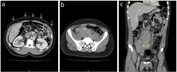
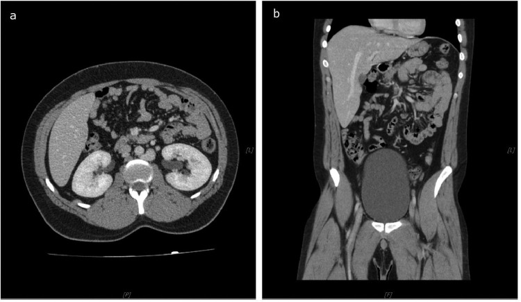
The inferior vena cava agenesis (IVCA) is a rare and often asymptomatic malformation due to the abundant development of the collateral circulation. However, it is frequently found in young people and carries a significant risk of deep venous thrombosis (DVT). It is estimated that about 5% of patients under 30 years of age presenting with DVT have this condition. We report a case of a previously healthy 23-year-old patient presenting with signs of acute abdomen and hydronephrosis due to the thrombophlebitis of an unusual iliocaval venous collateral, which developed secondary to IVCA. After treatment, the iliocaval collateral and hydronephrosis completely regressed on a 1-year follow-up. To our knowledge, this is the first such case reported in the literature.

Keywords: Acute abdomen; Collateral vein; Deep vein thrombosis; Hydronephrosis; Inferior vena cava agenesis; Thrombophlebitis.


Figures

Fig 1
Fig. 1
Postcontrast coronal (A, C, D) and axial (B) reformatted CT images of the thorax, abdomen, and pelvis in the portal-venous (PV) phase. (A) A blind-ending IVC (*) and partially thrombosed retroperitoneal collaterals are seen (arrow). (B) The enlarged azygous vein (*) is comparable in size to the aorta (Ao). (C) The hemiazygos vein drains into the azygos vein. Both vessels are significantly enlarged. (D) Markedly enlarged paravertebral veins are seen (arrows).
Fig 2
Fig. 2
Postcontrast coronal reformatted CT images of the abdomen and pelvis in the PV phase. (A) A strikingly enlarged, thrombosed, and serpiginous iliocaval venous collateral fills the large part of the abdomen, with the small intestine pushed into the left upper quadrant. Note the surrounding fat stranding and free fluid, indicating thrombophlebitis. (B) The arrow points to the origin point of the iliocaval collateral at the left CIV. (C) The arrow points to the termination point of the iliocaval collateral at the IVC, just above the blind end.
Fig 3
Fig. 3
Postcontrast axial (A, B) and coronal (C) CT images of the abdomen and the pelvis in the PV phase. (A, B) Extensive thrombosis is seen in the external (EIV) and internal iliac veins (IIV), as well as the common iliac veins (CIV). (C) A dilated and thrombosed left testicular vein (*) drains into the left renal vein (RV) at the level of the renal-hemiazygos vein junction. HaV – hemiazygos vein.
Fig 4
Fig. 4
Postcontrast axial (A,B) and coronal (C) CT images of the abdomen and pelvis. (A) Grade I hydronephrosis (*) with a delayed contrast uptake of the right kidney is seen. Note the prominent superficial abdominal wall veinous collaterals (arrows) and an enlarged hemiazygos vein (HaV). (B, C) The right ureter (*) is compressed by the thrombosed iliocaval collateral. Note the reactive inflammation of the ureteral wall.
Fig 5
Fig. 5
Postcontrast axial (A) and coronal (B) CT images of the abdomen in a PV phase on a 1-year follow-up. Surprisingly, iliocaval venous collateral completely regressed. Therefore, the right kidney shows no signs of hydronephrosis with prompt contrast uptake.

Similar articles

Cited by

References

    1. Tucker WD, Shrestha R, Anatomy Burns B. StatPearls. StatPearls Publishing; Treasure Island (FL): 2023. Abdomen and pelvis: inferior vena cava.https://www.ncbi.nlm.nih.gov/books/NBK482353/ [accessed July 25, 2022] - PubMed
    1. Gensas CS, Pires LM, Kruse ML, Leiria TL, Gomes DG, Lima GG. Agenesis of the inferior vena cava. Rev Bras de Cardiol Invasiva (English Edition) 2012;20(no. 4):427–430. doi: 10.1016/S2214-1235(15)30090-9. - DOI
    1. Gerges P, Mian A, Singh G, Aziz M, Guirguis S, Koteish A. Agenesis of the intrahepatic inferior vena cava: a case report and literature review. Cureus. 2023;15(2):e35589. doi: 10.7759/cureus.35589. - DOI - PMC - PubMed
    1. Knipp B, Knechtges P, Gest T, Wakefield T. Inferior vena cava: embryology and anomalies. Aortic Aneurysms. 2009;1:289–307. doi: 10.1007/978-1-60327-204-9_20. - DOI
    1. Ramos Aranda J, Ramírez Cerda C, Cohen Mussali S, Valdés Flores J. Inferior vena cava agenesis: an unusual cause of deep vein thrombosis and pulmonary embolism in young adult patients. EJVES Short Rep. 2018;39:12–15. doi: 10.1016/j.ejvssr.2018.03.005. - DOI - PMC - PubMed

Publication types

LinkOut - more resources