Case Reports
. 2023 Jun 21;18(9):3041-3045.
doi: 10.1016/j.radcr.2023.06.023. eCollection 2023 Sep.

Lymphoscintigraphy with single-photon emission computerized tomography/computed tomography for evaluating lymphatic leakage following pelvic and para-aortic lymphadenectomy

Affiliations
Case Reports

Lymphoscintigraphy with single-photon emission computerized tomography/computed tomography for evaluating lymphatic leakage following pelvic and para-aortic lymphadenectomy

Masato Tsuchiya et al. Radiol Case Rep. .

Abstract

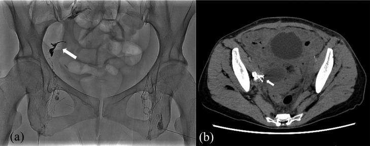
Lymphatic ascites following pelvic and para-aortic lymphadenectomy is a well-known complication. Surgical treatment and interventional radiology are required in a few cases. To determine the appropriate treatment strategy, it is important to preoperatively detect the presence and location of lymphatic leakage. However, the methods have yet to be established. We report a case in which lymphoscintigraphy with single-photon emission computerized tomography/computed tomography (SPECT/CT) was performed to evaluate pelvic lymphorrhea that occurred following total hysterectomy with pelvic and para-aortic lymphadenectomy for stage IIIA uterine sarcoma. Lymphoscintigraphy with SPECT/CT showed leakage of radioisotopes into the pelvic space, and intranodal lymphangiography was performed based on these findings. Following the procedure, the pelvic lymphorrhea improved, and no radioisotope leakage was confirmed by re-evaluation with lymphoscintigraphy with SPECT/CT. Our case indicates that lymphoscintigraphy with SPECT/CT may be useful for detecting the precise site of lymphatic leakage before interventional radiology or surgery.

Keywords: Interventional radiology; Lymphatic ascites; Lymphorrhea; Lymphoscintigraphy; SPECT/CT.


Figures

Fig 1
Fig. 1
Planar image of lymphoscintigraphy amd multiplanar reconstruction of SPECT/CT. (A) Ninety minutes after the injection, showing the reduced number of inguinal lymph nodes accumulating the isotope bilaterally and abnormal extranodal isotope distribution in the pelvis (black arrow). Fusion axial (B), coronal (C), and sagittal (D) SPECT/CT images of the pelvis demonstrate the extranodal hotspot in the pelvic cavity on the right side, 2 cm above the superior margin of the femoral head (thick white arrow). On axial imaging, another hotspot is observed in the midline pelvic cavity (narrow white arrow). SPECT/CT: Single-photon emission computerized tomography/computed tomography.
Fig 2
Fig. 2
Fluoroscopic image and a computerized tomography scan during intranodal lymphangiography. (A) Fluoroscopic image during intranodal lymphangiography. Lipiodol accumulated in the right pelvis (arrow), which showed a hotspot on single-photon emission computerized tomography/computed tomography. (B) A computerized tomography scan during intranodal lymphangiography. Extranodal accumulations of Lipiodol are observed in the right pelvic cavity but do not spread throughout the abdominal cavity (arrow).
Fig 3
Fig. 3
Planar image of lymphoscintigraphy and multiplanar reconstruction of SPECT/CT. (A) Six months after intranodal lymphangiography shows no abnormal extranodal isotope distribution in the pelvis. Fusion axial (B), coronal (C) and sagittal (D) SPECT/CT images of the pelvis also demonstrate no extranodal isotope distribution in the pelvic cavity. SPECT/CT: Single-photon emission computerized tomography/computed tomography.

References

    1. Chen L, Lin L, Li L, Xie Z, He H, Lin C, et al. Lymphatic leakage after pelvic lymphadenectomy for cervical cancer: a retrospective case-control study. BMC Cancer. 2021;21(1):1242. doi: 10.1186/s12885-021-08984-1. - DOI - PMC - PubMed
    1. Weniger M, D'Haese JG, Angele MK, Kleespies A, Werner J, Hartwig W. Treatment options for chylous ascites after major abdominal surgery: a systematic review. Am J Surg. 2016;211(1):206–213. doi: 10.1016/j.amjsurg.2015.04.012. - DOI - PubMed
    1. Kaas R, Rustman LD, Zoetmulder FAN. Chylous ascites after oncological abdominal surgery: incidence and treatment. Eur J Surg Oncol (EJSO) 2001;27(2):187–189. doi: 10.1053/ejso.2000.1088. - DOI - PubMed
    1. Favero G, Lanowska M, Schneider A, Marnitz S, Köhler C. Laparoscopic approach for correction of chylous fistula after pelvic and paraaortic lymphadenectomy. J Minim Invasive Gynecol. 2010;17(2):262–264. doi: 10.1016/j.jmig.2009.12.006. - DOI - PubMed
    1. Kawasaki R, Sugimoto K, Fujii M, Miyamoto N, Okada T, Yamaguchi M, et al. Therapeutic effectiveness of diagnostic lymphangiography for refractory postoperative chylothorax and chylous ascites: correlation with radiologic findings and preceding medical treatment. Am J Roentgenol. 2013;201(3):659–666. doi: 10.2214/AJR.12.10008. - DOI - PubMed

Publication types

LinkOut - more resources