Review
. 2023 Jun 26:14:1180848.
doi: 10.3389/fmicb.2023.1180848. eCollection 2023.

Interplay between chromosomal architecture and termination of DNA replication in bacteria

Affiliations
Review

Interplay between chromosomal architecture and termination of DNA replication in bacteria

Daniel J Goodall et al. Front Microbiol. .

Abstract

Faithful transmission of the genome from one generation to the next is key to life in all cellular organisms. In the majority of bacteria, the genome is comprised of a single circular chromosome that is normally replicated from a single origin, though additional genetic information may be encoded within much smaller extrachromosomal elements called plasmids. By contrast, the genome of a eukaryote is distributed across multiple linear chromosomes, each of which is replicated from multiple origins. The genomes of archaeal species are circular, but are predominantly replicated from multiple origins. In all three cases, replication is bidirectional and terminates when converging replication fork complexes merge and 'fuse' as replication of the chromosomal DNA is completed. While the mechanics of replication initiation are quite well understood, exactly what happens during termination is far from clear, although studies in bacterial and eukaryotic models over recent years have started to provide some insight. Bacterial models with a circular chromosome and a single bidirectional origin offer the distinct advantage that there is normally just one fusion event between two replication fork complexes as synthesis terminates. Moreover, whereas termination of replication appears to happen in many bacteria wherever forks happen to meet, termination in some bacterial species, including the well-studied bacteria Escherichia coli and Bacillus subtilis, is more restrictive and confined to a 'replication fork trap' region, making termination even more tractable. This region is defined by multiple genomic terminator (ter) sites, which, if bound by specific terminator proteins, form unidirectional fork barriers. In this review we discuss a range of experimental results highlighting how the fork fusion process can trigger significant pathologies that interfere with the successful conclusion of DNA replication, how these pathologies might be resolved in bacteria without a fork trap system and how the acquisition of a fork trap might have provided an alternative and cleaner solution, thus explaining why in bacterial species that have acquired a fork trap system, this system is remarkably well maintained. Finally, we consider how eukaryotic cells can cope with a much-increased number of termination events.

Keywords: DNA replication; DNA segregation; RecG helicase; Tus-ter complexes; bacterial chromosome dynamics; chromosomal architecture; termination of DNA replication.


Conflict of interest statement

The authors declare that the research was conducted in the absence of any commercial or financial relationships that could be construed as a potential conflict of interest.

Figures
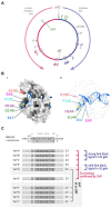
Figure 1
Figure 1
Chromosome structure and Tus-ter trap in Escherichia coli. (A) Schematic representation of the E. coli chromosome. Two replication forks are initiated at a single origin termed oriC and move in opposite directions along the DNA until they approach one another and fuse within the terminus region opposite oriC. A replication fork trap is formed in the terminus region via terminator sequences (terA–J) which are arranged as two opposed groups, with the pink terminators oriented to block movement of the clockwise replication fork and the blue terminators oriented to block the counter-clockwise fork. The locations of the dif chromosome dimer resolution site is marked. Locations of the rrn operons, which are particularly highly transcribed under fast growth conditions, are shown by green arrows, with the arrow pointing in the direction in which transcribing RNA polymerase molecules travel. ‘GRP’ indicates the location of a cluster of genes encoding ribosomal proteins, almost all of which are transcribed co-directionally with replication. (B) Structure of Tus-ter (PDB ID: 2I06) (Singleton et al., 2001). (Bi) Illustration of the “locked” conformation formed by DNA unwinding at the nonpermissive face. The cytosine base at position 6 of ter (C6), which flips into a specific binding site on the nonpermissive face of Tus to form the ‘lock’, is indicated, and important amino acid residues contributing to the locked conformation highlighted. (Bii) As above, but a transparent view is shown to allow easier visualisation of the DNA in general and the flipped C6 in particular. (C) Sequences of ter sites A–J from E. coli MG1655. The ter consensus sequence is shown at the top. Base pairs 1–5, 7, and 19–23 show a higher degree of variability and are shaded in a lighter grey. The highly conserved ter core (6, 8–18) is highlighted by a darker grey. Tus binding (Toft et al., 2021), in vivo fork blocking observed by 2D gel electrophoresis (Duggin and Bell, 2009) and analysis whether a ter site is part of an open reading frame (Duggin and Bell, 2009; Goodall et al., 2021) are indicated (see text for further details).
Figure 2
Figure 2
Altered replichore structure in E. coli cells with additional ectopic replication origins. (A) Schematic representation of E. coli chromosomes with additional ectopic replication origins oriX (left) and oriZ (right), respectively. Positions of oriC, ter sites as well as the dif site and rrn operons A–E, G, and H are shown. (Bi–iii) Marker frequency analysis (MFA) in MG1655, oriC+ oriX+ and oriC+ oriZ+ cells. The number of reads (normalized against reads for a stationary phase wild type control) is plotted against the chromosomal location. A schematic representation of the E. coli chromosome showing positions of oriC, oriX and oriZ (green lines) and ter sites (all above) as well as dif and rrn operons A–E, G, and H (all below) is shown above the plotted data. The MFA raw data were taken from Ivanova et al. (2015) and Dimude et al. (2018b) and re-plotted to allow axis scale changes, if necessary. The areas where forks are blocked by Tus-ter complexes are highlighted by circles and details magnified below to highlight the fork blocking ability of ter sites A, B, C, and D. (C) Over-replication in the termination area of ΔrecG cells growing in M9 minimal salts with glucose. The area where forks escaping the innermost ter sites are blocked by Tus-ter complexes are highlighted by a circle, and details magnified to the side to highlight the fork blocking ability of ter site G. The MFA raw data were taken from Midgley-Smith et al. (2018) and re-plotted.
Figure 3
Figure 3
Comparison of fork trap systems in various plasmids and bacterial chromosomes. (A) Replication dynamics and fork trap system in the plasmid R1. (B) Replication and fork trap features of the primary and secondary chromosome in Pseudoalteromonas haloplanctis TAC 125. The secondary chromosome contains a tus gene, which is located next to the origin of replication, oriC2. The presence of a ter site is only implicated and therefore marked with a question mark. (C) Type I fork trap system in Dickeya paradisiaca, as described in Toft et al. (2022). (D) Type II fork trap system in E. coli (Di) and the fork trap system in Bacillus subtilis (Dii). See main text for further details.
Figure 4
Figure 4
Replication fork fusion can be controlled in vitro to occur at Tus-terB. (A) pKJ1 replication assay template, indicating the location of replication initiation at oriC, the terB site and the lacO22 array. Replisome movement is shown pre (middle) and post (right) LacI-lacO block removal. (B) terB sequence indicating leading strand stop locations (bold and blue) and the Tus-binding site (shaded area). Mapping analysis of the leading strand products approaching Tus-terB in the non-permissive and permissive direction is shown below. Nicked products (stop sites) are indicated by arrows. Mapping data were reproduced from Jameson et al. (2021). (C) Schematic representation of the termination intermediates generated by a fork fusion event taking place at Tus-terB. We believe the most likely candidate for the removal of Tus from this super-complex in vivo is an as yet unidentified helicase.
Figure 5
Figure 5
Schematic illustrating how replication fork fusions might trigger over-replication in the termination area. (i) Two merging replication fork complexes. (ii) Successful termination event where the two replisomes are disassembled, synthesis of all strands is completed and all nascent strands are successfully sealed using DNA ligase. (iii) As part of the fork fusion reaction the helicase of one replication fork complex might displace the leading strand polymerase of the opposing fork, resulting in the formation of a 3’ flap structure, which might be degraded by proteins such as 3’ exonucleases, resulting in successful termination. Note that the formation of a 3’ flap can occur at both forks. However, for simplicity the schematic shows only one such reaction. (iv) A 3’ flap is one of the best substrates for RecG helicase, which would convert it into a 5’ flap. Upon degradation by 5’ exonucleases successful termination can be achieved. (v) If 3’ flaps persist they are also a substrate for the restart protein PriA, which will establish a replisome, thereby not only over-replicating an already fully replicated area of the genome, but also generating a double-stranded DNA end. (vi) The dsDNA end can engage in homologous recombination, resulting in the formation of a displacement or D-loops, which PriA will use to set up yet another replication fork complex, thereby exacerbating the problem of over-replication.
Figure 6
Figure 6
Over-replication at fork fusion sites can be modulated in E. coli if termination sites are artificially altered. (A) Schematic representation of the chromosome of oriC+ oriZ+ cells with additional ter sites integrated either side of the ectopic fork fusion area. (B) Chromosomal marker frequency analysis (MFA) of oriC+ oriZ+ and oriC+ oriZ+ ΔrecG cells with an ectopic replication fork trap in the presence and absence of RecG helicase. The numbers of reads (normalized against reads for a stationary phase wild type control) are plotted against the chromosomal location. A schematic representation of the E. coli chromosome showing positions of oriC, oriZ, native and ectopic ter sites (above) as well as dif and rrn operons A–E, G, and H (below) is shown above the plotted data. The data were re-plotted from Midgley-Smith et al. (2018). (C) Schematic representation of the linearization of the E. coli chromosome, as described in Cui et al. (2007) and Rudolph et al. (2013). The integrated bacteriophage N15 linearization site tos is highlighted. Processing by the N15 telomerase results in the formation of a linear chromosome with covalently-closed hairpin ends, which prevents forks meeting each other. (D) Effect of chromosome linearization on origin-independent synthesis in ΔrecG cells. Shown is the number of reads (normalized against the reads for a stationary-phase wild-type control) plotted against the chromosomal location. In panel (Di) data sets for recG and recG tos are plotted together for direct comparison, while panel (Dii) shows the data for the linearised construct. Data replotted from Rudolph et al. (2013).
Figure 7
Figure 7
Schematic illustrating how over-replication triggered by replication fork fusions might trigger problems for chromosome duplication and segregation, and how the presence of a fork trap area might help in resolving these issues. (i) Over-replication triggered in the termination area by fork fusion reactions will result in additional collisions with forks coming from oriC, thereby reiterating and exacerbating the problem. (ii) A fork fusion reaction by a freely moving fork encountering a second stably arrested at a Tus-ter complex is less likely to result in formation of a 3’ flap and subsequent over-replication, thereby facilitating successful termination. (iii) If termination induces over-replication and the chromosome dimer resolution site dif is duplicated, this can lead to problems of the chromosome dimer resolution mechanism. See text for further details.
Figure 8
Figure 8
Details of the inner replication fork trap architecture in genomes from three Shigella flexneri and two Shigella boydii genomes. The inner ter sites and the chromosome dimer resolution site dif are marked. The orientation of the ter sites are indicated by the direction of the triangle (forks encountering the tip of the triangle would get blocked). The orientation of the dif chromosome dimer resolution site is indicated by the marker pointing upwards (indicating the (+)-strand) or downwards (indicating the (−)-strand). The terD sites highlighted in red indicate a change of the G6 residue, which makes this ter site much less efficient at blocking a progressing replication fork.

Similar articles

Cited by

References

    1. Abhyankar M. M., Reddy J. M., Sharma R., Büllesbach E., Bastia D. (2004). Biochemical investigations of control of replication initiation of plasmid R6K. J. Biol. Chem. 279, 6711–6719. doi: 10.1074/jbc.M312052200 - DOI - PubMed
    1. Abhyankar M. M., Zzaman S., Bastia D. (2003). Reconstitution of R6K DNA replication in vitro using 22 purified proteins. J. Biol. Chem. 278, 45476–45484. doi: 10.1074/jbc.M308516200, PMID: - DOI - PubMed
    1. Abid Ali F., Costa A. (2016). The MCM helicase motor of the eukaryotic replisome. J. Mol. Biol. 428, 1822–1832. doi: 10.1016/j.jmb.2016.01.024 - DOI - PubMed
    1. Ahn K. S., Malo M. S., Smith M. T., Wake R. G. (1993). Autoregulation of the gene encoding the replication terminator protein of Bacillus subtilis. Gene 132, 7–13. doi: 10.1016/0378-1119(93)90508-Z - DOI - PubMed
    1. Alexander J. L., Orr-Weaver T. L. (2016). Replication fork instability and the consequences of fork collisions from rereplication. Genes Dev. 30, 2241–2252. doi: 10.1101/gad.288142.116 - DOI - PMC - PubMed

LinkOut - more resources