Review
. 2023 Jul 1;15(1):e2023038.
doi: 10.4084/MJHID.2023.038. eCollection 2023.

TP53-Mutated Myelodysplasia and Acute Myeloid Leukemia

Affiliations
Review

TP53-Mutated Myelodysplasia and Acute Myeloid Leukemia

Ugo Testa et al. Mediterr J Hematol Infect Dis. .

Abstract

TP53-mutated myelodysplastic syndrome (MDS) and acute myeloid leukemia (AML) form a distinct and heterogeneous group of myeloid malignancies associated with poor outcomes. Studies carried out in the last years have in part elucidated the complex role played by TP53 mutations in the pathogenesis of these myeloid disorders and in the mechanisms of drug resistance. A consistent number of studies has shown that some molecular parameters, such as the presence of a single or multiple TP53 mutations, the presence of concomitant TP53 deletions, the association with co-occurring mutations, the clonal size of TP53 mutations, the involvement of a single (monoallelic) or of both TP53 alleles (biallelic) and the cytogenetic architecture of concomitant chromosome abnormalities are major determinants of outcomes of patients. The limited response of these patients to standard treatments, including induction chemotherapy, hypomethylating agents and venetoclax-based therapies and the discovery of an immune dysregulation have induced a shift to new emerging therapies, some of which being associated with promising efficacy. The main aim of these novel immune and nonimmune strategies consists in improving survival and in increasing the number of TP53-mutated MDS/AML patients in remission amenable to allogeneic stem cell transplantation.

Keywords: Acute myeloid leukemias; Cytogenetic characterization; Gene sequencing; Molecular abnormalities; Myelodysplastic syndromes; TP53.


Conflict of interest statement

Competing interests: The authors declare no conflict of Interest.

Figures
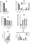
Figure 1
Figure 1
Main molecular properties of TP53-mutated MDS. A: proportion of MDS patients bearing 1, 2 or 3 TP53 mutations. B: MDS patients according to the number of TP53 abnormalities are classified as monoallelic or biallelic, following the involvement of one or both alleles: the types of TP53 mutations, defined as missense, truncated or other mutations, as well as the VAF of TP53 mutations and the frequency of complex karyotype are shown. C: proportion of TP53-mutant MDS patients classified into four subgroups following the presence of a single TP53 mutation (1mut) or of multiple TP53 mutations (>1 mut) or of TP53 mutations+chromosome 17 deletions at the level of TP53 locus (Mut+Del) or of TP53 mutations + cnLOH of TP53 detected only by NGS (Mut + cnLOH). D: Frequency of chromosome monosomies observed in MDS samples classified as above (mean±SEM). E: VAF of TP53 mutations (median value) observed in four subgroups of TP53-mutated MDS, classified as above. F: Frequency of different types of Chromosome 17 abormalities at TP53 locus into three subgroups of MDS with TP53 alterations: 0 TP53 mutations, a rare subgroup, with absent TP53 mutations but with structural alterations affecting TP53 expression, 1 and 2 TP53 mutations. The chromosome 17 status at TP53 locus is defined as normal, deleted, cnLOH or isoq17 (isochromosome 17q rearrangement). The data reported in this figure are issued from Bernard et al.
Figure 2
Figure 2
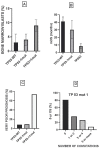
Association between molecular features of TP53-mutated MDSs and clinical parameters. A: Percentage of bone marrow blasts in MDS without TP53 mutations (TP53-WT) and with 1 or >1 TP53 mutations (mean value ± SEM); B: median OS in TP53-WT, TP53 1 mut and TP53 >1 mut (mean value ± SEM); C: frequency of MDS patients with very poor prognosis among TP53-WT, TP53-1 mut and TP53 >1 mut patients; D: 5-yr mean OS in TP53 1 mut patients subdivided into four subgroups according to the number of co-mutations.
Figure 3
Figure 3
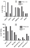
Molecular characterization of MDSs with complex karyotype (CK) in association (CK+TP53) or not (CK) with TP53 mutations. MDSs bearing CK were subdivided into two subgroups according to the presence or not of TP53 mutations. A: frequency of the most recurrent driver mutations observed in CK+TP53 and CK MDSs (mean value ± SEM). ASXL1, U2AF1 and RUNX1 mutations are significantly less frequent in CK+TP53 than in CK MDSs. B: frequency of some relevant chromosomal abnormalities in CK+TP53 and CK MDSs (mean value ± SEM): highly complex karyotype (HCK), monosomal abnormalities, del(5q), abnormal 3q, 13 and 17 chromosome are more frequent in CK+TP53 than in CK MDSs. The data present in this figure are issued from Haase et al.
Figure 4
Figure 4
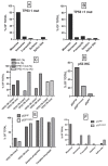
Main molecular properties of TP53-mutated AMLs. A: types of TP53 mutations present in TP53-mutated AMLs bearing 1 TP53 mutation; TP53 mutations were classified as missense, nonsense, frameshift, deletion and splice site. B: types of TP53 mutations present in TP53-mutant AMLs bearing >1 TP53 mutation. C: TP53-mutant AMLs were subclassified into two subgroups according to the presence or not of TP53 copy number alterations (TP53 copy number intact and TP53 CN loss): TP53 VAF was higher in TP53 CN loss than in TP53 CN intact AMLs; CK was markedly more frequent in TP53 CN loss AMLs than in TP53 CN intact; TP53 1 mutations are more frequent in TP53 CN loss than in TP53 CN intact; TP53 with >1 mutation are equally frequent in TP53 CN intact and TP53 CN loss AMLs. D: Immunohistochemical (IHC) classification of TP53-mutant AMLs, with the definition of three subgroups: p53high, p53truncated and p53WT. E: frequency of TP53 CN intact and of TP53 CN loss in p53high and p53truncated AMLs; frequency of AMLs with adverse prognosis and with complex karyotype in p53high and p53truncated AMLs. F: frequency of different types of TP53 mutants in p53high and p53truncated AMLs. The data present in this figure are issued from Takashori et al.
Figure 5
Figure 5
Comparison of the mutational profile (A) and chromosomal abnormalities (B) in TP53-mutated MDSs and AMLs. Top panel: the frequency of most recurrent driver mutations in reported. Bottom panel: the frequency of most recurrent chromosomal abnormalities is reported.
Figure 6
Figure 6
New therapeutic strategies under clinical investigation form the treatment of TP53-mutated MDS/AML.

References

    1. Shallis RM, Wang R, Davidoff A, Ma X, Zeidan AM. Epidemiology of acute myeloid leukemia: recent progress and enduring challenges. Blood Rev. 2019;36:70–87. doi: 10.1016/j.blre.2019.04.005. - DOI - PubMed
    1. Juliusson G, Abrahamsson J, Lazarevic V, Antonovic P, Derolf A, Gorelius H, Lehmanns S, Myhr-Eriksson K, Mollgord L, Uggla B, et al. P revalence and characteristics of survivors from acute myeloid leukemia in Sweden. Leukemia. 2017;31:728–731. doi: 10.1038/leu.2016.312. - DOI - PMC - PubMed
    1. Roman E, Smith A, Appleton S, Crouch S, Kelly R, Kinsey S, Corgo C, Patmore R. Myeloid malignancies in the real-world: occurrence, progression and survival in the UK’s population-based Haematological Malignancy Research Network 20094–15. Cancer Epidemiol. 2016;42:186–198. doi: 10.1016/j.canep.2016.03.011. - DOI - PMC - PubMed
    1. Vardiman JW, Harris NL, Brunning RD. The World Health Organization (WHO) classification of the myeloid neoplasms. Blood. 2002;100:2292–2302. doi: 10.1182/blood-2002-04-1199. - DOI - PubMed
    1. Vardiman JW, Thiele J, Arber DA, Brunning RD, Borowitrz MT, Porwit A, Harris NL, LeBeau MM, Hallstrom-Lindberg E, Tefferi A, et al. The 2008 revision of the World Health Organization (WHO) classification of myeloid neoplasms and acute leukemia: rationale and important changes. Blood. 2009;114:937–951. doi: 10.1182/blood-2009-03-209262. - DOI - PubMed