. 2023 Jun 30;14(3):1259-1278.
doi: 10.21037/jgo-23-240. Epub 2023 May 15.

Function of hsa_circ_0006646 as a competing endogenous RNA to promote progression in gastric cancer by regulating the miR-665-HMGB1 axis

Affiliations

Function of hsa_circ_0006646 as a competing endogenous RNA to promote progression in gastric cancer by regulating the miR-665-HMGB1 axis

Jing Qin et al. J Gastrointest Oncol. .

Abstract

Background: Mounting evidences indicate that circular RNAs (circRNAs) are a novel class of non-coding RNAs and play vital roles in the tumorigenesis and aggressiveness including gastric cancer (GC). Nevertheless, the precise functions and underlying mechanisms of circRNAs in GC remain largely unknown.

Methods: The Gene Expression Omnibus (GEO) data set GSE163416 was analyzed to screen the key circRNAs in GC. hsa_circ_0006646 was chosen for further study. GC tissues and matched adjacent normal gastric mucosal epithelial tissues were obtained from the Fourth Hospital of Hebei Medical University. The expressions of hsa_circ_0006646 was detected using quantitative real-time polymerase chain reaction (qRT-PCR). hsa_circ_0006646 was knocked down to identify its effects on GC cells. Bioinformatics algorithms were analyzed to predict the microRNA (miRNAs) potentially sponged by hsa_circ_0006646 and its target genes. Fluorescence in situ hybridization (FISH) was conducted to determine the subcellular location of hsa_circ_0006646 and the predicted miRNA. Then, qRT-PCR, luciferase reporter assay, radioimmunoprecipitation assay, Western blotting, and miRNA rescue experiments were used to confirm the hsa_circ_0006646-related regulatory axis in GC. Cell Counting Kit-8 (CCK-8), colony formation, wound healing, and Transwell experiments were performed to determine the effect of the hsa_circ_0006646-related regulatory axis on GC cells' malignant behaviors in vitro. The xenograft tumor mouse model was established to evaluate the effect of hsa_circ_0006646 in vivo.

Results: hsa_circ_0006646 exhibited a high expression in GC tissues as compared to corresponding adjacent normal gastric mucosal epithelial tissues and its high expression was positively correlated with TNM stage, lymph node invasion and poor prognosis (P<0.05). Knockdown of hsa_circ_0006646 suppressed the proliferation, colony formation, migration, and invasion in GC cells (all P<0.05). hsa_circ_0006646 upregulated high mobility group box 1 (HMGB1) by sponging miR-665 in GC cells (P<0.05). The hsa_circ_0006646-miR-665-HMGB1 axis promoted malignant behaviors and epithelial-mesenchymal transition (EMT) in GC cells by activating the Wnt/β-catenin pathway (P<0.05). The existence of hsa_circ_0006646-miR-665-HMGB1 axis was confirmed in GC specimens (P<0.05). Consequently, down-regulated hsa_circ_0006646 inhibited the progression and EMT of GC cells in vivo (P<0.05).

Conclusions: For the first time, we demonstrated that hsa_circ_0006646-miR-665-HMGB1 axis exerted its tumor-promoting effects in GC, which suggested that hsa_circ_0006646 could be potentially targeted for GC treatment.

Keywords: Gastric cancer (GC); epithelial-mesenchymal transition (EMT); high mobility group box 1 (HMGB1); hsa_circ_0006646; miR-665.


Conflict of interest statement

Conflicts of Interest: All authors have completed the ICMJE uniform disclosure form (available at https://jgo.amegroups.com/article/view/10.21037/jgo-23-240/coif). The authors have no conflicts of interest to declare.

Figures
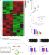
Figure 1
Figure 1
hsa_circ_0006646 was an upregulated circRNA in GC. (A) Heatmap according to the differentially expressed circRNAs between GC tissues and adjacent normal tissues in the GSE163416 data set. (B) The expression of hsa_circ_0006646 in GC tissues and matched normal gastric mucosal epithelium tissues. ***P<0.001. (C) Kaplan-Meier analysis of the relationship between expression of hsa_circ_0006646 and GC patients’ overall survival. hsa_circ_0006646 expression was negatively correlated with the OS of GC patients. (D) qRT-PCR analysis of hsa_circ_0006646 in the GC cell lines. Data are the mean ± SD. ***P<0.001 compared with GES-1 cells. (E) The head-to-tail splicing of hsa_circ_0006646 in the qRT-PCR product as detected with Sanger sequencing. (F) Combining PCR with an electrophoresis assay indicated the expression of hsa_circ_0006646 in AGS and HGC27 cells. (G) qRT-PCR analysis of hsa_circ_0006646 in GC cell lines after RNase R treatment. ***P<0.001 compared with MOCK. qRT-PCR, quantitative real-time polymerase chain reaction; GC, gastric cancer; OS, overall survival.
Figure 2
Figure 2
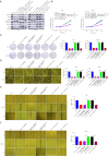
hsa_circ_0006646 knockdown suppressed proliferation, migration and invasion in GC cells in vitro. (A) The efficacy of si-RNAs for knocking down hsa_circ_0006646 in AGS and HGC27 cells. **P<0.01 and ***P<0.001 compared with si-NC. (B) The effect of hsa_circ_0006646 knockdown on proliferation in AGS and HGC27 cells as detected with CCK-8 assay. ***P<0.001 compared with sh-NC. (C) The effect of hsa_circ_0006646 knockdown on colony formation in AGS and HGC27 cells as detected with a colony formation experiment (magnification 200×, crystal violet staining). ***P<0.001 compared with sh-NC. (D) The effect of hsa_circ_0006646 knockdown on invasion in AGS and HGC27 cells as detected with transwell assay (magnification ×200, crystal violet staining). ***P<0.001 compared with sh-NC. (E) The effect of hsa_circ_0006646 knockdown on migration in AGS and HGC27 cells as detected with a wound healing assay (magnification ×200). **P<0.01 and ***P<0.001 compared with sh-NC. (F) The effect of hsa_circ_0006646 knockdown on EMT-related proteins in AGS and HGC27 cells as detected with Western blotting. qRT-PCR, quantitative real-time polymerase chain reaction; si, small interfering; sh, short hairpin; NC, negative control; OD, optical density; CCK-8, Cell Counting Kit-8; EMT, epithelial-mesenchymal transition.
Figure 3
Figure 3
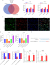
hsa_circ_0006646 served as a molecule sponge for miR-665 in GC. (A) Schematic illustration exhibiting the overlapping of the miRNAs that showed potential to be sponged by hsa_circ_0006646 as predicted by analyzing circBank and circInteractome. (B) The changes in the expression of miRNAs after knocking down hsa_circ_0006646 in AGS and HGC27 cells. *P<0.05 and ***P<0.001 compared with sh-NC. (C) FISH assay showed that hsa_circ_0006646 and miR-665 were predominantly localized in the cytoplasm. hsa_circ_0006646 and miR-665 were stained green and red, respectively. Nuclei were stained with DAPI (magnification 60×). (D) The effect of miR-665 mimic and inhibitor on the expression of hsa_circ_0006646. ***P<0.001 compared with miRNA mimic NC or miRNA inhibitor NC (E) The potential miR-665 target sites in hsa_circ_0006646 transcript. (F) The luciferase activities of the hsa_circ_0006646 luciferase reporter vector (WT or MUT) measured after transfection with miR-665 mimics or mimic NC into AGS and HGC27 cells. **P<0.01 and ***P<0.001 compared with hsa_circ_0006646-MUT. (G) Ago2-RIP assay to detect the enrichment of hsa_circ_0006646 and miR-665 in Ago2-immunoprecipitates. ***P<0.001 compared with IgG. Input was used as the internal control to normalize the expression of hsa_circ_0006646 and miR-665. sh, short hairpin; NC, negative control; FISH, Fluorescence in situ hybridization; RIP, RNA immunoprecipitation; DAPI, 4',6-diamidino-2-phenylindole; WT, wild-type; MUT, mutated-type.
Figure 4
Figure 4
The hsa_circ_0006646–miR-665–HMGB1 axis in GC. (A) Schematic illustration exhibiting the overlapping of the target genes of miR-665 predicted by FunRich, miRWalk, TargetScan, and miRDB. (B) The sequences of HMGB1 3'-UTR-WT and HMGB1 3'-UTR-MUT used in luciferase reporter assay for validating the binding 3'-UTR of HMGB1 mRNA and miR-665. (C) The luciferase activities of the HMGB1 luciferase reporter vector (WT or MUT) measured after transfection with miR-665 mimics or mimic NC into AGS and HGC27 cells. **P<0.01 and ***P<0.001 compared with hsa_circ_0006646-MUT. (D) The effect of miR-665 mimic on the expression of HMGB1 in AGS and HGC27 cells. (E) The hsa_circ_0006646–miR-665–HMGB1 axis in AGS and HGC27 cells. (F) The correlation between hsa_circ_0006646 and miR-665 in GC specimens (n = 35). (G) The presentative IHC staining of HMGB1 in GC tissues (magnification 200×, DAB and hematoxylin). (H) The expressions of hsa_circ_0006646 and miR-665 in HMGB1-positive and HMGB1-negative GC specimens. *P<0.05, ***P<0.001. GC, gastric cancer; NC, negative control; WT, wild-type; MUT, mutated-type; IHC, Immunohistochemistry; DAB, 3,3'-diaminobenzidine.
Figure 5
Figure 5
The hsa_circ_0006646–miR-665–HMGB1 axis promotes proliferation, migration, invasion, and EMT in GC cells in vitro. (A) The effect of the hsa_circ_0006646–miR-665–HMGB1 axis on the Wnt/β-catenin pathway and EMT-related proteins. (B) The effect of the hsa_circ_0006646–miR-665–HMGB1 axis on proliferation in AGS and HGC27 cells as detected with CCK-8 assay. (C) The effect of hsa_circ_0006646–miR-665–HMGB1 axis on colony formation in AGS and HGC27 cells as detected with colony formation assay (magnification 200×, crystal violet staining). (D) The effect of the hsa_circ_0006646–miR-665–HMGB1 axis on invasion in AGS and HGC27 cells as detected with Transwell assay (magnification 200×, crystal violet staining). (E,F) The effect of the hsa_circ_0006646–miR-665–HMGB1 axis on migration in AGS and HGC27 cells as detected with wound healing assay (magnification 200×). ***P<0.001. GC, gastric cancer; sh, short hairpin; NC, negative control; EMT, epithelial-mesenchymal transition; OD, optical density; CCK-8, Cell Counting Kit-8.
Figure 6
Figure 6
hsa_circ_0006646 promoted tumorigenesis and progression of GC in vivo. (A) The xenograft tumors derived from AGS cells transfected with sh-hsa_circ_0006646#1 or sh-NC. (B) The growth of GC xenograft tumor volumes. (C) The weights of GC xenograft tumor. (D) The expression of HMGB1 in xenograft tumors as detected with Western blotting. (E) Representative IHC staining of HMGB1 in xenograft tumors (magnification 200×, DAB and hematoxylin). (F) The expression of Wnt/β-catenin pathway and EMT-related proteins in xenograft tumors. (G) Representative IHC staining of β-catenin in xenograft tumors (magnification 200×, DAB and hematoxylin). ***P<0.001. sh, short hairpin; NC, negative control; GC, gastric cancer; EMT, epithelial-mesenchymal transition; IHC, Immunohistochemistry; DAB, 3,3'-diaminobenzidine.

Similar articles

Cited by

References

    1. Siegel RL, Miller KD, Fuchs HE, et al. Cancer Statistics, 2021. CA Cancer J Clin 2021;71:7-33. 10.3322/caac.21654 - DOI - PubMed
    1. Ilic M, Ilic I. Epidemiology of stomach cancer. World J Gastroenterol 2022;28:1187-203. 10.3748/wjg.v28.i12.1187 - DOI - PMC - PubMed
    1. Chen L, Shan G. CircRNA in cancer: Fundamental mechanism and clinical potential. Cancer Lett 2021;505:49-57. 10.1016/j.canlet.2021.02.004 - DOI - PubMed
    1. Norwood DA, Montalvan EE, Dominguez RL, et al. Gastric Cancer: Emerging Trends in Prevention, Diagnosis, and Treatment. Gastroenterol Clin North Am 2022;51:501-18. 10.1016/j.gtc.2022.05.001 - DOI - PubMed
    1. Cheng X, Ai K, Yi L, et al. The mmu_circRNA_37492/hsa_circ_0012138 function as potential ceRNA to attenuate obstructive renal fibrosis. Cell Death Dis 2022;13:207. 10.1038/s41419-022-04612-3 - DOI - PMC - PubMed