. 2023 Jun 30;9(2):166-175.
doi: 10.21037/jss-22-109. Epub 2023 Apr 13.

Transforaminal endoscopic thoracic discectomy: surgical technique

Affiliations

Transforaminal endoscopic thoracic discectomy: surgical technique

Albert E Telfeian et al. J Spine Surg. .

Abstract

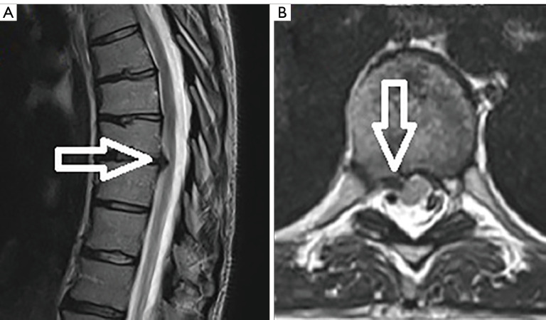
The major challenge inherent to the surgical treatment of thoracic disc herniations is that the disc herniation is often ventral to the spinal cord. Posterior approaches are difficult and dangerous due to the morbidity associated with retraction of the thoracic spinal cord. A ventral approach is not feasible due to the thoracic viscera. A lateral transcavitary approach is the standard for treating ventral thoracic disc pathology but is also quite morbid. Transforaminal endoscopic spine surgery has emerged as a minimally invasive technique for treating thoracic disc pathology and it can be performed in the outpatient setting even with the patient awake. Advances in endoscopic camera technologies as well as the availability of specialty instruments that can be used down a working channel endoscope has now made a myriad of spine pathologies accessible to the minimally invasive spine surgeon. The transforaminal approach and the angled endoscopic camera are an ideal combination for creating a technical advantage to accessing thoracic disc pathology in a minimally invasive fashion. The principal challenges to the approach are needle targeting and understanding the endoscopic visual anatomy. Many surgeons interested in pursuing this technique are often deterred by the burden of the cost and time it takes to become adept and performing the technique. Detailed here are the authors' step-by-step technique and illustrative video that demonstrate transforaminal endoscopic thoracic discectomy (TETD).

Keywords: Endoscopic discectomy; minimally invasive; thoracic disc; transforaminal.


Conflict of interest statement

Conflicts of Interest: Both authors have completed the ICMJE uniform disclosure form (available at https://jss.amegroups.com/article/view/10.21037/jss-22-109/coif). AET serves as an unpaid editorial board member of Journal of Spine Surgery from December 2022 to November 2024. The other author has no conflicts of interest to declare.

Figures

Figure 1
Figure 1
Right T7–8 foraminal disc herniation. (A) Pre-operative sagittal T2 MRI illustrating a large T7–8 foraminal herniated disc (open arrows) referred for endoscopic treatment. (B) Pre-operative axial T2 MRI demonstrates the right-sided T7–8-disc herniation (arrows) obliterating the foramen. (C) Lateral and (D) AP fluoroscopic views of the 18-gauge 25 cm spinal needle used for targeting. Needle is at the corner of the superior endplate of T8 on the lateral view and at the medial wall of the T8 pedicle on the AP view. (E) Lateral and (F) AP fluoroscopic images of the bevelled tubular retractor, after reaming, in the T7–8 foramen. (G) Endoscopic camera view of the T7–8 foramen after endoscopic drilling. The residual SAP is at 12 o’clock and the pedicle is at 3 o’clock. (H) Endoscopic camera view of the endoscopic grasper removing the disc herniation. MRI, magnetic resonance image; AP, anterior-posterior; SAP, superior articulating process.
Figure 2
Figure 2
Post-operative CT myelogram. (A) Sagittal CT reconstruction and (B) axial CT demonstrating the foraminotomy (open arrows) by the reamers and the endoscopic drill. CT, computerized tomography.
Figure 3
Figure 3
Right T8–9-disc herniation. (A) Pre-operative sagittal and (B) axial T2 MRI illustrating a right T8–9 herniated disc (open arrows) referred for endoscopic treatment. MRI, magnetic resonance image.
Figure 4
Figure 4
Needle targeting for right T8–9-disc herniation. (A) Lateral and (B) AP fluoroscopic images of Jamshidi needle entering before it is advance to its final position at the superior endplate of T9 on lateral fluoroscopy and the medial pedicle wall on AP fluoroscopy. AP, anterior-posterior.
Video 1
Video 1
Video depicts an awake, transforaminal endoscopic discectomy in a 31-year-old female who presented with a right-sided T8–9 herniated disc. MRI, magnetic resonance image.
Figure 5
Figure 5
Endoscopic camera views of right T8–9 discectomy. (A) Identifying SAP and epidural contents; (B) endoscopic drilling of dorsal SAP to enlarge foramen; (C) identifying SAP-pedicle junction; (D) drill caudal portion of foramen at SAP-pedicle junction; (E) visualizing the foramen; (F) endoscopic grasper performing discectomy; (G) identification of decompressed neural elements. SAP, superior articulating process.
Figure 6
Figure 6
Postoperative MRI after T8–9 TETD. (A) Post-operative sagittal T2 MRI and (B) axial T2 MRI demonstrating the thoracic spinal cord decompression at T8–9 after the endoscopic discectomy procedure. MRI, magnetic resonance image; TETD, transforaminal endoscopic thoracic discectomy.

Comment in

  • Expanding indications of full endoscopic spine surgery.
    Gadjradj PS, Fiani B, Sommer F, Ramirez RN, Harhangi BS. Gadjradj PS, et al. J Spine Surg. 2023 Sep 22;9(3):229-232. doi: 10.21037/jss-23-65. Epub 2023 Jun 27. J Spine Surg. 2023. PMID: 37841778 Free PMC article. No abstract available.
  • Full-endoscopic thoracic spine approaches.
    Amato MCM, Aprile BC, de Oliveira RS. Amato MCM, et al. J Spine Surg. 2023 Sep 22;9(3):238-241. doi: 10.21037/jss-23-73. Epub 2023 Jul 20. J Spine Surg. 2023. PMID: 37841798 Free PMC article. No abstract available.

Similar articles

Cited by

References

    1. Lewandrowski KU, Telfeian AE, Hellinger S, et al. Difficulties, Challenges, and the Learning Curve of Avoiding Complications in Lumbar Endoscopic Spine Surgery. Int J Spine Surg 2021;15:S21-37. 10.14444/8161 - DOI - PMC - PubMed
    1. Wang H, Huang B, Li C, et al. Learning curve for percutaneous endoscopic lumbar discectomy depending on the surgeon's training level of minimally invasive spine surgery. Clin Neurol Neurosurg 2013;115:1987-91. 10.1016/j.clineuro.2013.06.008 - DOI - PubMed
    1. Kerezoudis P, Rajjoub KR, Goncalves S, et al. Anterior versus posterior approaches for thoracic disc herniation: Association with postoperative complications. Clin Neurol Neurosurg 2018;167:17-23. 10.1016/j.clineuro.2018.02.009 - DOI - PubMed
    1. Hasan S, White-Dzuro B, Barber JK, et al. The Endoscopic Trans-Superior Articular Process Approach: A Novel Minimally Invasive Surgical Corridor to the Lateral Recess. Oper Neurosurg (Hagerstown) 2020;19:E1-E10. 10.1093/ons/opaa054 - DOI - PubMed
    1. Telfeian AE, Shen J, Ali R, et al. Incidence and Implications of Incidental Durotomy in Transforaminal Endoscopic Spine Surgery: Case Series. World Neurosurg 2020;134:e951-5. 10.1016/j.wneu.2019.11.045 - DOI - PubMed