. 2023 Jul 10;13(30):20512-20519.
doi: 10.1039/d3ra02812g. eCollection 2023 Jul 7.

Microfluidic fabrication of X-ray-visible sodium hyaluronate microspheres for embolization

Affiliations

Microfluidic fabrication of X-ray-visible sodium hyaluronate microspheres for embolization

Yang Shen et al. RSC Adv. .

Abstract

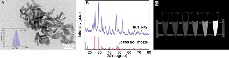
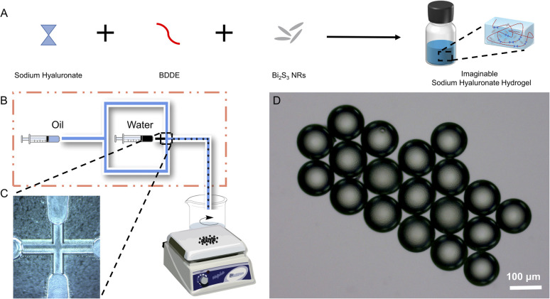
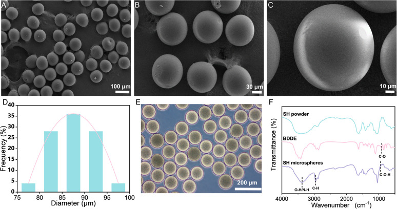
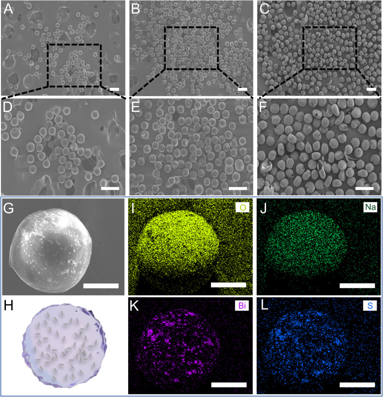
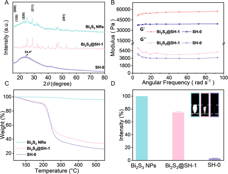
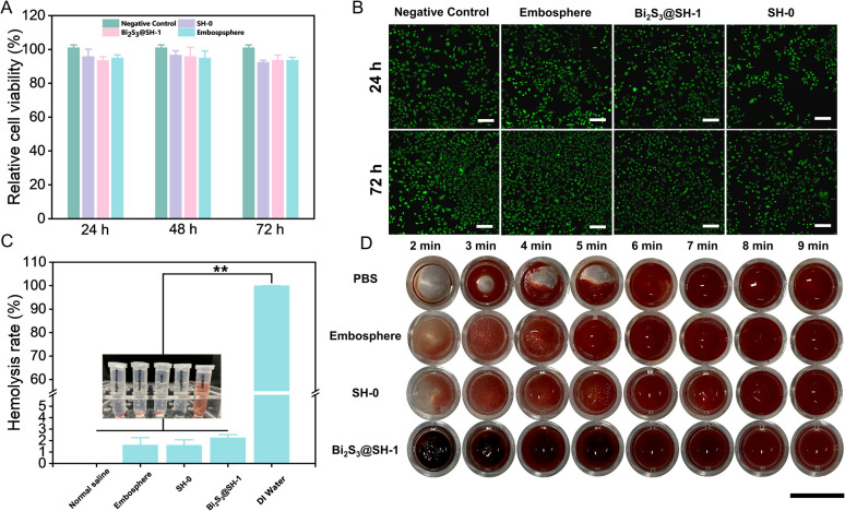
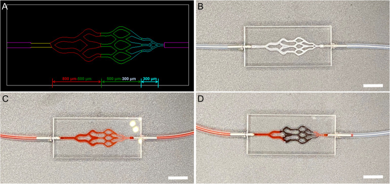
Catheter embolization is a minimally invasive technique that relies on embolic agents and is now widely used to treat various high-prevalence medical diseases. Embolic agents usually need to be combined with exogenous contrasts to visualize the embolotherapy process. However, the exogenous contrasts are quite simply washed away by blood flow, making it impossible to monitor the embolized location. To solve this problem, a series of sodium hyaluronate (SH) loaded with bismuth sulfide (Bi2S3) nanorods (NRs) microspheres (Bi2S3@SH) were prepared in this study by using 1,4-butaneglycol diglycidyl ether (BDDE) as a crosslinker through single-step microfluidics. Bi2S3@SH-1 microspheres showed the best performance among other prepared microspheres. The fabricated microspheres had uniform size and good dispersibility. Furthermore, the introduction of Bi2S3 NRs synthesized by a hydrothermal method as Computed Tomography (CT) contrast agents improved the mechanical properties of Bi2S3@SH-1 microspheres and endowed the microspheres with excellent X-ray impermeability. The blood compatibility and cytotoxicity test showed that the Bi2S3@SH-1 microspheres had good biocompatibility. In particular, the in vitro simulated embolization experiment results indicate that the Bi2S3@SH-1 microspheres had excellent embolization effect, especially for the small-sized blood vessels of 500-300 and 300 μm. The results showed the prepared Bi2S3@SH-1 microspheres have good biocompatibility and mechanical properties, as well as certain X-ray visibility and excellent embolization effects. We believe that the design and combination of this material has good guiding significance in the field of embolotherapy.


Conflict of interest statement

There are no conflicts to declare.

Figures

Fig. 1
Fig. 1. Characterizations of Bi2S3 NRs. (A) TEM image and histogram of the size distribution. (B) XRD spectra. (C) In vitro CT images of Bi2S3 NRs with different concentrations (2.5, 5, 10, 15, 30, and 60 mg mL−1, from left to right, respectively).
Fig. 2
Fig. 2. SH and Bi2S3@SH microspheres fabrication schematic: (A) a schematic depicting imaginable (sodium hyaluronate) SH gel fabrication. (B) Schematic depicting microspheres fabrication. (C) Local magnification image of a flow-focused droplet microfluidic chip. (D) Optical micrograph of hydrated SH microspheres.
Fig. 3
Fig. 3. Characterizations of SH microspheres: (A–C) SEM images of SH microspheres at various magnifications. (D) Histogram of the size distribution of SH microspheres. (E) Optical micrograph of SH microspheres post-swelling in PBS after 24 h. (F) FT-IR spectra of SH powder, BDDE, and SH microspheres.
Fig. 4
Fig. 4. Characterizations of Bi2S3@SH microspheres: (A–C) SEM images of the SH-0, Bi2S3@SH-1, and Bi2S3@SH-2 microspheres, respectively, scale bar: 200 μm. (D–F) The high magnifications of (A–C), scale bar: 200 μm. (G) The high magnification of a single Bi2S3@SH-1 microsphere, scale bar: 30 μm. (H) The schematic of (G) internal structure. (I–L) The EDS-mapping images of (G), scale bar: 30 μm.
Fig. 5
Fig. 5. Characterizations of Bi2S3@SH-1 microspheres: (A) XRD spectra. (B) Storage (G′) and loss (G′′) moduli were recorded for samples. (C) TGA curves of Bi2S3 NRs, SH-0, and Bi2S3@SH-1 microspheres. (D) In vitro CT images and the radiopacity values of the samples (n = 5).
Fig. 6
Fig. 6. In vitro biocompatibility: (A) the cell viability of HUVECs co-cultured with Bi2S3@SH-1 microsphere extracts at 20 mg mL−1 for 24 h, 48 h, and 72 h, respectively (n = 6). (B) Confocal microscopy images of HUVECs incubated with Bi2S3@SH-1 microspheres after 1 and 3 days, scale bar: 200 μm. (C) Hemolysis test of Bi2S3@SH-1 microspheres, the insert digital photograph of rabbit carotid blood supernatant and solution after centrifugation of samples, data were shown as mean ± standard deviation, **p < 0.01. (D) Blood coagulation test of samples for a different period, scale bar: 1 cm.
Fig. 7
Fig. 7. In vitro vascular model embolization: (A) the design image of the PMMA chip. (B) In vitro embolization model, scale bar: 1 cm. (C) Before embolization, scale bar: 1 cm. (D) After embolization, scale bar: 1 cm.

Similar articles

Cited by

References

    1. Hu J. J. Altun I. Zhang Z. F. Albadawi H. Salomao M. A. Mayer J. L. Hemachandra L. Rehman S. Oklu R. Adv. Mater. 2020;32:2002611. - PMC - PubMed
    1. Hu J. J. Albadawi H. Chong B. W. Deipolyi A. R. Sheth R. A. Khademhosseini A. Oklu R. Adv. Mater. 2019;31:1901071. - PMC - PubMed
    1. Wang Y. L. He X. L. Zhou C. Bai Y. W. Li T. Q. Liu J. C. Ju S. G. Wang C. Y. Xiang G. Y. Xiong B. Acta Biomater. 2022;154:536–548. - PubMed
    1. Perez Lopez A. Martin Sabroso C. Gomez Lazaro L. Torres Suarez A. I. Aparicio Blanco J. Acta Biomater. 2022;149:1–15. - PubMed
    1. Li X. H. Ullah M. W. Li B. S. Chen H. R. Adv. Healthcare Mater. 2023;41:2202787.