. 2023 Jun 26:14:1211473.
doi: 10.3389/fendo.2023.1211473. eCollection 2023.

Linsitinib, an IGF-1R inhibitor, attenuates disease development and progression in a model of thyroid eye disease

Affiliations

Linsitinib, an IGF-1R inhibitor, attenuates disease development and progression in a model of thyroid eye disease

Anne Gulbins et al. Front Endocrinol (Lausanne). .

Abstract

Introduction: Graves' disease (GD) is an autoimmune disorder caused by autoantibodies against the thyroid stimulating hormone receptor (TSHR) leading to overstimulation of the thyroid gland. Thyroid eye disease (TED) is the most common extra thyroidal manifestation of GD. Therapeutic options to treat TED are very limited and novel treatments need to be developed. In the present study we investigated the effect of linsitinib, a dual small-molecule kinase inhibitor of the insulin-like growth factor 1 receptor (IGF-1R) and the Insulin receptor (IR) on the disease outcome of GD and TED.

Methods: Linsitinib was administered orally for four weeks with therapy initiating in either the early ("active") or the late ("chronic") phases of the disease. In the thyroid and the orbit, autoimmune hyperthyroidism and orbitopathy were analyzed serologically (total anti-TSHR binding antibodies, stimulating anti TSHR antibodies, total T4 levels), immunohistochemically (H&E-, CD3-, TNFa- and Sirius red staining) and with immunofluorescence (F4/80 staining). An MRI was performed to quantify in vivo tissue remodeling inside the orbit.

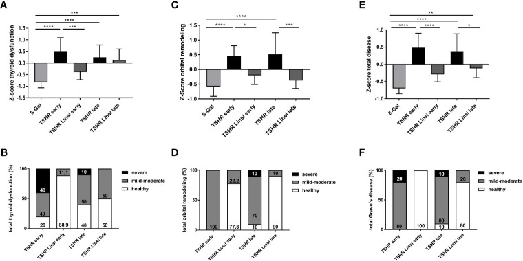
Results: Linsitinib prevented autoimmune hyperthyroidism in the early state of the disease, by reducing morphological changes indicative for hyperthyroidism and blocking T-cell infiltration, visualized by CD3 staining. In the late state of the disease linsitinib had its main effect in the orbit. Linsitinib reduced immune infiltration of T-cells (CD3 staining) and macrophages (F4/80 and TNFa staining) in the orbita in experimental GD suggesting an additional, direct effect of linsitinib on the autoimmune response. In addition, treatment with linsitinib normalized the amount of brown adipose tissue in both the early and late group. An in vivo MRI of the late group was performed and revealed a marked decrease of inflammation, visualized by 19F MR imaging, significant reduction of existing muscle edema and formation of brown adipose tissue.

Conclusion: Here, we demonstrate that linsitinib effectively prevents development and progression of thyroid eye disease in an experimental murine model for Graves' disease. Linsitinib improved the total disease outcome, indicating the clinical significance of the findings and providing a path to therapeutic intervention of Graves' Disease. Our data support the use of linsitinib as a novel treatment for thyroid eye disease.

Keywords: Graves’ disease (GD); IGF-1R; autoimmune disorder; inflammation; linsitinib; thyroid eye disease (TED).


Conflict of interest statement

Authors AS and RZ were employed by the company Sling Therapeutics Inc. The remaining authors declare that the research was conducted in the absence of any commercial or financial relationships that could be construed as a potential conflict of interest.

Figures

Figure 1
Figure 1
Experimental design of linsitinib treatment in a mouse model for Graves’ disease. In order to induce autoimmune hyperthyroidism and associated thyroid eye disease, female BALB/c mice at age of six weeks were immunized with a TSHR A-subunit encoding plasmid three times with three weeks intervals apart. THSR-immunized mice were either treated with linsitinib one week after the second immunization for four weeks (linsitinib “early”) to evaluate the effect of linsitinib in the active state of the disease, or treated with linsitinib two weeks after the second immunization for four weeks to evaluate the effect in a more chronic state of the disease (linsitinib “late”). Two groups of TSHR-immunized mice remained untreated. Female BALB/c mice immunized with a ß-Gal encoding control plasmid and served as healthy control mice (ß-Gal mice). The experiment was terminated six weeks after the last immunization and several investigations, which are also displayed in the figure, were performed on the different mouse groups.
Figure 2
Figure 2
Effect of linsitinib treatment on formation of autoantibodies against TSHR. (A, B) Total anti-TSHR binding antibody titers (TRAbs) in serum samples obtained from mice from each group, i.e., ß-Gal, TSHR early, TSHR early treated with linsitinib (TSHR Linsi early), TSHR late, TSHR late treated with linsitinib (TSHR Linsi late) (A) and inhibition of TSH binding to the TSHR by the autoantibodies (B) were determined by an ELISA. (C) Stimulating activity of anti-mTSHR antibodies (TSAbs) was evaluated by measuring cAMP production in CHO cells treated with autoantibodies. Shown are the mean ± SD, n=8 for ß-Gal, n=10 for TSHR early, TSHR late and TSHR Linsi late and n=9 for TSHR Linsi early. Statistical differences were determined using one-way-ANOVA; *p < 0.05, **p < 0.001, ***p < 0.001, ****p < 0.0001. The upper 99% CI of the TSHR-stimulating activity in the ß-Gal group is indicated by a dotted line.
Figure 3
Figure 3
Linsitinib treatment improves thyroid dysfunction. (A) Serum T4 values were measured by an ELISA. (B) The thyroid glands of all mice of each group were fixed, paraffin embedded and sections (1µm) of the middle thyroidal area were H&E stained. Representative images of normal, heterogeneous or hyperplastic thyroid morphology are shown (magnification x10). Arrows indicate hyperplastic parts within normal morphology. (C) Thyroid morphology was classified as normal, heterogeneous or hyperplastic and is given in % of each group. (D) Sections of the middle thyroidal area were immune histologically stained for CD3 and positive T-cells were counted. (E) Representative images of CD3 staining for each mouse group are shown (magnification x40). Arrows indicate CD3+ T-cells in the thyroid tissue. Given are the mean ± SD (A,D) and representative stainings (B, E), n=8 for ß-Gal, n=10 for TSHR early, TSHR late and TSHR Linsi late and n=9 for TSHR Linsi early. Statistical differences were determined using one-way-ANOVA; **p < 0.001. The upper 99% of the CD+ T-cell numbers and fT4 in the ß-Gal group is each indicated by a dotted line.
Figure 4
Figure 4
Physiological in vivo parameters in response to linsitinib treatment. (A) The weights of all mice were measured daily during the study. (B) Glucose was measured in blood using a blood glucose meter at the end of the experimental period. (A) Statistical differences were determined using the area under the curve (AUC), followed by analysis with a student t-test. (B) Shown are the mean ± SD, n=8 for ß-Gal, n=10 for TSHR early, TSHR late and TSHR Linsi late and n=9 for TSHR Linsi early. Statistical differences were determined using one-way-ANOVA.
Figure 5
Figure 5
Orbital T-cell infiltration and tissue remodeling improved upon linsitinib treatment. Orbitae were fixed, paraffin embedded and consecutive sections of the middle orbital area were stained for CD3+ cells. (A–C) The number of CD3+ T-cells was counted in the total orbita sections (A), in the orbital fat (B) and in the orbital muscle (C). (D) Representative images of the total orbita stained for CD3 (magnification x40) are shown in panel. Arrows indicate CD3+ T-cells within the orbital tissue. (E) The area of brown adipose tissue was determined in the sections and is given as percentage of the total area of fat. Displayed are the mean ± SD (A–C, E) and representative stainings (D), n=8 for ß-Gal, n=10 for TSHR early, TSHR late and TSHR Linsi late and n=9 for TSHR Linsi early. Statistical differences were determinedusing one-way-ANOVA; *p < 0.05,**p < 0.001, ***p < 0.001 .
Figure 6
Figure 6
Effect of linsitinib on orbital macrophage infiltration. Orbitae were removed, fixed, paraffin embedded and serial sections of the middle orbital area were stained with anti-F4/80 antibodies as a marker for macrophages. (A) Representative images of immunostainings for F4/80 are shown (magnification x40) (B) Arrows indicate F4/80 positive macrophages within the orbital tissue in early and late TSHR immunized mice. The macrophages can be found in different microscopic levels, which requires to focus through the tissue while counting the positive cells. For this reason, the background of the TSHR-late image appears darker. (B) Orbitae were removed, fixed, paraffin embedded and serial sections of the middle orbital area were stained for TNFα and positive cells in the orbit were counted. (C) The mice in the late group were divided in responder and non-responder. i.e., those under the dotted line, within the area of the ß-Gal mice, and the number of TNFα positive macrophages in the orbit was again determined. (D) Representative images of immunostainings for TNFα are shown (magnification x40). (E) Arrows indicate TNFα+-positive macrophages within the orbital tissue. Presented are the mean ± SD (A, C, D) and representative stainings (B, E), n=8 for ß-Gal, n=10 for TSHR early, TSHR late and TSHR Linsi late and n=9 for TSHR Linsi early. Statistical differences were determined using one-way-ANOVA; *p < 0.05, **p < 0.001, ***p < 0.001, ****p < 0.0001. The upper 99% CI of the number of macrophages in the ß-Gal group is indicated by a dotted line.
Figure 7
Figure 7
Effect of linsitinib on orbital fibrosis. Orbitae were removed, fixed, sectioned and stained with Sirius red. (A) The percentage of fibrosis was evaluated in the orbital sections using polarization filter and ImageJ after staining with Sirius red. (B) Representative images of staining for Sirius red are shown (magnification x4). Presented are the mean ± SD (A) and representative stainings (B), n=8 for ß-Gal, n=10 for TSHR early, TSHR late and TSHR Linsi late and n=9 for TSHR Linsi early. Statistical differences were determined using one-way-ANOVA; *p < 0.05. The upper 99% of Sirius red intensity in the ß-Gal group is indicated by a dotted line.
Figure 8
Figure 8
Effects of linsitinib on development of key features in the pathogenesis of TED, visualized in MR images of living mice. MRI was performed right before the sacrifice of the late TSHR immunized group, the late TSHR immunized + linsitinib treated group and the healthy control ß-Gal group. 8 mice per group were randomly selected for measurement. (A) 19F integral of PFC-loaded macrophages/monocytes are expressed in arbitrary units. (a.u.) (B) T2-weighted 1H MRI of the retro-orbital muscle is given in ms. (C) T2-weighted 1H MRI of the retro-orbital fat is given in ms. (D) Representative images of MR imaging of the mice. The images on the upper row, help to clarify the anatomical orientation. On the upper left side, the intersecting plane in which the images were created is visualized, whereas on the upper right side an image of the anatomical surroundings, including the optical nerve and the orbital muscles can be seen. (E) Representative images of 19F and T2 MR imaging of the mice. On the left side a 1H/19F merge image is displayed. On the right side a T2 map visualizes the effects in retro-orbital fat and muscle tissue. Arrows in the middle picture indicate to the surrounding fat (arrow on the left-hand side) and muscle tissue (arrow on the right-hand side). Presented are the mean ± SD (A–C) and representative images (D, E), n=8 for ß-Gal, n=10 for TSHR late and TSHR Linsi late. Statistical differences were determined using one-way-ANOVA; *p < 0.05, **p < 0.001, ***p < 0.001.
Figure 9
Figure 9
Total disease outcome of untreated and linsitinib treated, immunized mice. Results of different experiments were combined, normalized and analyzed using the Z-score method. The Z-score represents the value of standard deviation from the mean value of the total mouse population and is given in arbitrary units. (A) The Z-score for the thyroid dysfunction includes the stimulating activity of anti-TSHR autoantibodies ( Figure 2 ), total anti-TSHR binding antibody titers ( Figure 2 ), T4 values ( Figure 3 ), thyroidal CD3+ T-cell infiltration ( Figure 3 ) and weight change ( Figure 4 ). (B) Disease classification was based on Z-score values. The number of mice is given in %. Subclinical disease (Z-score <0): These mice did not significantly manifest autoimmune hyperthyroidism and/or TED although these TSHR A-subunit immunized mice might have developed TSHR antibodies. Clinical disease (Z-score >0): These mice manifested a clinical disease during the experiment. Clinical disease is classified in accordance with Z-score values as mild-moderate (Z-score 0>Z<1) or severe (Z-score >1). The number of mice is given in %. (C) The Z-score for the orbital remodeling includes the orbital CD3+ T-cell infiltration in the orbit ( Figure 5 ), brown adipose tissue enlargement ( Figure 5 ), orbital macrophage infiltration ( Figure 6 ) and fibrosis ( Figure 7 ). (D) Disease classification was based on Z-score values as above. (E) The Z-score for the total disease summarizes the Z-score for the thyroid dysfunction and the Z-score for the orbital remodeling and represents the disease as a whole. (F) Disease classification was based on Z-score values as above. Presented are the mean ± SD (A, C, E) and disease classifications in percent (B, D, F), n=8 for ß-Gal, n=10 for TSHR early, TSHR late and TSHR Linsi late and n=9 for TSHR Linsi early. Statistical differences were determined using one-way-ANOVA; *p < 0.05, **p < 0.001, ***p < 0.001, ****p < 0.0001.

References

    1. Davies TF, Andersen S, Latif R, Nagayama Y, Barbesino G, Brito M, et al. . Graves’ disease. Nat Rev Dis Primers (2020) 6(1):52. doi: 10.1038/s41572-020-0184-y - DOI - PubMed
    1. Bahn RS. Current insights into the pathogenesis of graves’ ophthalmopathy. Horm Metab Res (2015) 47(10):773–8. doi: 10.1055/s-0035-1555762 - DOI - PubMed
    1. Dik WA, Virakul S, van Steensel L. Current perspectives on the role of orbital fibroblasts in the pathogenesis of graves’ ophthalmopathy. Exp Eye Res (2016) 142:83–91. doi: 10.1016/j.exer.2015.02.007 - DOI - PubMed
    1. Pritchard J, Han R, Horst N, Cruikshank WW, Smith. Immunoglobulin activation of T cell chemoattractant expression in fibroblasts from patients with graves’ disease is mediated through the insulin-like growth factor I receptor pathway. J Immunol (2003) 170(12):6348–54. doi: 10.4049/jimmunol.170.12.6348 - DOI - PubMed
    1. Douglas RS, Gianoukakis AG, Kamat S, Smith TJ. Aberrant expression of the insulin-like growth factor-1 receptor by T cells from patients with graves’ disease may carry functional consequences for disease pathogenesis. J Immunol (2007) 178(5):3281–7. doi: 10.4049/jimmunol.178.5.3281 - DOI - PubMed

Publication types

Substances

LinkOut - more resources