. 2023 Aug 1;150(15):dev201853.
doi: 10.1242/dev.201853. Epub 2023 Aug 9.

DELLA proteins positively regulate seed size in Arabidopsis

Affiliations

DELLA proteins positively regulate seed size in Arabidopsis

Maria Dolores Gomez et al. Development. .

Abstract

Human and animal nutrition is mainly based on seeds. Seed size is a key factor affecting seed yield and has thus been one of the primary objectives of plant breeders since the domestication of crop plants. Seed size is coordinately regulated by signals of maternal and zygotic tissues that control the growth of the seed coat, endosperm and embryo. Here, we provide previously unreported evidence for the role of DELLA proteins, key repressors of gibberellin responses, in the maternal control of seed size. The gain-of-function della mutant gai-1 produces larger seeds as a result of an increase in the cell number in ovule integuments. This leads to an increase in ovule size and, in turn, to an increase in seed size. Moreover, DELLA activity promotes increased seed size by inducing the transcriptional activation of AINTEGUMENTA, a genetic factor that controls cell proliferation and organ growth, in the ovule integuments of gai-1. Overall, our results indicate that DELLA proteins are involved in the control of seed size and suggest that modulation of the DELLA-dependent pathway could be used to improve crop yield.

Keywords: Arabidopsis; DELLA; Gibberellin; Ovule; Seed.


Conflict of interest statement

Competing interests The authors declare no competing or financial interests.

Figures
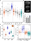
Fig. 1.
Fig. 1.
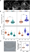
DELLA proteins act maternally to positively regulate seed size. (A) Seed area of Ler (wild type), gain-of-function della mutants rgaΔ17, rgl1Δ17, rgl2Δ17 and gai-1, and loss-of-function della mutants rga24, rgl1-1, rgl2-1 and gaiT6. (B) Scanning electron microscopy images of mature seeds of gai-1, Ler and gaiT6 plants. (C) Seed weight (mean±s.d. of 500 seeds) of Ler, gaiT6 and gai-1 plants. Significant differences (Student's t-test) are indicated (**P<0.01); ns, not statistically significant. (D) Seed length (Y) and width (X) of Ler, gai-1 and gaiT6 plants. The ratio of length to width (±s.d.) is shown in gray. (E) Seed area resulting from the indicated reciprocal crosses of Ler with gai-1 and gaiT6. The first genotype listed is the maternal parent (ovule) and the second is the paternal parent (pollen). (A,D,E) Dots represent data from individual seeds and horizontal lines represent the mean values (n>100 in A, n=30 in D and n≥30 in E). Different lowercase letters indicate a statistically significance difference, as determined by an ANOVA and a Bonferroni post-hoc test for multiple comparisons (P<0.05). Data that are not significantly different are marked with the same letter. Scale bar: 100 µm.
Fig. 2.
Fig. 2.
GAI promotes ovule growth by increasing cell proliferation in integuments. (A) Confocal images of representative mature ovules of Ler, gaiT6 and gai-1 plants. (B) Ovule area of Ler, gaiT6 and gai-1 plants (n≥20). (C) Cell number in the outer layer of the outer integument (oi2) and the inner layer of the inner integument (ii1) of Ler, gaiT6 and gai-1 mature ovules (n≥20). (D) Image of a Ler seed at 4 DAP observed by differential interference contrast (DIC) microscopy. (E) Cell number in the oi2 layer of Ler and gai-1 developing seeds at 4 DAP. (B,C,E) Dots represent data from individual ovules or cells and horizontal lines represent mean values. (B,E) Significant differences (Student's t-test) from the corresponding wild type are indicated (**P<0.01). (C) Different lowercase letters indicate a statistically significance difference, as determined by a one-way ANOVA and a Bonferroni post-hoc test for multiple comparisons (P<0.05). Data that are not significantly different are marked with the same letter. Scale bars: 20 µm in A; 100 µm in D.
Fig. 3.
Fig. 3.
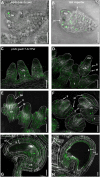
GAI is expressed in developing ovules and seeds. (A) Confocal images of GAI-3xYPet expression in ovules at stage 2-V. (B) GA-regulated reporter (GA HACR; Khakhar et al., 2018) expression in ovules at stage 2-V. Images correspond to an overlay of bright-field and fluorescence micrographs from fresh tissue. (C-H) Confocal images of the expression of gaiΔ17-3xYPet in the funiculus, chalaza and integuments of developing ovules (C, stage 2-I; D, 2-III; E, 2-IV; F, 2-V), mature ovules (G) and in the seed coat of developing seeds at 3 DAP (H). Ovules were cleared and cell walls were stained using Calcofluor White. The panels show the composite images of Calcofluor White and the z-stack projection of YPet fluorescence images. The dotted lines define the shape of the developing ovules. Confocal images are representative of several high-quality images obtained from three biological replicas. ch, chalaza; f, funiculus; ii, inner integument; oi, outer integument; nu, nucellus. Scale bars: 20 µm.
Fig. 4.
Fig. 4.
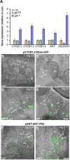
ANT and CYC gene expression is increased in gai-1 ovules. (A) Relative mRNA levels of CYCB1;1, CYCB1;2, CYCB1;4, ANT and GA20OX1 (At4g25420) in siliques at 3 DAP of Ler, gaiT6 and gai-1. Data are normalized to UBQ10 (At4g05320) in Ler. Data are mean±s.d. of three replicates. The expression of GA20OX1 was used to validate the result of the qPCR assay as it increases in gai-1 and decreases in gaiT6 (Rieu et al., 2008; Gallego-Bartolomé et al., 2011). (B-E) Confocal images of the localization of CYCB1;2-GFP in Ler (B,C) and gai-1 (D,E) ovules at stages 2-II (B,D) and 3-I (C,E). (F,G) Confocal images of the localization of ANT-YPet in Ler (F) and gai-1 (G) ovules at stage 2-IV. Panels show composite images of a bright-field image and a GFP (B-E) or YPet (F,G) z-stack projection. The dotted lines define the shape of the ovules. Confocal images are representative of several high-quality images obtained from three biological replicates. ch, chalaza; f, funiculus; nu, nucellus; ii, inner integument; oi, outer integument. Scale bars: 20 µm.
Fig. 5.
Fig. 5.
gai-1 seeds show no changes in morphological development or metabolic composition. (A) Embryo development in Ler and gai-1 plants. Ler and gai-1 cleared seeds at 4, 6, 8, 9 and 10 DAP were observed using DIC microscopy. The embryos were colored (orange for Ler and green for gai-1) to facilitate visualization. (B) Semi-thin sections of Ler and gai-1 dry seeds embedded in resin showing the external mucilage. (C) Fatty acid, sugar and amino acid content in dry seeds of Ler, gai-1 and Ler upon treatment with 1 µM PBZ. Dots represent individual values for each of the five biological replicates. One-way ANOVA and Tukey's multiple comparison tests were performed; no statistically significant differences were found. Scale bars: 50 μm in A; 100 μm in B.
Fig. 6.
Fig. 6.
Proposed working model of how DELLA activity regulates ovule and seed size in Arabidopsis. GAs mediate DELLA protein degradation, which is a positive factor in the promotion of cell proliferation in integumentary cell layers during ovule development. DELLA proteins interact with an unknown TF to positively regulate the expression of ANT (1). DELLA may also interact with other TFs to regulate other target genes (2). ANT upregulates GAI directly by binding to its promoter (3). In addition, other TFs, such as MEE45 or ARF2, can also directly regulate the expression of ANT (4). Altogether, the coordinated expression of cyclin and other genes regulate cell proliferation in ovule integuments (5). As a result, the cell number in integuments is increased, which leads to larger ovules and seeds.

References

    1. Achard, P., Gusti, A., Cheminant, S., Alioua, M., Dhondt, S., Coppens, F., Beemster, G. T. S. and Genschik, P. (2009). Gibberellin signaling controls cell proliferation rate in Arabidopsis. Curr. Biol. 19, 1188-1193. 10.1016/j.cub.2009.05.059 - DOI - PubMed
    1. Adamski, N. M., Anastasiou, E., Eriksson, S., O'Neill, C. M. and Lenhard, M. (2009). Local maternal control of seed size by KLUH/CYP78A5-dependent growth signaling. Proc. Natl. Acad. Sci. USA 106, 20115-20120. 10.1073/pnas.0907024106 - DOI - PMC - PubMed
    1. Barro-Trastoy, D., Carrera, E., Baños, J., Palau-Rodriguez, J., Ruiz-Rivero, O., Tornero, P., Alonso, J. M., Lopez-Diaz, I., Gomez, M. D. and Perez-Amador, M. A. (2020). Regulation of ovule initiation by gibberellins and brassinosteroids in tomato and Arabidopsis: two plant species, two molecular mechanisms. Plant J. 102, 1026-1041. 10.1111/tpj.14684 - DOI - PubMed
    1. Barro-Trastoy, D., Gomez, M. D., Blanco-Touriñán, N., Tornero, P. and Perez-Amador, M. A. (2022). Gibberellins regulate ovule number through a DELLA–CUC2 complex in Arabidopsis. Plant J. 110, 43-57. 10.1111/tpj.15607 - DOI - PubMed
    1. Brumos, J., Zhao, C., Gong, Y., Soriano, D., Patel, A. P., Perez-Amador, M. A., Stepanova, A. N. and Alonso, J. M. (2020). An improved recombineering toolset for plants. Plant Cell 32, 100-122. 10.1105/tpc.19.00431 - DOI - PMC - PubMed

Publication types

MeSH terms