. 2023 Dec;252(12):1428-1448.
doi: 10.1002/dvdy.642. Epub 2023 Jul 12.

Infigratinib, a selective FGFR1-3 tyrosine kinase inhibitor, alters dentoalveolar development at high doses

Affiliations

Infigratinib, a selective FGFR1-3 tyrosine kinase inhibitor, alters dentoalveolar development at high doses

Zachary D Michel et al. Dev Dyn. 2023 Dec.

Abstract

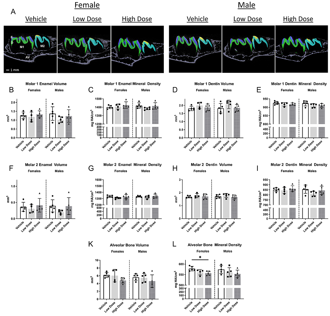
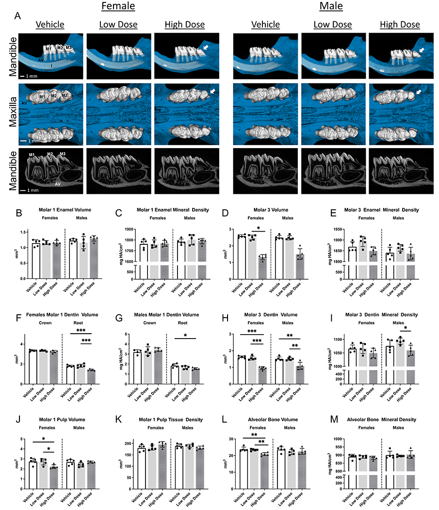
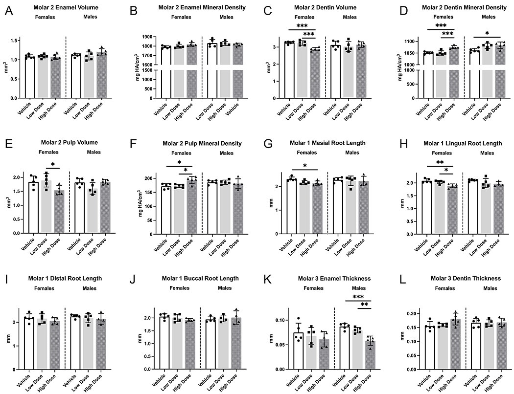
Background: Fibroblast growth factor receptor-3 (FGFR3) gain-of-function mutations are linked to achondroplasia. Infigratinib, a FGFR1-3 tyrosine kinase inhibitor, improves skeletal growth in an achondroplasia mouse model. FGFs and their receptors have critical roles in developing teeth, yet effects of infigratinib on tooth development have not been assessed. Dentoalveolar and craniofacial phenotype of Wistar rats dosed with low (0.1 mg/kg) and high (1.0 mg/kg) dose infigratinib were evaluated using micro-computed tomography, histology, and immunohistochemistry.

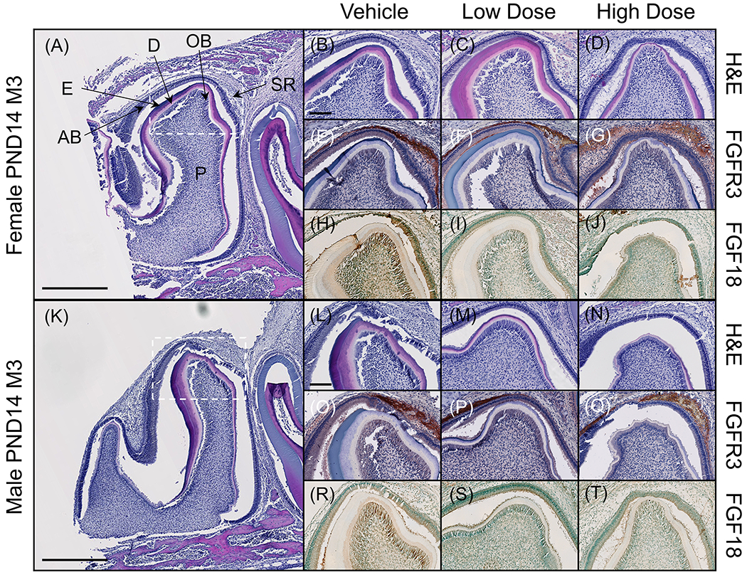
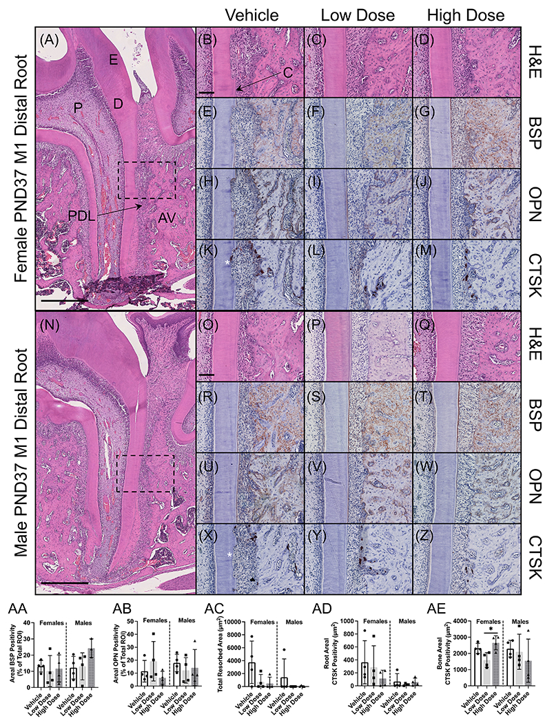
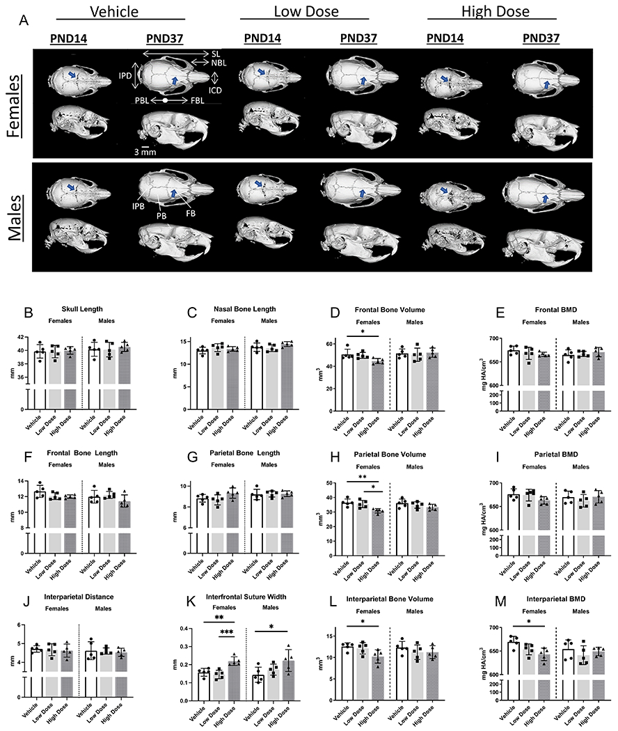
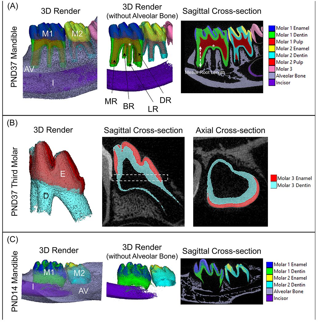
Results: Mandibular third molars were reduced in size and exhibited aberrant crown and root morphology in 100% of female rats and 80% of male rats at high doses. FGFR3 and FGF18 immunolocalization and extracellular matrix protein expression were unaffected, but cathepsin K (CTSK) was altered by infigratinib. Cranial vault bones exhibited alterations in dimension, volume, and density that were more pronounced in females. In both sexes, interfrontal sutures were significantly more patent with high dose vs vehicle.

Conclusions: High dose infigratinib administered to rats during early stages affects dental and craniofacial development. Changes in CTSK from infigratinib in female rats suggest FGFR roles in bone homeostasis. While dental and craniofacial disruptions are not expected at therapeutic doses, our findings confirm the importance of dental monitoring in clinical studies.

Keywords: bone; fibroblast growth factor receptor; mineralized tissue/development; tooth development.


Figures

Figure 1.
Figure 1.. Quantitative analysis of PND14 mandibular first and second molar enamel, dentin, and surrounding alveolar bone volumes and densities.
Alveolar bone mineral density was lower in female rats receiving high dose infigratinib compared to vehicle. In male rats, alveolar bone mineral density trended lower with high dose infigratinib compared to vehicle. One way ANOVA was performed, significance designated as p<0.05. * p<0.05. E=enamel, D=dentin, AV=alveolar bone, M1=mandibular first molar, M2=mandibular second molar.
Figure 2.
Figure 2.. Development of dentoalveolar tissues is affected by high dose infigratinib.
2D and 3D images of mandibles and maxillae from PND37 rats dosed with either vehicle, low dose infigratinib, or high dose infigratinib. Crown and root morphology altered in mandibular and maxillary third molars in high dose male and female rats, e.g., smaller crowns, loss of cusps, and peg shaped roots observed. Quantitative analysis was performed for mandibular first and third molars as well as surrounding alveolar bone. Statistical significance was designated as* p<0.05. ** p<0.01. ***P<0.001. ****p<0.0001. AV=alveolar bone, M1=mandibular first molar, M2=mandibular second molar, M3=mandibular third molar, I=incisor.
Figure 3.
Figure 3.. Further quantitative analysis of PND37 mandibular molars.
(A-F) Volumes and densities of second molar enamel, dentin, and pulp were obtained. Dentin volume was lower and dentin mineral density was higher in female rats receiving high dose infigratinib compared to vehicle and low dose infigratinib. (G-J) Mesial, distal, buccal, and lingual root lengths were obtained in PND37 mandibular first molars. Enamel and dentin thicknesses were obtained in PND37 mandibular third molars. Female rats exhibited shorter mesial and lingual roots with high dose infigratinib compared to vehicle. (K, L) Male rats exhibited reduced enamel thickness with high dose infigratinib vs vehicle. One way ANOVA was performed, significance designated as p<0.05. * p<0.05, ** p<0.01, *** p<0.001.
Figure 4.
Figure 4.. FGFR3 is expressed in the enamel organ and its ligand FGF18 is expressed in ameloblasts and odontoblasts of the developing third molar.
(A, K) Sectioned mandibles were stained for H&E to assess tissue morphology. Third molars of PND14 rats were determined to be in late bell stage and were chosen for further immunohistochemical analysis to investigate where in the developing tooth FGFR3 is expressed. Higher magnification micrographs correspond to the cusp of the third molar (dotted box). (B-D, L-N) At PND14, layers of enamel and dentin can be observed in the third molar and the enamel organ can be observed around the cusps. (E-G, O-Q) Immunostaining for FGFR3 revealed expression within the enamel organ, particularly within the stellate reticulum and stratum intermedium. Expression did not appear to be affected by infigratinib treatment. (H-J, R-T) FGF18, a ligand of FGFR3, was found to be expressed in ameloblasts and odontoblasts and, to a lesser extent, throughout the pulp chamber. Scale bars: 500 μm (A, K) and 100 μm (B-J, L-T). AB=ameloblasts D=dentin, E=enamel. OB=odontoblasts, P=pulp, SR=stellate reticulum.
Figure 5.
Figure 5.. Infigratinib does not alter expression of extracellular matrix proteins but may alter osteoclast activity.
(A, N) Sectioned mandibles were stained for H&E to assess tissue morphology. Higher magnification micrographs correspond to the mid-distal root of the first molar and adjacent alveolar bone (dotted boxes). (E-J, R-W) Given the changes in alveolar bone volume and density measured on microCT, we stained for bone sialoprotein (BSP) and osteopontin (OPN), two extracellular matrix proteins critical for bone formation. (K-M, X-Z) Upon histological evaluation, it was observed that several rats were exhibiting root resorption (asterisks). Osteoclast activity was assessed by staining for cathepsin K (CTSK). (AA, AB) Quantitation of BSP and OPN expression using QuPath revealed that expression was highly variable within and between groups, with no significant changes associated with infigratinib treatment. (AC) The area of resorption pits was measured using QuPath, revealing an insignificant trend toward decreased resorption in response to infigratinib. (AD, AE) CTSK expression was quantified along the distal root surface and alveolar bone surface, demonstrating a similar trend along the root surface. Scale bars: 500 μm (A, N) and 100 μm (B-M, O-Z). AV=alveolar bone, C=acellular cementum, D=dentin, E=enamel, P=pulp, PDL=periodontal ligament.
Figure 6.
Figure 6.. Examination of craniofacial skeletal phenotype of Wistar rats dosed with vehicle, low dose, or high dose infigratinib at PND14 and PND37.
Initiation of dosing was at postnatal day 7 (PND7). Coronal sutures appeared more patent in male and female rats receiving high dose infigratinib compared to vehicle and low dose (arrows). Craniofacial measurements were obtained (2D and 3D), and craniofacial development appeared to be more sensitive to infigratinib in females vs males. IPD=interparietal distance, SL=skull length, NBL=nasal bone length, ICD=inner canthal distance, PBL=parietal bone length, FBL=frontal bone length. IPB=interparietal bone, PB=parietal bone, FB=frontal bone.
Figure 7.
Figure 7.. Reconstructions of mandibles and third molars used for microCT analysis on AnalyzePro.
(A) The region of interest for PND37 mandibles consisted of the first and second molars, as well as the alveolar bone extending from 824 μm mesial to the first molar mesial height of contour and 824 um distal to the second molar distal height of contour. Enamel, dentin, and alveolar bone were segmented using density thresholds and the pulp was segmented using a combination of semiautomatic and manual methods. Volumes and densities of corresponding tissues were then obtained using the Measure tool with AnalyzePro. First molar root lengths were defined as the distance from the furcation to the apex of the root. (B) As PND37 third molars were not fully matured, the thickness of the developing enamel and dentin layers were analyzed. The region of interest was defined as mid-crown (dotted box), which was then run through the Bone Microarchitecture application on AnalyzePro to obtain the average thickness of the two layers. (C) First and second molars and alveolar bone of PND14 mandibles were analyzed in a similar fashion to PND37 mandibles, with lower density thresholds owing to the immaturity of the teeth. Root lengths were not able to be measured as root elongation had not yet begun. M1=mandibular first molar, M2=mandibular second molar, AV=alveolar bone, I=incisor, MR=mesial root, BR=buccal root, LR=lingual root, DR=distal root.

Similar articles

References

    1. Waller D k., Correa A, Vo TM, et al. The population-based prevalence of achondroplasia and thanatophoric dysplasia in selected regions of the US. Am J Med Genet A. 2008;146A(18):2385–2389. doi:10.1002/ajmg.a.32485 - DOI - PMC - PubMed
    1. Legare JM. Achondroplasia. In: Adam MP, Ardinger HH, Pagon RA, et al., eds. GeneReviews®. University of Washington, Seattle; 1993. Accessed March 1, 2022. http://www.ncbi.nlm.nih.gov/books/NBK1152/ - PubMed
    1. Kubota T, Adachi M, Kitaoka T, et al. Clinical Practice Guidelines for Achondroplasia. Clin Pediatr Endocrinol Case Rep Clin Investig Off J Jpn Soc Pediatr Endocrinol. 2020;29(1):25–42. doi:10.1297/cpe.29.25 - DOI - PMC - PubMed
    1. Ireland PJ, Pacey V, Zankl A, Edwards P, Johnston LM, Savarirayan R. Optimal management of complications associated with achondroplasia. Appl Clin Genet. 2014;7:117–125. doi:10.2147/TACG.S51485 - DOI - PMC - PubMed
    1. Rousseau F, Bonaventure J, Legeai-Mallet L, et al. Mutations in the gene encoding fibroblast growth factor receptor-3 in achondroplasia. Nature. 1994;371(6494):252–254. doi:10.1038/371252a0 - DOI - PubMed

Publication types

Substances

LinkOut - more resources