MRI enhances the understanding of critical anatomy during primary laparoscopic port placement
- PMID: 37436051
- PMCID: PMC10410659
- DOI: 10.52054/FVVO.15.2.061
MRI enhances the understanding of critical anatomy during primary laparoscopic port placement
Abstract
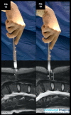
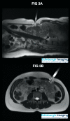
Despite the majority of laparoscopic visceral injuries occurring with primary entry, high-fidelity training models are lacking. Three healthy volunteers underwent non-contrast 3T MRI at Edinburgh Imaging. A direct entry 12mm trocar was filled with water to improve MR visibility, placed on the skin at entry points, then images were acquired in the supine position. Composite images were created, and distances from the trocar tip to the viscera were measured, demonstrating anatomical relationships during laparoscopic entry. With a BMI of 21 kg/m2, gentle downward pressure during skin incision or trocar entry reduced the distance to the aorta to less than the length of a No. 11 Scalpel blade (22mm). The need for counter-traction and stabilisation of the abdominal wall during incision and entry is demonstrated. With a BMI of 38 kg/m2, deviating from the vertical angle for trocar insertion can result in the entire trocar shaft being placed within the abdominal wall without entering the peritoneum, creating a 'failed entry.' At Palmer's point distance between the skin and bowel is only 20mm. Ensuring the stomach is not distended will minimise gastric injury risk. The use of MRI to provide visualisation of the critical anatomy during primary port entry allows the surgeon to gain better understanding of textually described best practice techniques.
Figures


References
-
- Merlin T, Jamieson G, Brown A, et al. A systematic review of the methods used to establish laparoscopic pneumoperitoneum In: Database of Abstracts of Reviews of Effects (DARE): Quality-assessed Reviews [Internet] 2001. York (UK): Centre for Reviews and Dissemination (UK), https://www.ncbi.nlm.nih.gov/books/NBK68860/
-
- Dingfelder JR. Direct laparoscopic trocar insertion without prior pneumoperitoneum. J Reprod Med. 1978;21:45–47. - PubMed
-
- Garry R. A consensus document concerning laparoscopic entry techniques. Gynaecol Endosc. 1999;8:403–406.
-
- Garry R. Towards evidence-based laparoscopic entry techniques: clinical problems and dilemmas. Gynaecol Endosc. 2001;8:315–326.
LinkOut - more resources
Full Text Sources
