. 2023 Jul 14;9(28):eadg2955.
doi: 10.1126/sciadv.adg2955. Epub 2023 Jul 12.

GRL-142 binds to and impairs HIV-1 integrase nuclear localization signal and potently suppresses highly INSTI-resistant HIV-1 variants

Affiliations

GRL-142 binds to and impairs HIV-1 integrase nuclear localization signal and potently suppresses highly INSTI-resistant HIV-1 variants

Manabu Aoki et al. Sci Adv. .

Abstract

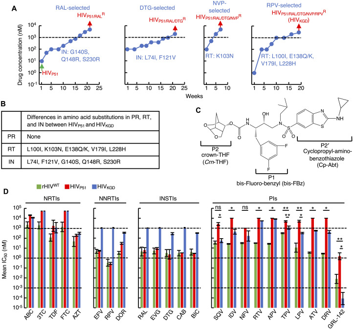
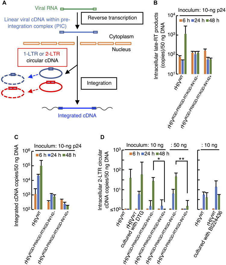
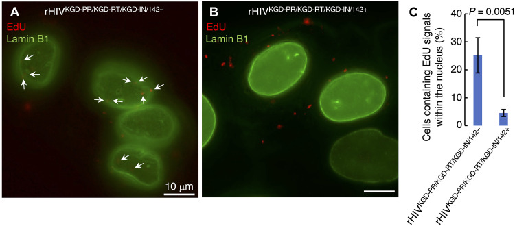
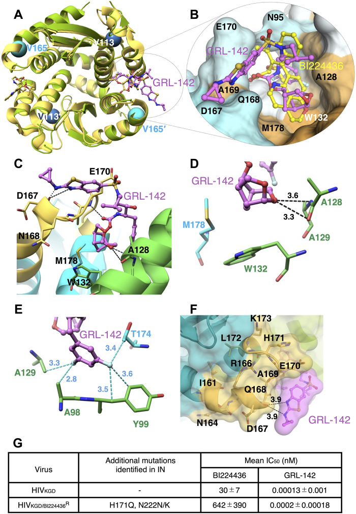
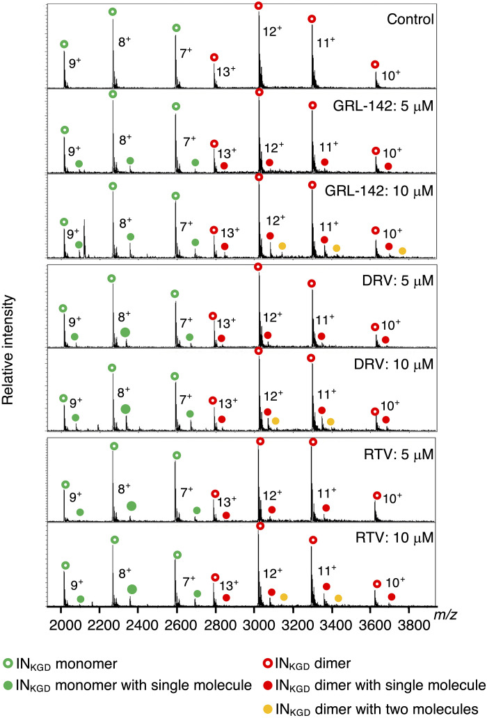
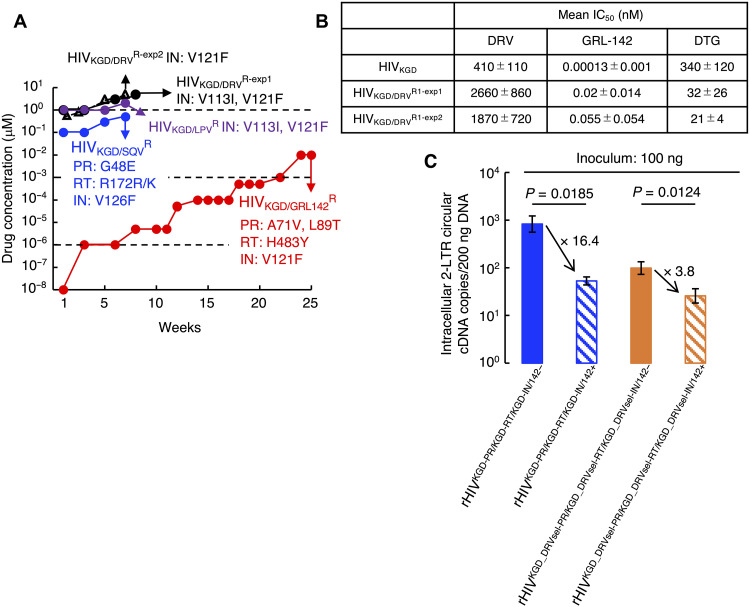
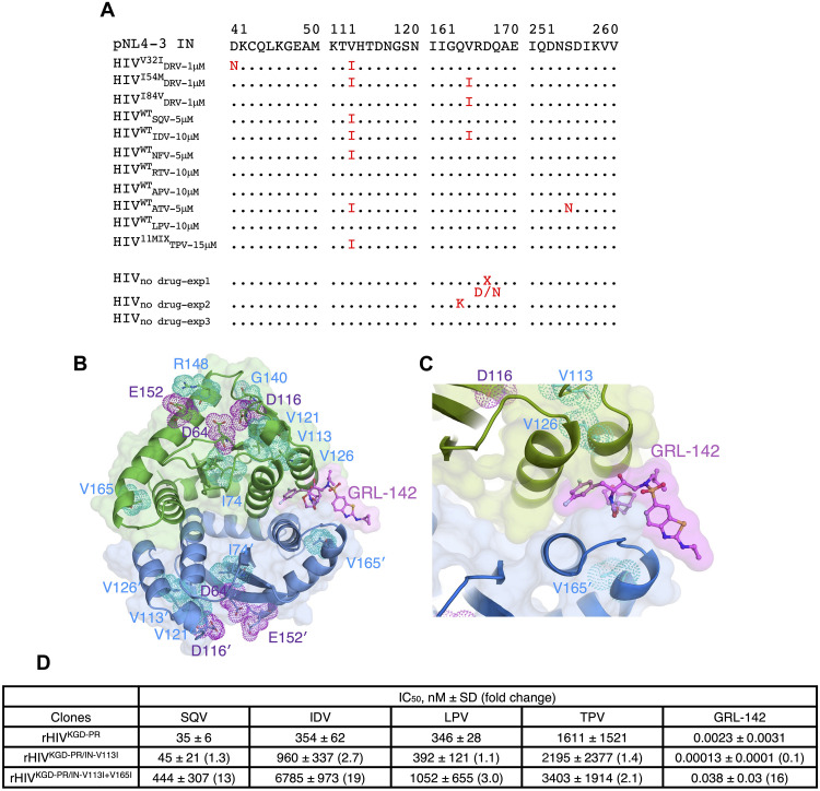
Nuclear localization signal (NLS) of HIV-1 integrase (IN) is implicated in nuclear import of HIV-1 preintegration complex (PIC). Here, we established a multiclass drug-resistant HIV-1 variant (HIVKGD) by consecutively exposing an HIV-1 variant to various antiretroviral agents including IN strand transfer inhibitors (INSTIs). HIVKGD was extremely susceptible to a previously reported HIV-1 protease inhibitor, GRL-142, with IC50 of 130 femtomolar. When cells were exposed to HIVKGD IN-containing recombinant HIV in the presence of GRL-142, significant decrease of unintegrated 2-LTR circular cDNA was observed, suggesting that nuclear import of PIC was severely compromised by GRL-142. X-ray crystallographic analyses revealed that GRL-142 interacts with NLS's putative sequence (DQAEHLK) and sterically blocks the nuclear transport of GRL-142-bound HIVKGD's PIC. Highly INSTI-resistant HIV-1 variants isolated from heavily INSTI-experienced patients proved to be susceptible to GRL-142, suggesting that NLS-targeting agents would serve as salvage therapy agents for highly INSTI-resistant variant-harboring individuals. The data should offer a new modality to block HIV-1 infectivity and replication and shed light on developing NLS inhibitors for AIDS therapy.


Figures

Fig. 1.
Fig. 1.. Generation of a four-class antiretroviral-resistant HIV-1 variant (HIVKGD) and its susceptibility to antiretrovirals.
(A) HIVP51, a highly NRTI/PI-resistant HIV-1 variant previously obtained through in vitro selection (21), indicated by a green arrow, was cultured in the presence of gradually increasing concentrations of RAL. After 22 weeks, a highly RAL-resistant HIV-1 variant (HIVP51/RALR), indicated by a red arrow in the very left, well replicated in the presence of 5 μM RAL and had acquired three mutations (G140S, Q148R, and S230R) in IN. HIVP51/RALR was further propagated in the presence of DTG, well replicated in the presence of 2 μM DTG in 21 weeks, and had acquired additional two mutations (L74I and F121V) in IN. The resultant highly DTG-resistant HIV-1 variant is referred to as HIVP51/RAL/DTGR, indicated by a red arrow in the second from the left. HIVP51/RAL/DTGR was further propagated in the presence of 5 μM NVP and acquired K103N in RT in 7 weeks, referred to as HIVP51/RAL/DTG/NVPR, indicated by a red arrow in the third from left. HIVP51/RAL/DTG/NVPR was further selected with RPV. After 18 weeks, HIVP51/RAL/DTG/NVP/RPVR was obtained and referred to as HIVKGD, indicated by a red arrow in the very right. (B) Amino acid substitutions in the polymerase region of HIVP51 and HIVKGD identified through cloning (see details in table S2) are shown. Note that differences in amino acid substitutions in PR, RT, and IN between HIVP51 and HIVKGD are illustrated. (C) Chemical structure of GRL-142. (D) Mean IC50 values of 5 NRTIs, 3 NNRTIs, 5 INSTIs, and 10 PIs including an experimental drug, GRL-142 (19), against rHIVWT, HIVP51, and HIVKGD are shown. Notably, IC50s of GRL-142 were 1.8 nM against HIVP51 and 0.00013 nM against HIVKGD. P values were determined using the Student’s t test (EZR software). ns, not significant; *P < 0.01 and **P < 0.05.
Fig. 2.
Fig. 2.. GRL-142 significantly decreases 2-LTR circular DNA production.
(A) Schematic representation of early stages of HIV-1 replication. The viral RNA genome is converted to a linear double-stranded viral cDNA through reverse transcription. The viral cDNA is subsequently transported into the nucleus and integrated into the cellular DNA, while unintegrated viral cDNA is circularized and form 1-LTR or 2-LTR circular cDNA. (B and C) rHIVKGD-PR/KGD-RT/KGD-IN/142− and rHIVKGD-PR/KGD-RT/KGD-IN/142+ were produced through transfection in the absence or presence of 0.5 nM GRL-142, respectively, and served as a source of infectious clones, together with rHIVWT. MT-4 cells were infected with each recombinant clone (calibrated with 10 ng of p24 Gag protein), cultured for 6, 24, and 48 hours, high–molecular weight DNA was purified, and the amounts of late reverse transcription products and integrated cDNA copies in 50 ng of the DNA were determined using real-time PCR. Note that rHIVWT showed robust infectivity, increasingly produced daughter virions, and showed an increase in late–reverse transcription cDNA products and integrated cDNA. While rHIVKGD-PR/KGD-RT/KGD-IN/142− had moderated levels of intracellular and integrated cDNA, rHIVKGD-PR/KGD-RT/KGD-IN/142+ had decreased intracellular and integrated cDNA. (D) MT-4 cells were exposed to rHIVWT, rHIVKGD-PR/KGD-RT/KGD-IN/142−, or rHIVKGD-PR/KGD-RT/KGD-IN/142+ (calibrated with 10 or 50 ng of p24 Gag protein), cultured for 6, 24, and 48 hours, high–molecular weight DNA was purified, and the amounts of intracellular 2-LTR circular cDNA in 50 ng of the DNA were determined using real-time PCR. Note that in MT-4 cells infected with rHIVWT and cultured with 1 μM DTG, large amounts of 2-LTR circular cDNA were seen; however, in MT-4 cells exposed to rHIVKGD-PR/KGD-RT/KGD-IN/142+ had significantly decreased 2-LTR circular cDNA compared to those exposed to rHIVKGD-PR/KGD-RT/KGD-IN/142−. Values were obtained from three independent experiments, and the values below 1 were treated as 1 copy since the detection limit was 1 copy. Geometric means with 1 SD values shown. *P = 0.000189 and **P = 0.00108.
Fig. 3.
Fig. 3.. Determination of localization of viral cDNA using click labeling with EdU.
Thymidine-blocked TZM-b1 cells were infected with (A) rHIVKGD-PR/KGD-RT/KGD-IN/142− and (B) rHIVKGD-PR/KGD-RT/KGD-IN/142+ and cultured for 12 hours in the presence of thymidine and EdU, followed by fixation, click labeling, and immunofluorescence staining with anti–Lamin B1 antibody. EdU signals found within the nucleus are indicated by white arrows (C). The number of cells containing EdU signals only within the nucleus was divided by the number of cells positive for EdU signals. Data shown represent mean values derived from the results of three independent experiments.
Fig. 4.
Fig. 4.. Molecular interactions of GRL-142 with INKGD.
(A) X-ray crystallographic superimposition of INKGD CCD (green) in complex with GRL-142 (pink: 7UE1) and INWT CCD in complex with BI224436 (yellow: 6NUJ). Although structural comparison exhibits no major differences, one GRL-142 molecule binds per dimer in contrast to two BI-224436 molecules (built by symmetry operation). (B) Close-up view focused on the dimer interface where GRL-142 and BI-224436 are overlaid. Note that GRL-142 occupies the binding “groove” due to its larger molecule size. (C) Hydrogen bond interactions of GRL-142 with INKGD. Five hydrogen bonds are identified. A presumed functional NLS region (I161-K173) is shown in yellow, and GRL-142 conformation lays parallel to NLS and contacts D167 and Q168 on the exterior. (D) Crn-THF moiety of GRL-142 is deeply inserted into the hydrophobic cavity stacked on one side, while nonpolar portion of GRL-142 forms van der Waals interactions with the aromatic ring of W132. On the other side, oxygen atoms of the crn-THF moiety forms two hydrogen bonds with the main chains of A128 and A129. (E) Close-up view of fluorine atom interactions. Essential fluorine-mediated interactions are shown with cyan dashed lines. While one fluorine atom from GRL-142 engages in halogen interactions with the main chains of A98 and A129, the other fluorine atom forms halogen bonds with Y99 ring and T174. (F) The cyclopropylbenzothiazole group of GRL-142 protrudes from the external part of the binding pocket but forms weak hydrogen bond interactions with the main chain oxygen of D167 and Q168. The conformation of GRL-142 covers the large segment of 167-DQAEHLK-173 sequence of the NLS, thereby it is thought that GRL-142 sterically blocks the nuclear transport of INKGD contained in the HIV-1–PIC into the nucleus. (G) Mean IC50 values of BI224436 or GRL-142 against HIVKGD/BI224436R.
Fig. 5.
Fig. 5.. Native MS analysis of GRL-142–, DRV-, or RTV-bound INKGD.
INKGD (5 μM) was treated with 5 or 10 μM of each PI in 10 mM ammonium acetate. Relative native mass spectra of the INKGD with or without GRL-142, DRV, or RTV show signals of INKGD monomer (green open circles), INKGD monomer with a single PI molecule (green closed circles), INKGD dimer (red open circles), INKGD dimer with a single PI molecule (red closed circles), and INKGD dimer with two PI molecules (orange closed circles). Charge states 7+, 8+, and 9+ are annotated to mass spectra corresponding to INKGD monomer species, and charge states 10+, 11+, 12+, and 13+ are annotated to mass spectra corresponding to INKGD dimer species. m/z, mass/charge ratio.
Fig. 6.
Fig. 6.. V121F in HIVKGD reverts to wild-type phenylalanine after reselection with DRV and the resultant HIVKGD/DRVR is DTG-sensitive and GRL-142–resistant.
(A) HIVKGD was propagated in the presence of 1 μM DRV (black solid circles and black open triangles), 1 μM LPV (green solid circles), 0.1 μM SQV (blue solid circles), or 10 fM GRL-142 (red solid circles) at the first week, and viruses lastly obtained were as follows: HIVKGD/DRVR-exp1, indicated by a black arrow, was well-replicating in the presence of 5 μM DRV in 7 weeks and had V121F in IN; HIVKGD/DRVR-exp2, indicated by black solid arrows, was well-replicating in 5 μM DRV in 8 weeks and had V113I and V121F in IN; HIVKGD/LPVR, indicated by a green arrow, was replicating in 2 μM LPV in 7 weeks and had V113I and V121F in IN; HIVKGD/SQVR, indicated by a blue arrow, was replicating in the presence of 0.5 μM SQV in 7 weeks and had G48E in PR, R172R/K in RT, and V126F in IN; and HIVKGD/GRL-142R, indicated by a red arrow, was replicating in the presence of 0.01 μM GRL-142 in 25 weeks and had A71V and L89T in PR, H483Y in RT, and V113I in IN. (B) Mean IC50 values of DTG, DRV, or GRL-142 against two HIVKGD/DRVRs obtained are shown. (C) MT-4 cells were infected with rHIVKGD-PR/KGD-RT/KGD-IN/142−, rHIVKGD-PR/KGD-RT/KGD-IN/142+, rHIVKGD_DRVsel-PR/KGD_DRVsel-RT/KGD_DRVsel-IN-142−, or rHIVKGD_DRVsel-PR/KGD_DRVsel-RT/KGD_DRVsel-IN/142+ (calibrated with 100 ng of p24 Gag protein), cultured for 24 hours, high–molecular weight DNA was purified, and the amounts of intracellular 2-LTR circular cDNA in 200 ng of the DNA were determined using real-time PCR. Values were obtained from three independent experiments. Geometric means with 1 SD values shown.
Fig. 7.
Fig. 7.. Amino acid substitutions identified in the IN-encoding gene of various in vitro PI-selected HIV-1 variants and susceptibility of recombinant clones containing selected amino acid substitutions.
(A) Selected amino acid sequences of IN of 11 highly PI-resistant HIV-1 variants obtained through in vitro selection are shown. HIVDRVV32I-1μM, HIVDRVI54M-1μM, and HIVDRVI84V-1μM were selected with DRV for 29 to 30 weeks using rHIVWT carrying V32I, I54M, or I84V in PR. HIVSQV-5μM, HIVIDV-10μM, HIVNFV-5μM, HIVRTV-10μM, HIVAPV-10μM, HIVATV-5μM, and HIVLPV-10μM were generated by selecting rHIVWT with SQV, IDV, NFV, RTV, APV, ATV, or LPV. HIVTPV-15μM was selected with TPV using a mixture of 11 multidrug-resistant clinical HIV-1 isolates as a starting viral population. Amino acid substitutions which occur upon passaging without PIs over 50 weeks are shown for rHIVWTno drug-1, rHIVWTno drug-2, and rHIVWTno drug-3, serving as controls. (B and C) X-ray crystal structures of the CCD of IN of HIVKGD (INKGD residues 50 to 212) in complex with GRL-142. Figure 5B shows the ribbon representation of the dimerized INKGD (each protomer is shown in blue or green) complexed with GRL-142 (pink), which binds to the dimer interface. The three catalytic residues (D64, D116, and E152) are shown in purple. The locations of seven amino acid substitutions (L74I, V113I, V121F, V126I, G140S, Q148R, and V165I) identified following the in vitro selection with DTG or PIs in the present work are shown in light blue. (D) Susceptibility of rHIV-1 containing KGD’s PR plus V113I in IN (rHIVKGD-PR/IN-V113I) and rHIV-1 containing KGD’s PR plus V113I/V165I in IN (rHIVKGD-PR/IN-V113I+V165I) to various PIs is shown. Note that the addition of V165I substitution decreases the susceptibility of rHIVKGD-PR/IN-V113I to all PIs examined.

Similar articles

Cited by

References

    1. J. L. Marcus, C. R. Chao, W. A. Leyden, L. Xu, C. P. Quesenberry Jr., D. B. Klein, W. J. Towner, M. A. Horberg, M. J. Silverberg, Narrowing the gap in life expectancy between HIV-infected and HIV-uninfected individuals with access to care. J. Acquir. Immune Defic. Syndr. 73, 39–46 (2016). - PMC - PubMed
    1. Antiretroviral Therapy Cohort ollaboration , Survival of HIV-positive patients starting antiretroviral therapy between 1996 and 2013: A collaborative analysis of cohort studies. Lancet HIV 4, e349–e356 (2017). - PMC - PubMed
    1. D. Armenia, D. Di Carlo, P. Flandre, Y. Bouba, V. Borghi, F. Forbici, A. Bertoli, C. Gori, L. Fabeni, W. Gennari, C. Pinnetti, A. Mondi, S. Cicalini, R. Gagliardini, A. Vergori, R. Bellagamba, V. Malagnino, F. Montella, M. Colafigli, A. Latini, R. Marocco, M. Licthner, M. Andreoni, C. Mussini, F. Ceccherini-Silberstein, A. Antinori, C. F. Perno, M. M. Santoro, HIV MDR is still a relevant issue despite its dramatic drop over the years. J. Antimicrob. Chemother. 75, 1301–1310 (2020). - PubMed
    1. R. M. Kagan, K. J. Dunn, G. P. Snell, R. E. Nettles, H. W. Kaufman, Trends in HIV-1 drug resistance mutations from a U.S. Reference Laboratory from 2006 to 2017. AIDS Res. Hum. Retroviruses 35, 698–709 (2019). - PubMed
    1. D. Grover, A. Copas, H. Green, S. G. Edwards, D. T. Dunn, C. Sabin, A. Phillips, E. Allen, D. Pillay; UK Collaborative Group on HIV Drug Resistance and UK Collaborative HIV Cohort Study (UK CHIC) , What is the risk of mortality following diagnosis of multidrug-resistant HIV-1? J. Antimicrob. Chemother. 61, 705–713 (2008). - PubMed

Substances