. 2023 Jul 12;15(704):eabq6225.
doi: 10.1126/scitranslmed.abq6225. Epub 2023 Jul 12.

RNF41 orchestrates macrophage-driven fibrosis resolution and hepatic regeneration

Affiliations

RNF41 orchestrates macrophage-driven fibrosis resolution and hepatic regeneration

Alazne Moreno-Lanceta et al. Sci Transl Med. .

Abstract

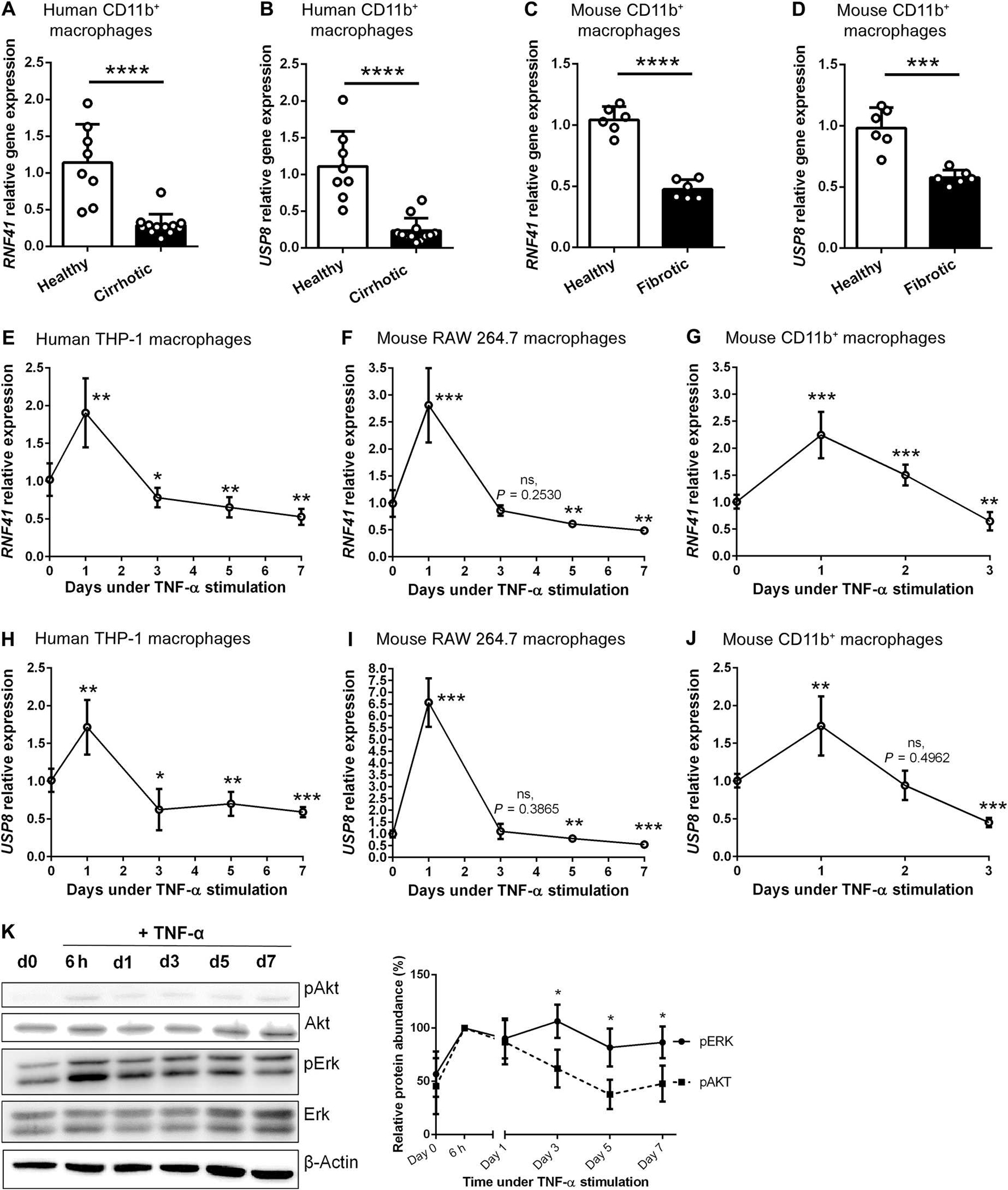
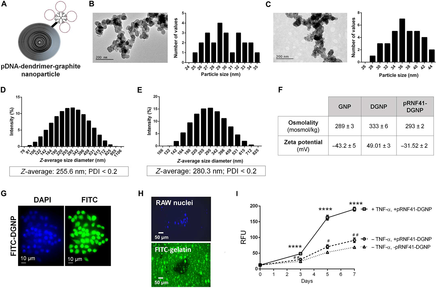
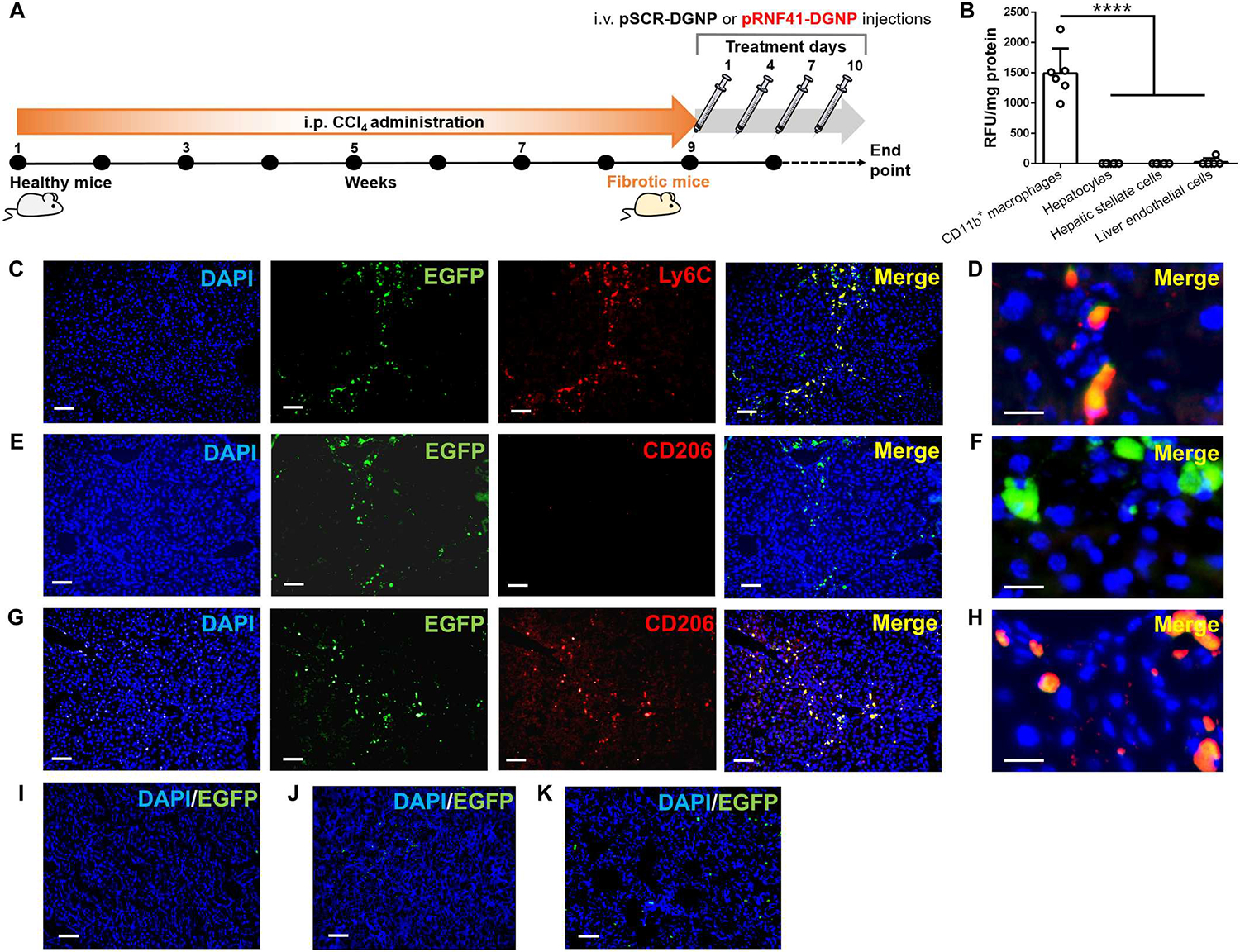
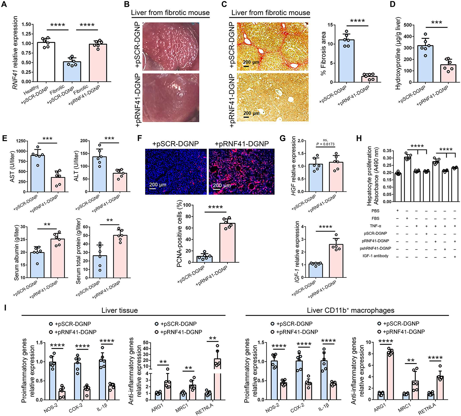
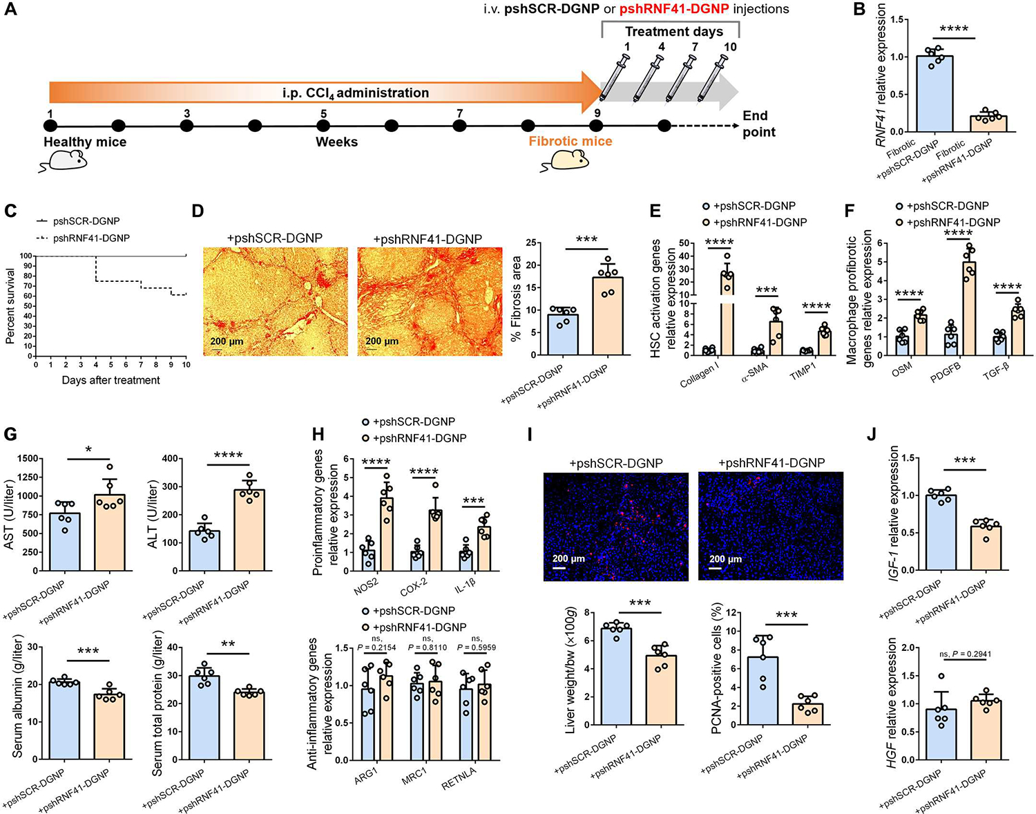
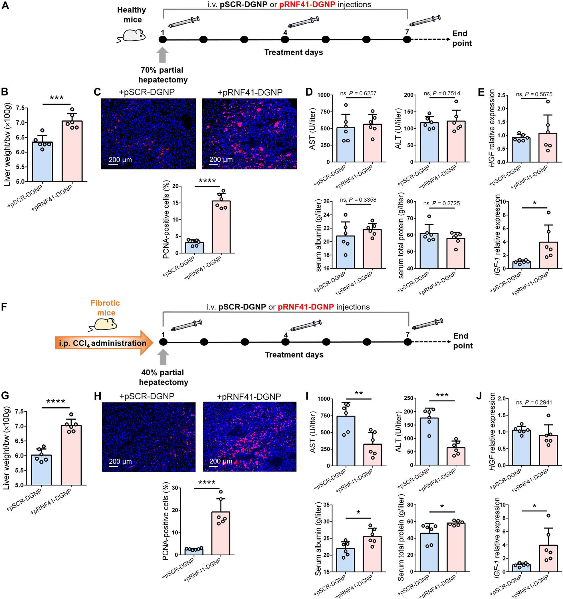
Hepatic inflammation is a common trigger of chronic liver disease. Macrophage activation is a predictive parameter for survival in patients with cirrhosis. Ring finger protein 41 (RNF41) negatively regulates proinflammatory cytokines and receptors; however, the precise involvement of macrophage RNF41 in liver cirrhosis remains unknown. Here, we sought to understand how RNF41 dictates macrophage fate in hepatic fibrosis and repair within the inflammatory milieu. We found that RNF41 expression is down-regulated in CD11b+ macrophages recruited to mouse fibrotic liver and to patient cirrhotic liver regardless of cirrhosis etiology. Prolonged inflammation with TNF-α progressively reduced macrophage RNF41 expression. We designed a macrophage-selective gene therapy with dendrimer-graphite nanoparticles (DGNPs) to explore the influence of macrophage RNF41 restoration and depletion in liver fibrosis and regeneration. RNF41 expression induced in CD11b+ macrophages by DGNP-conjugated plasmids ameliorated liver fibrosis, reduced liver injury, and stimulated hepatic regeneration in fibrotic mice with or without hepatectomy. This therapeutic effect was mainly mediated by the induction of insulin-like growth factor 1. Conversely, depletion of macrophage RNF41 worsened inflammation, fibrosis, hepatic damage, and survival. Our data reveal implications of macrophage RNF41 in the control of hepatic inflammation, fibrosis, and regeneration and provide a rationale for therapeutic strategies in chronic liver disease and potentially other diseases characterized by inflammation and fibrosis.


Figures

Fig. 1.
Fig. 1.. Macrophage RNF41 and its stabilizer USP8 are down-regulated in cirrhotic liver in part due to chronic inflammation.
(A) RNF41 expression in CD11b+ macrophages isolated from the livers of patients with liver cirrhosis (n = 12) and healthy participants (n = 8). (B) USP8 expression in CD11b+ macrophages isolated from the livers of patients with liver cirrhosis (n = 12) and healthy participants (n = 8). (C) RNF41 expression in CD11b+-macrophages isolated from the livers of healthy and fibrotic mice (n = 6 per group). (D) USP8 expression in CD11b+-macrophages isolated from the livers of healthy and fibrotic mice (n = 6 per group). (E) RNF41 expression in THP-1 macrophages stimulated with TNF-α for 7 days. (F) RNF41 expression in RAW 264.7 macrophages stimulated with TNF-α for 7 days. (G) RNF41 expression in freshly isolated primary hepatic CD11b+ macrophages stimulated with TNF-α for 3 days. (H) USP8 expression in THP-1 macrophages stimulated with TNF-α for 7 days. (I) USP8 expression in RAW 264.7 macrophages stimulated with TNF-α for 7 days. (J) USP8 expression in freshly isolated primary hepatic CD11b+ macrophages stimulated with TNF-α for 3 days. Experiments in (E) and (F) to (J) were performed in triplicates in two independent experiments. (K) Western blot analysis of phospho-Akt, total Akt, phospho-Erk, total Erk, and β-actin in RAW 264.7 macrophages stimulated with TNF-α for 7 days and relative protein abundance (%) of phospho-Akt and phospho-Erk relative to β-actin (n = 3). For (A) to (D), Student’s t test. For (E) to (J), versus day 0 using Student’s t test with Benjamini-Hochberg correction for multiple comparisons. For (K), comparison between pAKT protein abundance and pERK protein abundance in each time point using Student’s t test. Data are shown as means ± SD. *P ≤ 0.05, **P ≤ 0.01, ***P ≤ 0.001, and ****P ≤ 0.0001. ns, not significant.
Fig. 2.
Fig. 2.. Dendrimer-graphite nanoparticles are macrophage-selective plasmid-delivery vectors for effective gene therapy.
(A) Structure of graphite nanoparticles linked to dendrimer and plasmid DNA. (B) Particle size (in nanometers) of graphite nanoparticles measured using TEM images. (C) Particle size (in nanometers) of dendrimer-graphite nanoparticles measured using TEM images. (D) Z-average size and polydispersity index (PDI) of graphite nanoparticles measured using dynamic light scattering. (E) Z-average size and PDI of dendrimer-graphite nanoparticles measured using dynamic light scattering. (F) Osmolality and zeta potential of every graphite nanoparticle composite. (G) RAW 264.7 macrophage intracellular distribution of FITC-dendrimer-graphite nanoparticles. (H) Fluorescence images of RAW 264.7 macrophages seeded on FITC-gelatin–coated plates and treated with dendrimer-graphite nanoparticles linked to pRNF41 (pRNF41-DGNP) for 5 days displaying a black halo indicating collagen digestion and green nuclear staining indicating EGFP expression. (I) Time-course quantitative analysis of FITC released to the medium in the gelatinase activity assay in the presence or absence of TNF-α or pRNF41-DGNP for 7 days (n = 3 to 5). For (I), ****P ≤ 0.0001 versus macrophages without TNF-α and with or without pRNF41-DGNP, #P ≤ 0.05 versus macrophages without TNF-α and pRNF41-DGNP, and ##P ≤ 0.01 versus macrophages without TNF-α and pRNF41-DGNP at the same time point using a one-way analysis of variance (ANOVA) with posthoc Newman-Keuls test. RFU, relative fluorescence units. Data are shown as means ± SD.
Fig. 3.
Fig. 3.. Dendrimer-graphite nanoparticles efficiently and selectively transfect a RNF41-encoding plasmid into macrophages recruited to mouse fibrotic liver.
(A) Schematic figure illustrating the time points of fibrosis induction with CCl4 and the administration schedule of dendrimer-graphite nanoparticles linked to plasmid pRNF41 (pRNF41-DGNPs) or scrambled pSCR (pSCR-DGNPs). (B) Relative fluorescence units (RFU) of EGFP per milligram of protein of liver isolated CD11b+ macrophages, hepatocytes, hepatic stellate cells, and liver endothelial cells from mice treated with pSCR-DGNPs (n = 6). (C) Immunofluorescence (IF) staining for Ly6C and simultaneous detection of EGFP fluorescence in the livers of fibrotic mice treated with pSCR-DGNPs. (D) High-power image for Ly6C and simultaneous detection of EGFP fluorescence in the livers of fibrotic mice treated with pSCR-DGNPs. (E) IF staining for CD206 and simultaneous detection of EGFP fluorescence in the livers of fibrotic mice treated with pSCR-DGNPs. (F) High-power image for CD206 and simultaneous detection of EGFP fluorescence in the liver of fibrotic mice treated with pSCR-DGNPs. (G) IF staining for CD206 and simultaneous detection of EGFP fluorescence in the livers of fibrotic mice treated with pRNF41-DGNPs. (H) High-power image for CD206 and simultaneous detection of EGFP fluorescence in the livers of fibrotic mice treated with pRNF41-DGNPs. (I) EGFP fluorescence in the kidneys of fibrotic animals treated with pSCR-DGNPs. (J) EGFP fluorescence in the spleens of fibrotic animals treated with pSCR-DGNPs. (K) EGFP fluorescence in the lungs of fibrotic mice treated with pSCR-DGNPs. All plasmids constitutively expressed EGFP under the control of a CMV promoter. Scale bars, 200 μm (C, E, G, I, J, and K) and 20 μm (D, F, and H). For (B), ****P ≤ 0.0001 versus CD11b+ macrophages using a one-way analysis of variance (ANOVA) with the posthoc Newman-Keuls test. i.p., intraperitoneal; i.v., intravenous.
Fig. 4.
Fig. 4.. RNF41 restoration in macrophages located into the fibrotic liver orchestrates fibrosis and inflammation regression and reduction of hepatic injury in mice.
(A) RNF41 abundance in healthy and fibrotic mice treated with dendrimer-graphite nanoparticles linked to plasmid pRNF41 (pRNF41-DGNPs) or scrambled pSCR (pSCR-DGNPs). (B) Macroscopic aspect of fibrotic liver after treatment with pRNF41-DGNPs. (C to G) Sirius Red staining and quantification of liver fibrosis area (C), hydroxyproline measurements (D), serum liver injury parameters [ALT (alanine aminotransferase), AST (aspartate aminotransferase), serum albumin, and serum total protein] (E), hepatic PCNA immunofluorescence staining (F), and hepatic expression of HGFand IGF-1 (G) in fibrotic mice treated with pSCR-DGNPs or pRNF41-DGNPs. (H) Cell proliferation in isolated mouse hepatocytes treated for 24 hours with conditioned medium from RAW 264.7 cultures treated with FBS, TNF-α, pSCR-DGNPs, pRNF41-DGNPs, or IGF-1 antibody for 3 days. Experiments were performed in triplicates in two independent experiments. (I) Expression of proinflammatory and anti-inflammatory genes in liver tissue and in CD11b+ macrophages isolated from the livers of fibrotic mice treated with pSCR-DGNPs or pRNF41-DGNPs. n = 6 animals per group. Student’s t test was used for (A), (C) to (G), and (I), and a one-way ANOVA with posthoc Newman-Keuls test was used for (H). Data are shown as means ± SD. **P ≤ 0.01, ***P ≤ 0.001, and ****P ≤ 0.0001.
Fig. 5.
Fig. 5.. Depletion of macrophage RNF41 worsens fibrosis, inflammation, and hepatic damage in mice with liver fibrosis.
(A) Schematic figure illustrating time points of fibrosis induction with CCl4 and administration schedule of dendrimer-graphite nanoparticles linked to pshSCR (pshSCR-DGNPs) or pshRNF41 (pshRNF41-DGNPs). (B and C) Hepatic gene expression of RNF41 (B) and survival rate (C) of fibrotic mice treated with pshSCR-DGNPs or pshRNF41-DGNPs. (D to J) Sirius red staining (D), gene expression related to hepatic stellate cell (HSC) activation (E) or profibrogenic agents produced by liver-resident macrophages (F), serum parameters of liver injury (ALT, AST, serum albumin, and serum total protein) (G), expression of inflammatory and anti-inflammatory macrophage genes (H), PCNA immunofluorescence staining (I), and HGF and IGF-1 expression (J) in liver of fibrotic mice treated with pshSCR-DGNPs or pshRNF41-DGNPs. n = 6 per group of animals. *P ≤ 0.05, **P ≤ 0.01, ***P ≤ 0.001, and ****P ≤ 0.0001 using Student’s t test. Data are shown as means ± SD.
Fig. 6.
Fig. 6.. Macrophage RNF41 induces liver regeneration after hepatectomy.
(A) Schematic figure illustrating 70% hepatectomy in healthy mice and administration schedule of dendrimer-graphite nanoparticles linked to pRNF41 (pRNF41-DGNPs) or pSCR (pSCR-DGNPs). (B to E) Liver restoration rate (B), PCNA immunofluorescence staining (C), serum liver injury parameters [ALT, AST, serum albumin, and serum total protein] (D), and HGF and IGF-1 expression (E) in healthy mice undergoing 70% hepatectomy and treated with pSCR-DGNPs or pRNF41-DGNPs. (F) Schematic figure illustrating the time points of fibrosis induction with CCl4, 40% hepatectomy in fibrotic mice, and the administration schedule of pSCR-DGNPs or pRNF41-DGNPs. (G to J) Liver restoration rate (G), PCNA immunofluorescence staining (H), serum liver injury parameters (ALT, AST, serum albumin, and serum total protein) (I), and expression of HGF and IGF-1 in the livers of fibrotic mice undergoing 40% hepatectomy and treated with pSCR-DGNPs or pRNF41-DGNPs. n = 6 animals per group. *P ≤ 0.05, **P ≤ 0.01, ***P ≤ 0.001, and ****P ≤ 0.0001 using Student’s t test. Data are shown as means ± SD.

Comment in

  • RNF41 reverses liver fibrosis.
    Crunkhorn S. Crunkhorn S. Nat Rev Drug Discov. 2023 Sep;22(9):697. doi: 10.1038/d41573-023-00125-6. Nat Rev Drug Discov. 2023. PMID: 37528208 No abstract available.

References

    1. Asrani SK, Devarbhavi H, Eaton J, Kamath PS, Burden of liver diseases in the world. J. Hepatol. 70, 151–171 (2019). - PubMed
    1. Melgar-Lesmes P, Edelman ER, Monocyte-endothelial cell interactions in the regulation of vascular sprouting and liver regeneration in mouse. J. Hepatol. 63, 917–925 (2015). - PMC - PubMed
    1. Melgar-Lesmes P, Balcells M, Edelman ER, Implantation of healthy matrix-embedded endothelial cells rescues dysfunctional endothelium and ischaemic tissue in liver engraftment. Gut 66, 1297–1305 (2017). - PMC - PubMed
    1. Oro D, Yudina T, Fernandez-Varo G, Casals E, Reichenbach V, Casals G, González de la Presa B, Sandalinas S, Carvajal S, Puntes V, Jiménez W, Cerium oxide nanoparticles reduce steatosis, portal hypertension and display anti-inflammatory properties in rats with liver fibrosis. J. Hepatol. 64, 691–698 (2016). - PubMed
    1. Cordoba-Jover B, Arce-Cerezo A, Ribera J, Pauta M, Oró D, Casals G, Fernández-Varo G, Casals E, Puntes V, Jiménez W, Morales-Ruiz M, Cerium oxide nanoparticles improve liver regeneration after acetaminophen-induced liver injury and partial hepatectomy in rats. J. Nanobiotechnol. 17, 112 (2019). - PMC - PubMed

Publication types