. 2023 Aug 15;62(16):2503-2515.
doi: 10.1021/acs.biochem.3c00165. Epub 2023 Jul 12.

Computational Exploration of Potential CFTR Binding Sites for Type I Corrector Drugs

Affiliations

Computational Exploration of Potential CFTR Binding Sites for Type I Corrector Drugs

Anna Lester et al. Biochemistry. .

Abstract

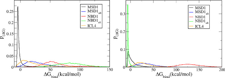
Cystic fibrosis (CF) is a recessive genetic disease that is caused by mutations in the cystic fibrosis transmembrane conductance regulator (CFTR) protein. The recent development of a class of drugs called "correctors", which repair the structure and function of mutant CFTR, has greatly enhanced the life expectancy of CF patients. These correctors target the most common disease causing CFTR mutant F508del and are exemplified by the FDA-approved VX-809. While one binding site of VX-809 to CFTR was recently elucidated by cryo-electron microscopy, four additional binding sites have been proposed in the literature and it has been theorized that VX-809 and structurally similar correctors may engage multiple CFTR binding sites. To explore these five binding sites, ensemble docking was performed on wild-type CFTR and the F508del mutant using a large library of structurally similar corrector drugs, including VX-809 (lumacaftor), VX-661 (tezacaftor), ABBV-2222 (galicaftor), and a host of other structurally related molecules. For wild-type CFTR, we find that only one site, located in membrane spanning domain 1 (MSD1), binds favorably to our ligand library. While this MSD1 site also binds our ligand library for F508del-CFTR, the F508del mutation also opens a binding site in nucleotide binding domain 1 (NBD1), which enables strong binding of our ligand library to this site. This NBD1 site in F508del-CFTR exhibits the strongest overall binding affinity for our library of corrector drugs. This data may serve to better understand the structural changes induced by mutation of CFTR and how correctors bind to the protein. Additionally, it may aid in the design of new, more effective CFTR corrector drugs.


Conflict of interest statement

The authors declare no competing financial interest.

Figures

Figure 1
Figure 1
(Left) Pictorial representation of the CFTR structure with key structural elements labeled. (Right) Starting structure for molecular dynamics simulations conducted (water is omitted for clarity). The MSD1 and NBD1 domains are colored green, MSD2 and NBD2 domains are colored blue, the R domain is red, and the lipid bilayer is gray. The sidechain of F508 is shown in an orange van der Waals rendering (between yellow and green circles). The colored circles indicate the general location of the five potential binding sites investigated in this study. The sites are colored as such: MSD1 is yellow, MSD1alt is cyan, NBD1 is red, NBD1alt is blue, and CL4 is green.
Figure 2
Figure 2
Structures of CFTR modulators that are FDA-approved or in clinical trials for the treatment of CF. The 1-(2,2-difluorobenzo[d][1,3]dioxol-5-yl)cyclopropane-1-carboxamide moiety shared by VX-809, VX-661, and ABBV-2222 is highlighted in red.
Figure 3
Figure 3
F508del mutant was created from (a) the wild-type CFTR. (b) Residue F508 was removed from the sequence, (c) a new peptide bond was created between residues I507 and G509, and (d) the new bond was relaxed with a short minimization.
Figure 4
Figure 4
Representative structures of the (a) VX-809 structural class reported in the primary and patent literature, (b) VX-661, (c) ABBV-2222 structural class, (d) VX-809 thiazole analog structural class developed via docking studies of VX-809 to NBD1, and (e) ARN structural series hypothesized to share the VX-809 binding site.
Figure 5
Figure 5
(Left) Root-mean-square deviation of the WT (black) and the MT (red) over the course of the simulation. (Right) Average b-factor of each residue for each system. Major structural regions are marked using the colored bar at the bottom.
Figure 6
Figure 6
(Left) Superimposition of the final structures of the wild type (green) and the mutant (gray). (Right) Calculated RMSF between the same two structures. Major structural regions are marked using the colored bar at the bottom.
Figure 7
Figure 7
Distribution of binding scores for each binding site for (left) the WT and (right) the mutant. Here, the MSD1 site is black, MSD1alt site is blue, NBD1 is red, NBD1alt site is green, and ICL4 is yellow.
Figure 8
Figure 8
VX-809 bound to the MSD1 site of the wild type (top) and the mutant (bottom). (Left) Entire protein is shown with the calculated electrostatic surface and the ligand is shown in a green surface rendering. (Middle) Left figure is rotated ∼90° and ligand is shown in a licorice rendering. (Right) Zoomed-in version of the middle figure. For all electrostatic surfaces, negative regions are indicated by red, white is neutral, and blue is positive.
Figure 9
Figure 9
Top pose of VX-809 bound to the NBD1alt site of the wild type (top) and the F508del mutant (bottom). (Left) Protein is shown in gray cartoon rendering and ligand is shown in green surface rendering. (Middle) Calculated electrostatic surface of the protein is shown in a semi-transparent rendering (for clarity) along with the ligand. (Right) Zoomed-in version of the middle figure with the ligand shown in a licorice rendering. For all electrostatic surfaces, negative regions are indicated by red, white is neutral, and blue is positive.
Figure 10
Figure 10
Interactions found in the binding pocket for the best binding poses of VX-809 for each system. The ligand is shown bound to (top left) the WT at the MSD1 site, (top right) the MT at the MSD1 site, (bottom left) the WT at the NBD1alt site, and (bottom right) the MT at the NBD1alt site. The coloring scheme is as follows: carbon (black), oxygen (red), nitrogen (blue), and fluorine (pink). Note that hydrogens are not shown here for clarity. Red dashes indicate nearby residues and the atoms they interact with, green dashes indicate hydrogen bonds and their length. Purple lines indicate a bond between ligand and protein (note, these are an artifact of poor docking conditions).

References

    1. Dagenais R. V. E.; Su V. C.; Quon B. S. Real-World Safety of CFTR Modulators in the Treatment of Cystic Fibrosis: A Systematic Review. J. Clin. Med. 2021, 10, 23.10.3390/jcm10010023. - DOI - PMC - PubMed
    1. Middleton P. G.; Mall M. A.; Dřevínek P.; Lands L. C.; McKone E. F.; Polineni D.; Ramsey B. W.; Taylor-Cousar J. L.; Tullis E.; Vermeulen F.; Marigowda G.; McKee C. M.; Moskowitz S. M.; Nair N.; Savage J.; Simard C.; Tian S.; Waltz D.; Xuan F.; Rowe S. M.; Jain R. Elexacaftor–Tezacaftor–Ivacaftor for Cystic Fibrosis with a Single Phe 508del Allele. N. Engl. J. Med. 2019, 381, 1809–1819. 10.1056/NEJMoa1908639. - DOI - PMC - PubMed
    1. Zhang Z.; Liu F.; Chen J. Molecular Structure of the ATP-Bound, Phosphorylated Human CFTR. Proc. Natl. Acad. Sci. U. S. A. 2018, 115, 12757–12762. 10.1073/pnas.1815287115. - DOI - PMC - PubMed
    1. Mornon J.-P.; Lehn P.; Callebaut I. Molecular Models of the Open and Closed States of the Whole Human CFTR Protein. Cell. Mol. Life Sci. 2009, 66, 3469–3486. 10.1007/s00018-009-0133-0. - DOI - PMC - PubMed
    1. Okiyoneda T.; Veit G.; Dekkers J. F.; Bagdany M.; Soya N.; Xu H.; Roldan A.; Verkman A. S.; Kurth M.; Simon A.; Hegedus T.; Beekman J. M.; Lukacs G. L. Mechanism-Based Corrector Combination Restores ΔF508-CFTR Folding and Function. Nat. Chem. Biol. 2013, 9, 444–454. 10.1038/nchembio.1253. - DOI - PMC - PubMed

Publication types

LinkOut - more resources