. 2023 Aug 17;186(17):3642-3658.e32.
doi: 10.1016/j.cell.2023.06.013. Epub 2023 Jul 11.

Engineering RNA export for measurement and manipulation of living cells

Affiliations

Engineering RNA export for measurement and manipulation of living cells

Felix Horns et al. Cell. .

Abstract

A system for programmable export of RNA molecules from living cells would enable both non-destructive monitoring of cell dynamics and engineering of cells capable of delivering executable RNA programs to other cells. We developed genetically encoded cellular RNA exporters, inspired by viruses, that efficiently package and secrete cargo RNA molecules from mammalian cells within protective nanoparticles. Exporting and sequencing RNA barcodes enabled non-destructive monitoring of cell population dynamics with clonal resolution. Further, by incorporating fusogens into the nanoparticles, we demonstrated the delivery, expression, and functional activity of exported mRNA in recipient cells. We term these systems COURIER (controlled output and uptake of RNA for interrogation, expression, and regulation). COURIER enables measurement of cell dynamics and establishes a foundation for hybrid cell and gene therapies based on cell-to-cell delivery of RNA.

Keywords: RNA; delivery; export; extracellular vesicles; monitoring; non-destructive; reporter; virus-like particles.


Conflict of interest statement

Declaration of interests Patent applications have been filed by the California Institute of Technology related to this work (US application numbers 17/820,232 and 17/820,235). M.B.E. is a scientific advisory board member or consultant at TeraCyte, Primordium, and Spatial Genomics.

Figures
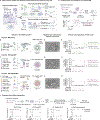
Figure 1.
Figure 1.. Engineered viral RNA exporters package, secrete, and protect RNA.
(A) RNA export enables non-destructive tracking of cell populations and cell-to-cell delivery of RNA. For delivery, exporter nanoparticles incorporate fusogens (yellow dots). (B, F, and I) Designs of viral RNA exporters, in which a capsid fused to an RNA binding domain packages RNA cargo bearing an export tag into virus-like particles (VLPs) that are secreted from cells. (C, G, and J) Cells expressing RNA exporters secreted VLPs (marked by arrowheads), as visualized by electron microscopy of purified culture supernatant. (D) Schematic of assay for abundance of exported RNA. (E, H, and K) Engineered RNA exporters secreted RNA bearing export tags efficiently with varying specificity. Each dot represents one technical replicate; solid line indicates mean of replicates. Black dashed line indicates lower limit of quantification. (L) Top: Schematic of RNase protection assay. Bottom: Exporters protected RNA from degradation by RNases. Unpackaged mRNA was not protected (right). Data for (L) are mean and standard deviation of three technical replicates. See also Figure S1 and Figure S4.
Figure 2.
Figure 2.. Engineered protein nanocages package, secrete, and protect RNA.
(A) Design model of nanocage (PDB 5KP9) has cavities that accommodate the RNA binding protein MCP (PDB 1MSC). N- and C-termini of nanocage protomer (marked by spheres) are surface-exposed and oriented towards the cavity. (B) Design of nanocage-based RNA exporter, in which self-assembling protomers fused to an RNA binding domain package RNA cargo bearing the MS2 export tag (8 MS2 hairpins, denoted MS2×8) into vesicles that are secreted from cells (schematic). (C) Architectures of candidate nanocage-based exporters. (D) Cells expressing nanocage-based exporters efficiently and specifically secreted RNA bearing export tags into culture supernatant, as measured by RT-qPCR. Each dot represents one technical replicate and bar indicates their mean. (E) Cells expressing EPN24-MCP secreted vesicles (marked by arrowheads), as visualized by electron microscopy of purified culture supernatant. (F) Nanocage-based exporters protected RNA from degradation by RNase. Data for (F) are mean and standard deviation of three technical replicates. See also Figure S2 and Figure S4.
Figure 3.
Figure 3.. Genome-scale characterization of RNA export.
(A) Workflow for sequencing-based analysis of RNA export efficiency, specificity, and bias. (B) Abundance of target RNA and non-target (endogenous) RNA in culture supernatant with and without expression of exporters. Iterative engineering (left to right) of RNA binding properties of exporters reduced non-target RNA enrichment, reflecting improved specificity, while efficient export of target RNA bearing export tags (star) was maintained. (C) Target RNA was enriched in culture supernatant in the presence of each exporter (compared to without the exporter). In (B) and (C), CPMS denotes counts per million of spike-in standard. (D) Engineering improved the specificity of exporters, thereby increasing the fractional abundance of target RNA among total supernatant RNA. (E) Transcript abundances were strongly correlated in the cellular and supernatant transcriptomes, indicating that exporters secreted unbiased samples of the cellular transcriptome. One notable exception was mtRNA, which was depleted in secreted RNA, likely due to its compartmentalization within the mitochondrial lumen. CPM, counts per million (not normalized to spike-in standard); ND, not detected. See also Figure S3.
Figure 4.
Figure 4.. RNA exporters are portable across cell types and species.
Human lymphoblastoid (A), human T lymphocyte (B), mouse fibroblast (C), and Chinese hamster ovary (D) cell lines efficiently exported cargo RNA bearing export tags, as measured by RT-qPCR. Each dot represents one technical replicate; colors represent biological replicates (distinct culture wells); bar indicates mean of replicates. Dashed line indicates lower limit of quantification. In (C) and (D), RNA abundances were normalized to account for varying transfection efficiency across cell lines.
Figure 5.
Figure 5.. RNA export enables monitoring of mammalian cell population dynamics.
(A) Cell population dynamics, including growth and death, can be monitored by longitudinal sampling of exported clonal barcode RNA. (B) Cells were engineered to inducibly express an RNA exporter (Gag-MCP), and genetically barcoded (rainbow) by transduction with a diverse lentiviral library at low multiplicity of infection (MOI < 0.1). Barcode transcripts contained the MS2 export tag. (C) Cells were barcoded, sorted, and cultured for 6 days in the presence of growth-altering drugs. Exported barcodes were collected from supernatant, sequenced, and used to reconstruct population dynamics. (D) The reporter system accurately measured clone abundances, as indicated by strong correlation between barcode abundances in exported and cellular RNA. (E) The reporter system reproducibly measured clone abundances, as indicated by strong correlation of technical replicates. (F) Collective dynamics of drug-resistant and -sensitive populations. Traces show the total abundance of puromycin- and zeocin-resistant cells (purple and green, respectively). CPMS, counts per million of standard. (G) Number of clones detected within each population. (H) Population dynamics of individual clones were resolved by tracking clone barcodes. Relative abundances of 100 randomly selected clones from each population are shown. (I) Distributions of growth rates of clones grown under puromycin (left) or zeocin (right) selection, as determined by fitting an exponential growth equation. Dashed line indicates population-average growth rate determined independently by cell counting. See also Figure S5, Figure S6, and Methods.
Figure 6.
Figure 6.. Cell-to-cell delivery and expression of RNA cargo.
(A) EPN24-MCP RNA delivery system incorporates a fusogen to enable cell entry and cargo transfer (schematic). (B) HEK293T sender cells were transfected with the delivery system and Cre-expressing mRNA cargo. Conditioned media containing secreted particles was transferred to receiver cells harboring a Cre-activatable RFP cassette. (C) The EPN24-MCP delivery system delivered Cre mRNA cargo to reporter cells. Each dot represents one replicate culture well; solid line indicates mean of replicates. Dashed line indicates maximum activity observed with saturating doses of Cre mRNA transfected into reporter cells. (D) Assay for testing simultaneous dual cargo delivery. (E) Optimized system delivered two mRNA cargos encoding distinct fluorescent proteins to receiver cells (upper right). See also Figure S7.
Figure 7.
Figure 7.. Delivery and expression of RNA in a co-culture setting.
(A) To test direct cell-to-cell RNA delivery, we co-cultured sender cells, expressing the optimized delivery system and Cre mRNA cargo, and Cre reporter receiver cells. (B) More delivery was observed throughout the receiver cell population with the full system present, compared to when either the exporter or both exporter and fusogen were omitted. Large pink filaments and blobs are autofluorescent non-cell material. (C) Delivery was independent of the distance from a receiver cell to the nearest sender cell population. Data for (C) are mean and standard deviation of 10 resamplings of pixels within each distance bin.

Comment in

References

    1. Aldridge S, and Teichmann SA (2020). Single cell transcriptomics comes of age. Nat. Commun 11, 4307. - PMC - PubMed
    1. Tabula Sapiens Consortium*, Jones RC, Karkanias J, Krasnow MA, Pisco AO, Quake SR, Salzman J, Yosef N, Bulthaup B, Brown P, et al. (2022). The Tabula Sapiens: A multiple-organ, single-cell transcriptomic atlas of humans. Science 376, eabl4896. - PMC - PubMed
    1. Schaum N, Karkanias J, Neff NF, May AP, Quake SR, Wyss-Coray T, Darmanis S, Batson J, Botvinnik O, Chen MB, et al. (2018). Single-cell transcriptomics of 20 mouse organs creates a Tabula Muris. Nature 562, 367–372. - PMC - PubMed
    1. Eng C-HL, Lawson M, Zhu Q, Dries R, Koulena N, Takei Y, Yun J, Cronin C, Karp C, Yuan G-C, et al. (2019). Transcriptome-scale super-resolved imaging in tissues by RNA seqFISH+. Nature 568, 235. - PMC - PubMed
    1. Koh W, Pan W, Gawad C, Fan HC, Kerchner GA, Wyss-Coray T, Blumenfeld YJ, El-Sayed YY, and Quake SR (2014). Noninvasive in vivo monitoring of tissue-specific global gene expression in humans. Proceedings of the National Academy of Sciences 111, 7361–7366. - PMC - PubMed

Publication types