. 2023 Aug 21:526:290-304.
doi: 10.1016/j.neuroscience.2023.07.009. Epub 2023 Jul 10.

Effects of Corticosterone on the Excitability of Glutamatergic and GABAergic Neurons of the Adolescent Mouse Superficial Dorsal Horn

Affiliations

Effects of Corticosterone on the Excitability of Glutamatergic and GABAergic Neurons of the Adolescent Mouse Superficial Dorsal Horn

Kyle Harbour et al. Neuroscience. .

Abstract

Stress evokes age-dependent effects on pain sensitivity and commonly occurs during adolescence. However, the mechanisms linking adolescent stress and pain remain poorly understood, in part due to a lack of information regarding how stress hormones modulate the function of nociceptive circuits in the adolescent CNS. Here we investigate the short- and long-term effects of corticosterone (CORT) on the excitability of GABAergic and presumed glutamatergic neurons of the spinal superficial dorsal horn (SDH) in Gad1-GFP mice at postnatal days (P)21-P34. In situ hybridization revealed that glutamatergic SDH neurons expressed significantly higher mRNA levels of both glucocorticoid receptors (GR) and mineralocorticoid receptors (MR) compared to adjacent GABAergic neurons. The incubation of spinal cord slices with CORT (90 min) evoked select long-term changes in spontaneous synaptic transmission across both cell types in a sex-dependent manner, without altering the intrinsic firing of either Gad1-GFP+ or GFP- neurons. Meanwhile, the acute bath application of CORT significantly decreased the frequency and amplitude of miniature excitatory postsynaptic currents (mEPSCs), as well as the frequency of miniature inhibitory postsynaptic currents (mIPSCs), in both cell types leading to a net reduction in the balance of spontaneous excitation vs. inhibition (E:I ratio). This CORT-induced reduction in the E:I ratio was not prevented by selective antagonists of either GR (mifepristone) or MR (eplerenone), although eplerenone blocked the effect on mEPSC amplitude. Collectively, these data suggest that corticosterone modulates synaptic function within the adolescent SDH which could influence the overall excitability and output of the spinal nociceptive network.

Keywords: glucocorticoid; intrinsic firing; mineralocorticoid; spinal cord; stress; synapse.


Conflict of interest statement

Declaration of Competing Interest The authors declare that they have no known competing financial interests or personal relationships that could have appeared to influence the work reported in this paper.

Figures

Figure 1:
Figure 1:. Higher expression of glucocorticoid and mineralocorticoid receptor mRNA in Slc17a6+ (excitatory) neurons of the SDH compared to Gad1-GFP+ (inhibitory) neurons.
A, Representative section of spinal dorsal horn showing Gad1-GFP (green), Slc17a6 mRNA encoding VGLUT2 (purple) and Nr3c1 transcripts encoding the glucocorticoid receptor (white). Scale bar = 50 μm. B, High magnification (x40) of region outlined in yellow in panel A. Solid arrows: colocalization of Nr3c1 with Gad1-GFP, and open arrows: colocalization of Nr3c1 with Slc17a6 mRNA. Scale bar = 20 μm. C, Nr3c1 was significantly more prevalent in excitatory neurons compared to Gad1+ inhibitory neurons in the SDH (n = 16, t = 9.796, df =30, ****p < 0.0001, nested unpaired t test). D, GR transcript was significantly higher in excitatory neurons compared to inhibitory neurons in the SDH (n = 16, t = 3.983, df = 30; ***p < 0.0004; nested unpaired t-test). E, Representative image of in situ hybridization in a dorsal horn section illustrating Gad1-GFP (green), Slc17a6 (purple) and Nr3c2 transcripts encoding the mineralocorticoid receptor (white). Scale bar = 50 μm. F, High magnification (x40) of region outlined in yellow in panel E. Solid arrows: colocalization of Nr3c2 with Gad1-GFP, and open arrows: colocalization of Nr3c2 with Slc17a6 mRNA. Scale bar = 20 μm. G, Nr3c2 was significantly more prevalent in excitatory neurons compared to Gad1+ inhibitory neurons (n = 16, t = 6.198, df = 30, ****p < 0.0001, nested unpaired t test). H, Nr3c2 transcript expression was significantly higher in excitatory neurons compared to inhibitory neurons in the SDH (n = 16, t = 5.686, df = 30, ****p < 0.0001; nested unpaired t-test).
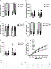
Figure 2:
Figure 2:. CORT persistently modulates select intrinsic membrane properties of adolescent glutamatergic and GABAergic SDH neurons.
A, Resting membrane potential was unchanged by the 90-minute CORT exposure in either cell type (n = 23 - 30 cells per group; Drug: F(1,107) = 0.001, p = 0.9754; Drug x Cell Type Interaction: F(1,107) = 0.1815, p = 0.6709; Two-way ANOVA). B, Membrane resistance was also unchanged by CORT incubation (n =22 - 30; Drug: F(1,105) = 0.429, p = 0.5139, Two-way ANOVA) regardless of cell type (Drug x Cell Type Interaction: F(1,105) = 0.3298, p = 0.567). C, CORT significantly enhanced action potential (AP) amplitude (n = 23 - 30; Drug: F(1,106) = 4.291, *p = 0.0407, Two-way ANOVA) irrespective of cell type (Drug x Cell Type Interaction: F(1,106) = 3.595, p = 0.0607). D, AP threshold in both neuronal populations was significantly reduced with 90-minute application of corticosterone (Drug: F(1,106) = 11.24, **p = 0.0011; Drug x Cell Type Interaction: F(1,106) = 0.9245, p = 0.3385, Two-way ANOVA). E, F, Rheobase (n = 23 - 30, Drug: F(1,106) = 0.4907, p = 0.4852, Two-way ANOVA, E) and repetitive firing frequency (n = 15 - 25, Drug: F(1,73) = 0.1176, p = 0.7326; Cell type x Stimulus Interaction: F(12,876) = 8.866, p < 0.001, RM Three-way ANOVA; F) were unaffected by CORT incubation. G, Representative examples of repetitive firing at stimulus intensities of 20 pA (left) and 50 pA (right). Scale bars = 40 ms, 40 mV.
Figure 3:
Figure 3:. Prolonged exposure to corticosterone (CORT) does not evoke persistent alterations in spontaneous synaptic transmission in the adolescent SDH.
A, Representative traces of mIPSCs (top) and mEPSCs (bottom) recorded in a SDH neuron. Scale bars = 100 ms, 40 pA for mIPSCs and 20 ms, 20 pA for mEPSCs. B, C, The incubation of spinal cord slices with CORT (100 nM for 90 min) failed to significantly change mEPSC frequency (n = 14 - 22 cells per group, Drug: F(1,71) = 2.224, p = 0.1403, Two-way ANOVA, B) or mEPSC amplitude (Drug: F(1,71) = 0.091, p = 0.7638, Two-way ANOVA, C) compared to treatment with a vehicle control at 2 - 6 hours after initial CORT exposure. D, E, mIPSC frequency (n = 15 - 22 cells per group, Drug: F(1,74) = 0.60, p = 0.4410, Two-way ANOVA, D) and mIPSC amplitude (Drug: F(1,74) = 0.2726, p = 0.6031, Two-way ANOVA, E) were also similar between SDH neurons sampled from CORT- and vehicle-treated slices. F, There was also no effect of CORT on the ratio between spontaneous excitation and inhibition (E:I ratio) in adolescent SDH neurons (Drug: F(1,67) = 3.305, p = 0.0735, Two-way ANOVA).
Figure 4:
Figure 4:. Acute corticosterone reduces the balance between spontaneous excitation vs. inhibition in both presumed glutamatergic neurons and GABAergic neurons of the adolescent SDH.
A, mEPSC frequency is significantly higher in presumed glutamatergic neurons compared to GABAergic neurons (n = 15 - 16 cells per group, Cell Type: F(1,29) = 8.875, **p = 0.0058, RM Two-way ANOVA) and is significantly reduced by bath-application of CORT compared to baseline (i.e. in presence of DMSO vehicle) independently of cell type (Drug: F(1,29) = 58.52, ****p < 0.0001; Drug x Cell Type Interaction: F(1,29) = 3.152, p= 0.0863). B, mEPSC amplitude is significantly reduced by acute application of CORT irrespective of neuronal population (Drug: F(1,29) = 9.808, **p = 0.0039; Drug x Cell Type Interaction: F(1,29) = 0.4445, p = 0.5102, RM Two-Way ANOVA). C, mIPSC frequency is also significantly reduced in both neuronal populations by bath application of CORT (n = 14 - 16, Drug: F(1,28) = 12.18, **p = 0.0016; Drug x Cell Type Interaction: F(1,28) = 1.952, p = 0.1733, RM Two-way ANOVA). D, mIPSC amplitude is unchanged by the application of corticosterone (Drug: F(1,28) = 0.8247, p = 0.3716, RM Two-way ANOVA). E, CORT significantly reduces the E:I ratio in both neuronal populations within the SDH (n = 13 - 16, Drug: F(1,27) = 5.906, *p = 0.022; Drug x Cell Type Interaction: F(1,27) = 0.6081, p = 0.4423, RM Two-way ANOVA).
Figure 5:
Figure 5:. Mineralocorticoid receptors (MR) are required for the reduction in mEPSC amplitude in the adolescent SDH during acute CORT exposure.
A, Bath application of the selective GR antagonist mifepristone (Mif) or the selective MR antagonist eplerenone (Epl) failed to prevent the reduction in mEPSC frequency by acute CORT application (n = 26 - 37 cells per group, Drug: F(1,91) = 90.53, ****p < 0.0001; Drug x Antagonist Interaction: F(2,91) = 0.9660, p = 0.3845, RM Two-way ANOVA). B, The CORT-evoked decrease in mEPSC amplitude was blocked by eplerenone (Drug x Antagonist Interaction: F(2,91) = 4.467, p = 0.0141; p = 0.9996, Šidák’s post-test) but not by mifepristone (*p = 0.0339, ***p = 0.0006, Šidák’s post-test). C, Bath application of mifepristone or eplerenone alone was unable to prevent the acute reduction in mIPSC frequency in response to CORT (n = 28 - 46, Drug: F(1,101) = 11.83, ***p = 0.0008; Drug x Antagonist Interaction: F(2,101) = 1.797, p = 0.171, RM Two-way ANOVA). D, mIPSC amplitude was not significantly different across groups (Drug: F(1,101) = 0.0005, p = 0.982, RM Two-way ANOVA). E, The E:I Ratio was significantly reduced by CORT in the presence of mifepristone or eplerenone (n = 26 - 36, Drug: F(1,88) = 35.69, ****p < 0.0001; Drug x Antagonist Interaction: F(2,88) = 1.632, p = 0.2013, RM Two-way ANOVA). In all panels, for the Vehicle group, the same data shown in Fig. 4 were pooled across the two cell types (i.e. Gad1-GFP+ and GFP−).

References

    1. Abbott FV, Franklin KB, Connell B (1986) The stress of a novel environment reduces formalin pain: possible role of serotonin. European journal of pharmacology 126:141–144. - PubMed
    1. Alexander JK, DeVries AC, Kigerl KA, Dahlman JM, Popovich PG (2009) Stress exacerbates neuropathic pain via glucocorticoid and NMDA receptor activation. Brain, behavior, and immunity 23:851–860. - PMC - PubMed
    1. Alvarez P, Green PG, Levine JD (2013) Stress in the adult rat exacerbates muscle pain induced by early-life stress. Biological psychiatry 74:688–695. - PMC - PubMed
    1. Baba H, Goldstein PA, Okamoto M, Kohno T, Ataka T, Yoshimura M, Shimoji K (2000a) Norepinephrine facilitates inhibitory transmission in substantia gelatinosa of adult rat spinal cord (part 2): effects on somatodendritic sites of GABAergic neurons. Anesthesiology 92:485–492. - PubMed
    1. Baba H, Shimoji K, Yoshimura M (2000b) Norepinephrine facilitates inhibitory transmission in substantia gelatinosa of adult rat spinal cord (part 1): effects on axon terminals of GABAergic and glycinergic neurons. Anesthesiology 92:473–484. - PubMed

Publication types

LinkOut - more resources