

How Cryo-EM Has Expanded Our Understanding of Membrane Transporters
- PMID: 37438132
- PMCID: PMC10353158
- DOI: 10.1124/dmd.122.001004
How Cryo-EM Has Expanded Our Understanding of Membrane Transporters
Abstract
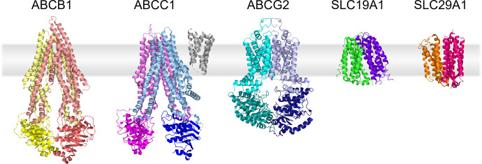
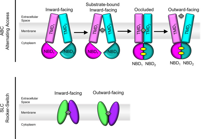
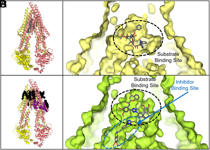
Over the past two decades, technological advances in membrane protein structural biology have provided insight into the molecular mechanisms that transporters use to move diverse substrates across the membrane. However, the plasticity of these proteins' ligand binding pockets, which allows them to bind a range of substrates, also poses a challenge for drug development. Here we highlight the structure, function, and transport mechanism of ATP-binding cassette/solute carrier transporters that are related to several diseases and multidrug resistance: ABCB1, ABCC1, ABCG2, SLC19A1, and SLC29A1. SIGNIFICANCE STATEMENT: ATP-binding cassette transporters and solute carriers play vital roles in clinical chemotherapeutic outcomes. This paper describes the current understanding of the structure of five pharmacologically relevant transporters and how they interact with their ligands.
Copyright © 2023 by The Author(s).
Figures
Similar articles
-
Prescription of Controlled Substances: Benefits and Risks.2025 Jul 6. In: StatPearls [Internet]. Treasure Island (FL): StatPearls Publishing; 2025 Jan–. 2025 Jul 6. In: StatPearls [Internet]. Treasure Island (FL): StatPearls Publishing; 2025 Jan–. PMID: 30726003 Free Books & Documents.
-
Structure and Mechanism of Human ABC Transporters.Annu Rev Biophys. 2023 May 9;52:275-300. doi: 10.1146/annurev-biophys-111622-091232. Epub 2023 Feb 3. Annu Rev Biophys. 2023. PMID: 36737602 Review.
-
A systems framework for investigating the roles of multiple transporters and their impact on drug resistance.Integr Biol (Camb). 2024 Jan 23;16:zyae007. doi: 10.1093/intbio/zyae007. Integr Biol (Camb). 2024. PMID: 38537223
-
Characterization of three B-cell lymphoma cell lines from chemotherapy resistant patients with respect to in vitro sensitivity to 21 antitumor agents, ABC-transporter expression and cellular redox status.J Cancer Res Clin Oncol. 2007 Dec;133(12):957-67. doi: 10.1007/s00432-007-0241-x. Epub 2007 Jun 12. J Cancer Res Clin Oncol. 2007. PMID: 17562080 Free PMC article.
-
Fabricating mice and dementia: opening up relations in multi-species research.In: Jenkins N, Jack-Waugh A, Ritchie L, editors. Multi-Species Dementia Studies. Bristol (UK): Bristol University Press; 2025 Feb 25. Chapter 2. In: Jenkins N, Jack-Waugh A, Ritchie L, editors. Multi-Species Dementia Studies. Bristol (UK): Bristol University Press; 2025 Feb 25. Chapter 2. PMID: 40690569 Free Books & Documents. Review.
Cited by
-
The uniqueness of ABCB5 as a full transporter ABCB5FL and a half-transporter-like ABCB5β.Cancer Drug Resist. 2024 Aug 7;7:29. doi: 10.20517/cdr.2024.56. eCollection 2024. Cancer Drug Resist. 2024. PMID: 39267923 Free PMC article. Review.
-
Investigations of membrane protein interactions in cells using fluorescence microscopy.Curr Opin Struct Biol. 2024 Jun;86:102816. doi: 10.1016/j.sbi.2024.102816. Epub 2024 Apr 21. Curr Opin Struct Biol. 2024. PMID: 38648680 Free PMC article. Review.
-
Cadmium transport by mammalian ATP-binding cassette transporters.Biometals. 2024 Jun;37(3):697-719. doi: 10.1007/s10534-024-00582-5. Epub 2024 Feb 6. Biometals. 2024. PMID: 38319451 Free PMC article. Review.
-
Integration of co-culture and transport engineering for enhanced metabolite production.Plant Biotechnol (Tokyo). 2024 Sep 25;41(3):195-202. doi: 10.5511/plantbiotechnology.24.0312b. Plant Biotechnol (Tokyo). 2024. PMID: 40115766 Free PMC article.
-
Prenatal opioid exposure significantly impacts placental protein kinase C (PKC) and drug transporters, leading to drug resistance and neonatal opioid withdrawal syndrome.Front Neurosci. 2024 Aug 19;18:1442915. doi: 10.3389/fnins.2024.1442915. eCollection 2024. Front Neurosci. 2024. PMID: 39238930 Free PMC article.
References
-
- Abdul-Ghani R, Serra V, Györffy B, Jürchott K, Solf A, Dietel M, Schäfer R (2006) The PI3K inhibitor LY294002 blocks drug export from resistant colon carcinoma cells overexpressing MRP1. Oncogene 25:1743–1752. - PubMed
-
- Allikmets R, Schriml LM, Hutchinson A, Romano-Spica V, Dean M (1998) A human placenta-specific ATP-binding cassette gene (ABCP) on chromosome 4q22 that is involved in multidrug resistance. Cancer Res 58:5337–5339. - PubMed
-
- Ambudkar SV, Kim IW, Xia D, Sauna ZE (2006) The A-loop, a novel conserved aromatic acid subdomain upstream of the Walker A motif in ABC transporters, is critical for ATP binding. FEBS Lett 580:1049–1055. - PubMed
Publication types
MeSH terms
Substances
Grants and funding
LinkOut - more resources
Full Text Sources
Miscellaneous
