. 2023 Jul 12;14(1):4085.
doi: 10.1038/s41467-023-39545-8.

External globus pallidus input to the dorsal striatum regulates habitual seeking behavior in male mice

Affiliations

External globus pallidus input to the dorsal striatum regulates habitual seeking behavior in male mice

Matthew Baker et al. Nat Commun. .

Abstract

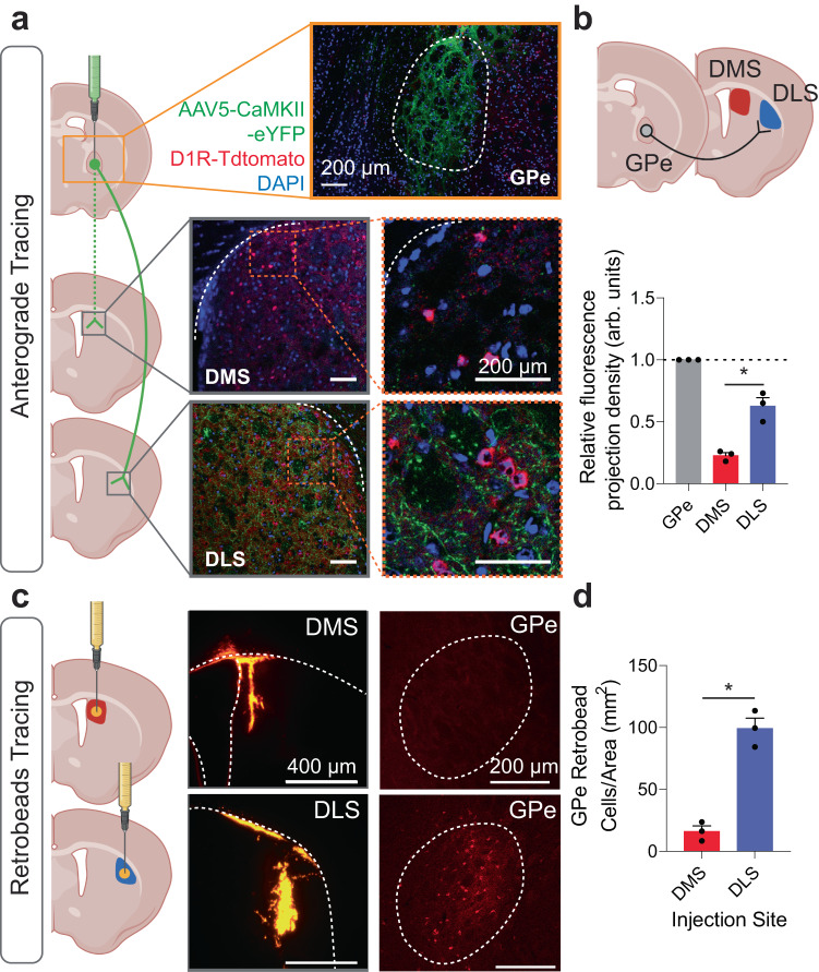
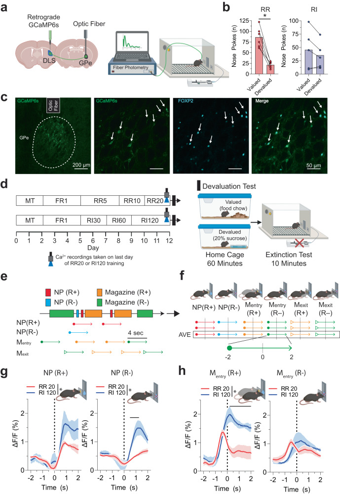
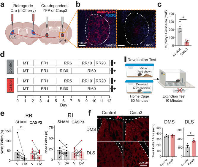
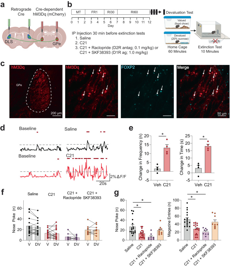
The external globus pallidus (GPe) coordinates action-selection through GABAergic projections throughout the basal ganglia. GPe arkypallidal (arky) neurons project exclusively to the dorsal striatum, which regulates goal-directed and habitual seeking. However, the role of GPe arky neurons in reward-seeking remains unknown. Here, we identified that a majority of arky neurons target the dorsolateral striatum (DLS). Using fiber photometry, we found that arky activities were higher during random interval (RI; habit) compared to random ratio (RR; goal) operant conditioning. Support vector machine analysis demonstrated that arky neuron activities have sufficient information to distinguish between RR and RI behavior. Genetic ablation of this arkyGPe→DLS circuit facilitated a shift from goal-directed to habitual behavior. Conversely, chemogenetic activation globally reduced seeking behaviors, which was blocked by systemic D1R agonism. Our findings reveal a role of this arkyGPe→DLS circuit in constraining habitual seeking in male mice, which is relevant to addictive behaviors and other compulsive disorders.


Conflict of interest statement

D.S.C. is a scientific advisory board member to Peptron Inc. Peptron had no role in the preparation, review, or approval of the manuscript; nor the decision to submit the manuscript for publication. The remaining authors declare no competing interests.

Figures

Fig. 1
Fig. 1. Anterograde and retrograde tracing of external globus pallidus (GPe) projections.
a Injection schematic of AAV5-CaMKII-eYFP virus into the GPe in D1R-TdTomato mice and projection densities in the dorsomedial striatum (DMS) and dorsolateral striatum (DLS). Scale: 200 µm. Anterograde virus (Green), D1R (Red), DAPI (Blue). b A schematic circuit showing a majority of GPe projections target the DLS, compared to the DMS (p = 0.0012). n = 3 images/group. F(2,6) = 87.60, p < 0.0001. c Retrobead tracing of DMS and DLS afferents and representative IHC images. Scale: 400 µm (left), 200 µm (right). d DLS retrobead injections showed more GPe projection cells compared to DMS. n = 3 images/group. One-way ANOVA with Tukey’s posthoc tests were used for (c). Two-tailed t tests were used for (d). *p < 0.05. Data represent mean ± SEM. See Supplementary Table 2 for full statistical information. Source data are provided as a Source Data file. (ac) were created with BioRender.com.
Fig. 2
Fig. 2. Fiber photometry Ca2+ signal of external globus pallidus (GPe) arkypallidal neurons during random ratio (RR) and interval (RI) operant conditioning.
a Schematic of retrograde GCaMP6s virus injection into the DLS, fiber placement over the GPe, and Ca2+ recordings in the operant chamber. b RR-trained mice (n = 6) showed a significant decrease in nose pokes during extinction testing for the devalued state, suggesting of goal-directed behavior (p = 0.03). RI-trained mice (n = 5) showed no differences between valued and devalued states (p = 0.62), suggesting of habitual behavior. c IHC images showing representative GCaMP6s expression in the GPe, optic fiber placement, and coexpression with arkypallidal cellular marker FOXP2 from n = 3 mice. Scale: 200 µm (left), 50 µm (rest). d Operant RR and RI behavior schedules to establish goal-directed and habitual seeking, respectively. Ca2+ signal recordings were collected on the last day of training, followed by evaluation testing to determine reward-outcome valuation testing. e Behavioral and arkypallidal neuron Ca2+ signal alignment in RR and RI-trained mice surrounding rewarded and unrewarded nose poke. f Ca2+ signal was recorded 2 s before and after each behavioral event. Solid lines indicate mean Ca2+ signal for rewarded (R+) and unrewarded (R−) nose poke (NP). g Ca2+ signaling in the nose poke. For the rewarded nose poke (NP+), F(1,7) = 5.73, p = 0.048 for group (RR vs RI), F(120,840) = 11.40, p < 0.0001 for time, F(120,840) = 1.62, p = 0.0001 for interaction. For the non-rewarded nose poke (NP-), F(1,7) = 12.34, p = 0.0098 for group (RR vs RI), F(120,840) = 12.52, p < 0.0001 for time, F(120,840) = 5.52, p < 0.0001 for interaction. h Ca2+ signaling in the magazine entry. For the rewarded magazine entry [Mentry (R+)], F(1,7) = 25.84, p = 0.0014 for group (RR vs RI), F(120,840) = 14.48, p < 0.0001 for time, F(120,840) = 5.73, p < 0.0001 for interaction. For the non-rewarded magazine entry [Mentry (R−)], F(1,7) = 0.05, p = 0.83 for group (RR vs RI), F(120,840) = 5.60, p < 0.0001 for time, F(120,840) = 1.74, p < 0.0001 for interaction. Wilcoxon test was used for (b). Two-way repeated measures ANOVA with Tukey’s posthoc tests were used for (g, h). n = 5 (RR) and 4 (RI) mice. The solid line indicates the time range of significant posthoc difference between RR and RI Ca2+ traces. *p < 0.05. Data represent mean ± SEM. See Supplementary Table 2 for full statistical information and Supplementary Table 3 for specific significant time ranges of fiber photometry. Source data are provided as a Source Data file. (a, d, and fh) were created with BioRender.com.
Fig. 3
Fig. 3. SVM modeling predicts goal-directed and habitual reward seeking using external globus pallidus (GPe) arkypallidal neuron Ca2+ dynamics.
a Averaged receiver operator characteristic (ROC) curves for prediction of goal-directed or habitual reward-seeking around behavioral events (nose-poke, magazine entry, and magazine exit; −2 – 2 s input range). b Comparison of accuracy, sensitivity, and specificity to random chance among each behavioral event. Two-tailed one sample t tests were used for (b). For accuracy NP versus 50%, t = 116.1, p < 0.0001, for accuracy Mentry versus 50%, t = 136.4, p < 0.0001, for accuracy Mexit versus 50%, t = 105. 7, p < 0.0001. For sensitivity NP versus 50%, t = 109.1, p < 0.0001, for sensitivity Mentry versus 50%, t = 126.9, p < 0.0001, for sensitivity Mexit versus 50%, t = 101.4, p < 0.0001. For specificity NP versus 50%, t = 104.8, p < 0.0001, for specificity Mentry versus 50%, t = 118.9, p < 0.0001, for specificity Mexit versus 50%, t = 94.35, p < 0.0001. *p < 0.05 for comparison to random chance for accuracy, sensitivity, and specificity. See Supplementary Table 1 for AUC values for additional time input ranges. See Supplementary Table 2 for full statistical information. Source data are provided as a Source Data file.
Fig. 4
Fig. 4. Effects of Caspase 3 (casp3)-dependent ablation of external globus pallidus (GPe) arkypallidal neurons on goal-directed and habitual-seeking.
a Schematic of virus injections for Cre-dependent ablation of GPe arkypallidal neurons. b Representative GPe images of sham control and caspase mice (Scale: 100 µm) from n = 5 mice/group. c Caspase mice showed a significant reduction of mCherry-positive neurons in the GPe (t = 6.90, p = 0.001). n = 5/group. d Operant RR and RI behavior schedules in sham control and caspase mice to establish goal-directed and habitual seeking, respectively. e RR-trained (Goal-directed) sham mice reduced nose poke responses in the devalued state, confirming goal-directed behavior (p = 0.0039). However, caspase mice showed no changes in nose poke responses between valued and devalued states, typical of habitual behavior (p = 0.73). No differences in nose poke rates between the valued and devalued state for RI-trained sham (p = 0.38) and caspase mice (p = 0.83) indicated habitual reward-seeking. n = 10 mice/group. f Representative IHC images of cFos expression in the dorsomedial (DMS) and dorsolateral (DLS) striatum for sham and casp3 mice (Scale: 200 µm). GPe arkypallidal neuron ablation increased the number of cFos-positive cells in the DLS (t = 3.99, p = 0.004), but not DMS (t = 1.02, p = 0.34). n = 5 mice/group. Two-tailed t tests were used for (c, f). Wilcoxon test was used for (e). Data represent mean ± SEM. *p < 0.05. See Supplementary Table 2 for full statistical information. Source data are provided as a Source Data file. (a, d) were created with BioRender.com.
Fig. 5
Fig. 5. Effects of chemogenetic activation of external globus pallidus (GPe) arkypallidal neurons on habitual reward-seeking.
a Schematic of virus injection strategy for Cre-dependent expression of the excitatory DREADDs hM3Dq. b RI operant schedule followed by evaluation tests with IP injection groups. c Representative images showing hM3Dq expression in the GPe and coexpression with arkypallidal neuron marker FOXP2 from n = 3 mice. Scale: 200 µm (left), 50 µm (rest). d Representative calcium traces for hM3Dq-expressing GPe arkypallidal neurons at baseline and following C21 or vehicle administration in freely moving mice. e C21 administration caused a notable increase in the frequency and duration of Ca2+ events (frequency: t = 4.47, p = 0.021, time: t = 11.86, p = 0.0013). n = 4 mice/group. f There were no differences between the valued and devalued states for IP injection treatment. g The effects of DREADDs activation and IP injection groups on nose poke and magazine-entry seeking behaviors during the extinction test. Noke poke with drug treatment (p = 0.0004), z = 2.71, p = 0.04 for saline versus C21, z = 3.96, p = 0.0005 for saline versus C21+Raclopride, z = 0.83, p = 0.99 for saline versus C21 + SKF383393. Magazine entry with drug treatment (p < 0.00001), z = 3.59, p = 0.002 for saline versus C21, z = 4.13, p = 0.0002 for saline versus C21+Raclopride, z = 0.69, p = 0.99 for saline versus C21 + SKF383393. Two-tailed t tests were used for (e). Wilcoxon test was used for (f). Kruskal–Wallis test with Dunn’s posthoc tests were used for (g). n = 17 (Saline, C21), 8 (C21 + Raclopride), 9 (C21 + SKF38393) for (f, g). Data represent mean ± SEM. *p < 0.05. See Supplementary Table 2 for full statistical information. Source data are provided as a Source Data file. (a, b) were created with BioRender.com.

Comment in

  • Arky neurons and reward seeking.
    Mejia LA. Mejia LA. Nat Neurosci. 2023 Aug;26(8):1319. doi: 10.1038/s41593-023-01412-6. Nat Neurosci. 2023. PMID: 37537348 No abstract available.

References

    1. Dong J, Hawes S, Wu J, Le W, Cai H. Connectivity and functionality of the globus pallidus externa under normal conditions and Parkinson’s disease. Front. Neural Circuits. 2021;15:645287. doi: 10.3389/fncir.2021.645287. - DOI - PMC - PubMed
    1. Schwab BC, et al. Synchrony in Parkinson’s disease: importance of intrinsic properties of the external globus pallidus. Front. Syst. Neurosci. 2013;7:60. doi: 10.3389/fnsys.2013.00060. - DOI - PMC - PubMed
    1. Wichmann T, Dostrovsky JO. Pathological basal ganglia activity in movement disorders. Neuroscience. 2011;198:232–244. doi: 10.1016/j.neuroscience.2011.06.048. - DOI - PMC - PubMed
    1. Bevan MD, Magill PJ, Terman D, Bolam JP, Wilson CJ. Move to the rhythm: oscillations in the subthalamic nucleus-external globus pallidus network. Trends Neurosci. 2002;25:525–531. doi: 10.1016/S0166-2236(02)02235-X. - DOI - PubMed
    1. Chazalon M, et al. GAT-3 dysfunction generates tonic inhibition in external globus pallidus neurons in parkinsonian rodents. Cell Rep. 2018;23:1678–1690. doi: 10.1016/j.celrep.2018.04.014. - DOI - PubMed

Publication types