. 2023 Jul 12;10(1):449.
doi: 10.1038/s41597-023-02330-9.

Magnetic resonance imaging datasets with anatomical fiducials for quality control and registration

Affiliations

Magnetic resonance imaging datasets with anatomical fiducials for quality control and registration

Alaa Taha et al. Sci Data. .

Abstract

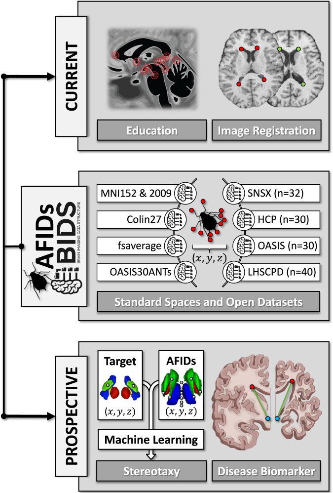
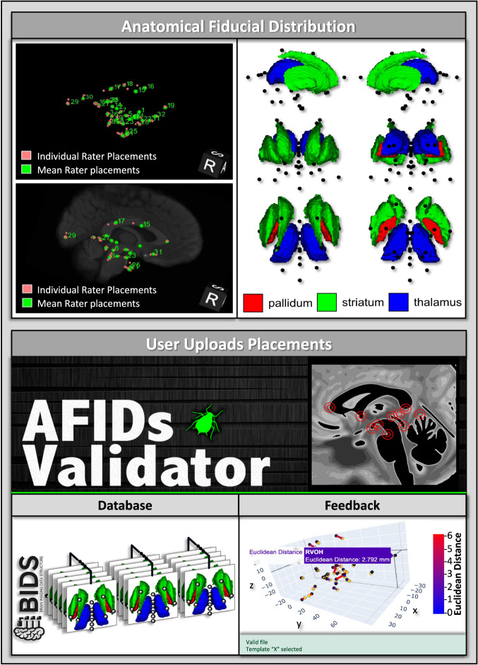
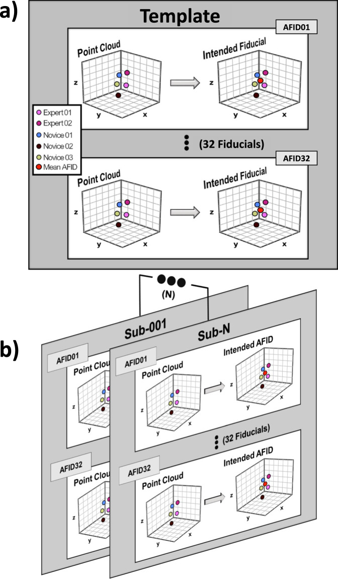
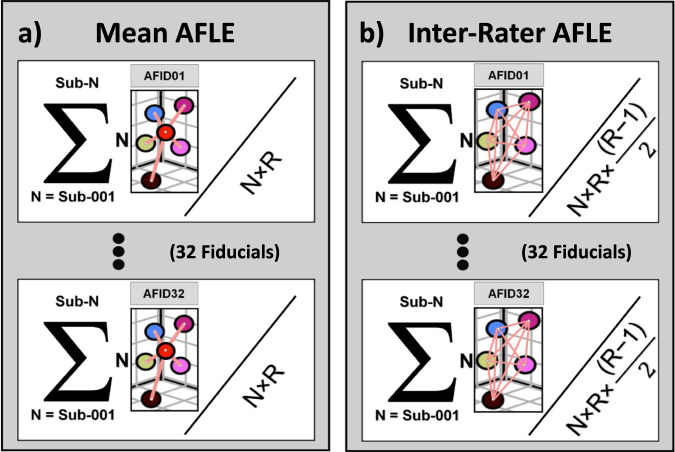
Tools available for reproducible, quantitative assessment of brain correspondence have been limited. We previously validated the anatomical fiducial (AFID) placement protocol for point-based assessment of image registration with millimetric (mm) accuracy. In this data descriptor, we release curated AFID placements for some of the most commonly used structural magnetic resonance imaging datasets and templates. The release of our accurate placements allows for rapid quality control of image registration, teaching neuroanatomy, and clinical applications such as disease diagnosis and surgical targeting. We release placements on individual subjects from four datasets (N = 132 subjects for a total of 15,232 fiducials) and 14 brain templates (4,288 fiducials), totalling more than 300 human rater hours of annotation. We also validate human rater accuracy of released placements to be within 1 - 2 mm (using more than 45,000 Euclidean distances), consistent with prior studies. Our data is compliant with the Brain Imaging Data Structure allowing for facile incorporation into neuroimaging analysis pipelines.


Conflict of interest statement

The authors declare no competing interests.

Figures

Fig. 1
Fig. 1
Current and prospective applications of curated anatomical fiducial (AFID) placements. Top Panel: Current applications in neuroanatomy education and image registration. Middle Panel: Released healthy and pathologic datasets and templates (detailed descriptions can be found in text). Bottom Panel: Prospective applications of AFIDs in stereotactic targeting and as a disease biomarker.
Fig. 2
Fig. 2
Curated AFID locations within the brain and usage of the AFIDs validator website. Top Panel: Distribution of AFIDs overlayed on one of the released templates. We also show major subcortical structures with AFIDs (black points) in various anatomical views. Bottom Panel: Uses and outputs from the AFIDs validator website (https://validator.afids.io). The user decides whether to upload their placements to our database and will receive summary metrics regarding their placements in an interactive 3D coordinate system and tabular format (not shown).
Fig. 3
Fig. 3
Ground truth anatomical fiducial (AFID) placement on templates and datasets. (a,b) show the process of computing the intended AFID placement on a neuroimaging template or dataset, respectively. It is the mean of the rater point cloud at each AFID, referred to as “ground truth” in the text.
Fig. 4
Fig. 4
The technical validation computations for our anatomical fiducial (AFID) placements on templates and datasets. (a,b) show the equations used to compute mean and inter-rater anatomical localization error, respectively. N = number of subjects in a dataset. If calculating for a template, N would be 1. R = the number of raters per image. In (a) Euclidean distances (shown in pink) represent distance from rater placement to the ground truth (red). The mean AFLE was calculated by dividing the sum of all Euclidean distances across all subjects with the total number of Euclidean distances in the dataset (N × R) for each AFID. In (b) Euclidean distances (shown in pink) represent the pairwise distances between all rater placements on a scan. Inter-rater AFLE was calculated by dividing the sum of the pairwise distances (shown by the sigma notation) by the total number of rater pairwise distances across a dataset per AFID N×R×R12.

References

    1. Rohlfing T. Image similarity and tissue overlaps as surrogates for image registration accuracy: widely used but unreliable. IEEE Trans. Med. Imaging. 2012;31:153–163. doi: 10.1109/TMI.2011.2163944. - DOI - PMC - PubMed
    1. Lau JC, et al. A framework for evaluating correspondence between brain images using anatomical fiducials. Hum. Brain Mapp. 2019;40:4163–4179. doi: 10.1002/hbm.24693. - DOI - PMC - PubMed
    1. Abbass M, et al. Application of the anatomical fiducials framework to a clinical dataset of patients with Parkinson’s disease. Brain Struct. Funct. 2022;227:393–405. doi: 10.1007/s00429-021-02408-3. - DOI - PMC - PubMed
    1. Chakravarty MM, et al. Comparison of piece-wise linear, linear, and nonlinear atlas-to-patient warping techniques: analysis of the labeling of subcortical nuclei for functional neurosurgical applications. Hum. Brain Mapp. 2009;30:3574–3595. doi: 10.1002/hbm.20780. - DOI - PMC - PubMed
    1. Chakravarty MM, Sadikot AF, Germann J, Bertrand G, Collins DL. Towards a validation of atlas warping techniques. Med. Image Anal. 2008;12:713–726. doi: 10.1016/j.media.2008.04.003. - DOI - PubMed