. 2023 Aug;30(8):1957-1972.
doi: 10.1038/s41418-023-01190-5. Epub 2023 Jul 12.

N6-methyladenosine hypomethylation of circGPATCH2L regulates DNA damage and apoptosis through TRIM28 in intervertebral disc degeneration

Affiliations

N6-methyladenosine hypomethylation of circGPATCH2L regulates DNA damage and apoptosis through TRIM28 in intervertebral disc degeneration

Zhenhao Chen et al. Cell Death Differ. 2023 Aug.

Abstract

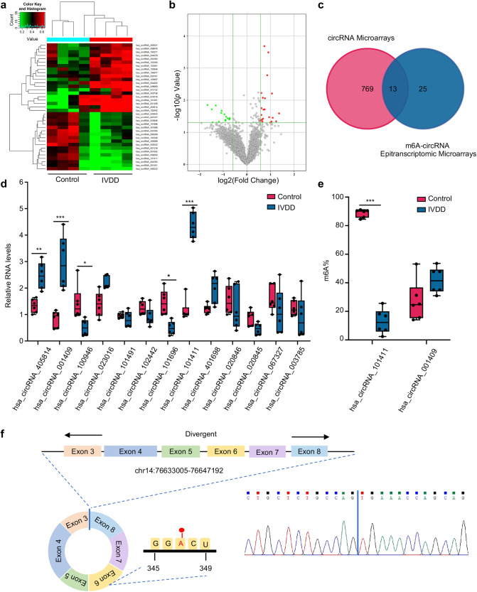
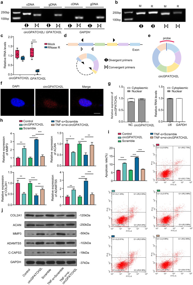
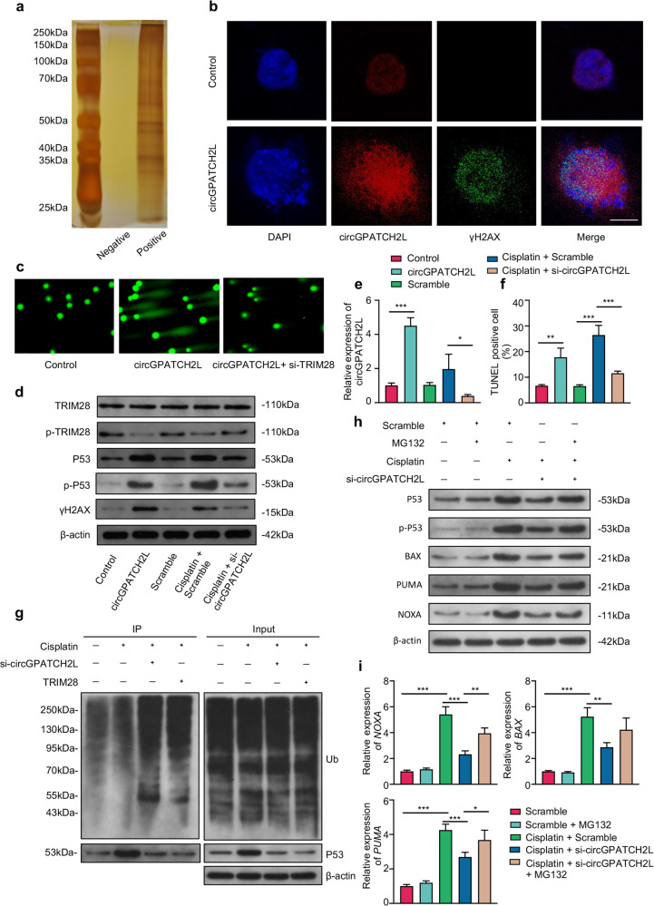
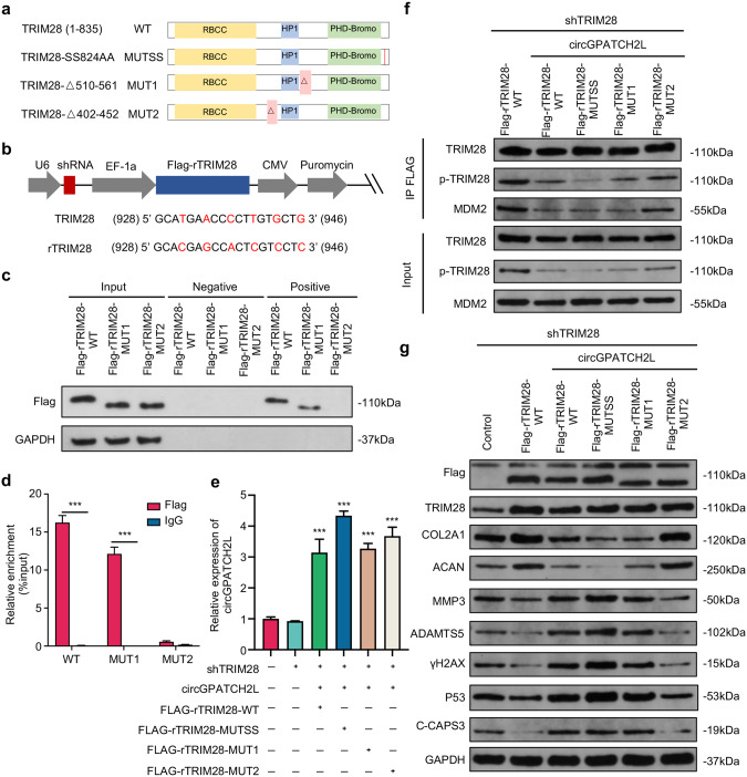
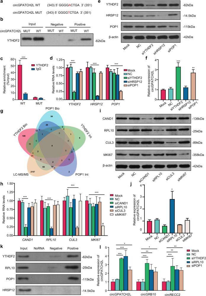
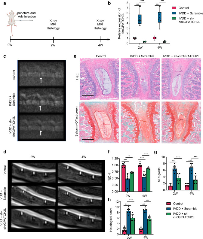
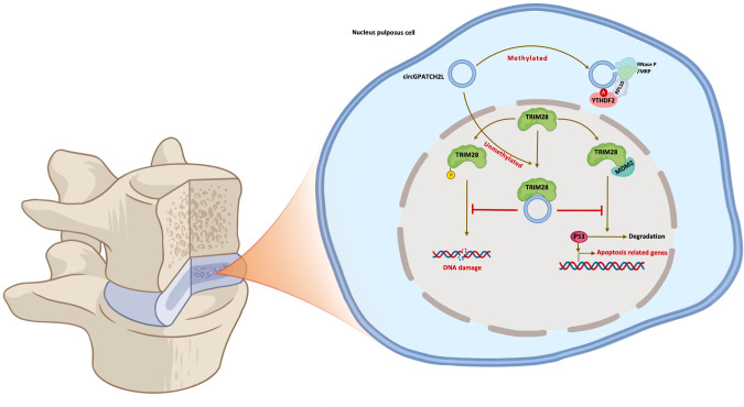
Circular RNAs (circRNAs) are a class of noncoding RNAs that have been found to be involved in intervertebral disc degeneration (IVDD) progression, and N6-methyladenosine (m6A) broadly exists in circRNAs. Here, we identified circGPATCH2L with a low m6A methylation level to be upregulated in degenerative nucleus pulposus tissues. Mechanistically, as a protein decoy for tripartite motif containing 28 (TRIM28) within aa 402-452 region, circGPATCH2L abrogates the phosphorylation of TRIM28 and inhibits P53 degradation, which contributes to DNA damage accumulation and cellular apoptosis and leads to IVDD progression. Moreover, m6A-methylated circGPATCH2L is recognised and endoribonucleolytically cleaved by a YTHDF2-RPL10-RNase P/MRP complex to maintain the physiological state of nucleus pulposus cells. Thus, our data show the physiological significance of m6A modification in regulating circRNA abundance and provide a potentially effective therapeutic target for the treatment of IVDD.


Conflict of interest statement

The authors declare no competing interests.

Figures

Fig. 1
Fig. 1. circGPATCH2L is hypomethylated and upregulated in IVDD.
a Hierarchical clustering for circRNAs with differential m6A methylation levels in the IVDD and control groups. The red–green gradient colour scheme represents the high and low m6A methylation relative levels as referenced in the Colour Key. b Volcano plot showing 38 differentially m6A-methylated circRNAs in NP tissues of the IVDD and control groups. The cut off is |fold change| ≥ 1.5, p ≤ 0.05. The red and green points in the plot indicate the upregulated and downregulated circRNAs with statistical significance, respectively. c Venn diagram identifying 13 differentially expressed circRNAs with altered methylation in IVDD samples based on the overlap of circRNA microarray and m6A-circRNA epitranscriptomic microarray data. d qRT–PCR analysis of the expression of 13 circRNAs in NP tissues. n = 6. e The m6A ratio of hsa_circRNA_101411 and hsa_circRNA_001409 was calculated using a T3 DNA ligase assay. n = 6. f Schematic of the genomic loci of circGPATCH2L and the m6A site. The back-splice junction site of circGPATCH2L was confirmed by Sanger sequencing. NP nucleus pulposus, m6A N6-methyladenosine, qRT–PCR quantitative real-time polymerase chain reaction, IVDD intervertebral disc degeneration. Data are shown as the mean ± S.D. ***p < 0.001; **p < 0.01; *p < 0.05.
Fig. 2
Fig. 2. Characterisation of circGPATCH2L and its function in NPCs.
a PCR analysis of circGPATCH2L using divergent and convergent primers in cDNA and gDNA. GAPDH was used as a control. b PCR analysis of circGPATCH2L using divergent and convergent primers after treatment with RNase R. c qRT–PCR showing resistance of circGPATCH2L to RNase R digestion. n = 6. d A schematic diagram representing divergent (black arrow) and convergent (white arrow) primers. e A schematic diagram of a probe targeting the back-spliced site of circGPATCH2L. f RNA-FISH analysis of circGPATCH2L in NPCs. The circGPATCH2L probe was labelled with Cy3. Cell nuclei were stained with DAPI. Scale bar = 10 μm. g Relative RNA levels of circGPATCH2L in the nuclear and cytoplasmic fractions of NPCs. circGPATCH2L was mainly localised in the nucleus. Relative RNA levels of U6 and GAPDH were tested as quality controls. n = 3. h qRT–PCR analysis of COL2A1, ACAN, MMP3, and ADAMTS5 mRNA expression in each group. The control group was transfected with a blank vector with flanking introns containing complementary Alu elements. NPCs transfected with sequence-scrambled DNA oligos were named Scramble. TNF-α (10 ng/ml) was used to induce an in vitroIVDD model. The circGPATCH2L overexpression plasmid successfully upregulated circGPATCH2L expression and siRNA for circGPATCH2L downregulated circGPATCH2L expression. n = 3. i Representative flow cytometry dot plots of apoptosis after Annexin V-FITC/PI dual staining. Histograms showing the apoptosis rate in the control, circGAPTCH2L (circGAPTCH2L overexpression), Scramble, TNF-α + Scramble, and TNF-α + si-circGAPTCH2L-2 groups. n = 3. j Western blot analysis of COL2, ACAN, MMP3, ADAMTS5, and C-CASP3 protein levels. circGPATCH2L overexpression induced degeneration of NPCs, and circGPATCH2L knockdown rescued the degeneration phenotype created by TNF-α (10 ng/ml) treatment. NPCs nucleus pulposus cells, M mock, R RNase R, RNase R ribonuclease R, PCR polymerase chain reaction, cDNA complementary DNA, gDNA genomic DNA, FISH fluorescent in situ hybridisation, DAPI 4,6-diamidino-2-phenylindole, qRT–PCR quantitative real-time PCR, GAPDH glyceraldehyde 3-phosphate dehydrogenase, COL2A1 collagen type II alpha 1 chain, ACAN aggrecan, MMP3 matrix metallopeptidase 3, ADAMTS5 a disintegrin and metalloproteinase with thrombospondin type 1 motif 5, IVDD intervertebral disc degeneration, C-CASP3 cleaved Caspase 3, TNF-α tumour necrosis factor-α. Data are shown as the mean ± S.D. ***p < 0.001; **p < 0.01; *p < 0.05.
Fig. 3
Fig. 3. circGPATCH2L promotes DNA damage and apoptosis in NPCs through TRIM28.
a Silver staining of the precipitates from circGPATCH2L pulldown with positive and negative probes after SDS–PAGE. b RNA-FISH assay of circGPATCH2L (red) and immunofluorescence staining of γH2AX (green) in NPCs with and without circGPATCH2L overexpression. The circGPATCH2L probe was labelled with Cy3. Cell nuclei were stained with DAPI (blue). Scale bar = 10 μm. c The comet assay was performed to detect cellular DNA damage (original magnification, 200×). The circGPATCH2L overexpression plasmid and siTRIM28 were used. d Western blot analysis of TRIM28, p-TRIM28, γH2AX, P53, p-P53 (Ser392) and β-actin in NPCs. Gain- and loss-of-function experiments showed that circGPATCH2L abrogated TRIM28 phosphorylation and induced DNA damage, P53 phosphorylation and accumulation. The control group was transfected with a blank vector with flanking introns containing complementary Alu elements. NPCs transfected with sequence-scrambled DNA oligos were named Scramble. Cisplatin (50 μM) was used to induce DNA damage in NPCs in vitro. The circGPATCH2L overexpression plasmid and si-circGPATCH2L were used to regulate circGPATCH2L expression. e qRT–PCR analysis of circGPATCH2L expression in different groups. n = 3. f Quantification of TUNEL-positive cells. n = 3. g Ubiquitination of p53 was analysed by immunoprecipitation with p53 antibody and followed by western blot analysis in NPCs with circGPATCH2L or TRIM28 depletion in the presence of Cisplatin (50 μM). h Western blot analysis of P53, p-P53 (Ser392), BAX, PUMA, NOXA and β-actin expression in NPCs. Scramble and Scramble with MG132 (5 µM, 24 h) were used as control. Cisplatin (50 μM) was used to induce DNA damage in NPCs. MG132 and si-circGPATCH2L were used to regulate total P53. i qRT–PCR analysis confirmed the expression of BAX, PUMA and NOXA mRNA in different groups. n = 3. NPCs nucleus pulposus cells, qRT–PCR quantitative real-time polymerase chain reaction, TRIM28 tripartite motif containing 28, p-TRIM28 TRIM28 phosphorylation, MDM2 E3 ubiquitin-protein ligase Mdm2, γH2AX H2A.X variant histone phosphorylation, P53 cellular tumour antigen p53, p-P53 phosphorylation of p53, β-actin actin beta. Data are shown as the mean ± S.D. ***p < 0.001; **p < 0.01; *p < 0.05.
Fig. 4
Fig. 4. circGPATCH2L functions in NPCs by binding the aa 402–452 region of TRIM28.
a Schematic representation of the full-length, truncated or point mutant TRIM28. TRIM28 wild type (TRIM28-WT) was full-length and contained three main TRIM28 domains. TRIM28-MUT1 and TRIM28-MUT2 lacked the aa 510–561 and aa 402–452 regions, respectively. Each serine was mutated to alanine at aa 823/824 (TRIM28-MUTSS) to avoid the possibility that Ser823 becomes phosphorylated when Ser824 is mutated. b Schematic diagrams of the lentiviral plasmids used in this study. The modified plasmid was designed for simultaneous expression of shTRIM28, Flag-tagged shTRIM28-resistant wild-type or mutant TRIM28 cDNA (rTRIM28), and the puromycin resistance gene. The shRNA-targeted sequences and the silencing mutations in shTRIM28-resistant cDNAs are shown below. The numbers correspond to the nt positions in the open reading frames of TRIM28, and the changed bases are marked in red. c, d RNA pulldown and RIP assays confirmed the interaction between circGPATCH2L and TRIM28 within the site at residues 402–452. c Coprecipitation of circGPATCH2L and wild-type or mutant TRIM28 in an RNA pulldown assay. Total protein (Input), precipitates with biotinylated probes targeting the circGPATCH2L back-spliced site (positive) or negative biotinylated probes (negative) were analysed via western blot with Flag antibody. d RIP experiments were performed using Flag or negative IgG antibody. Purified RNA was used for qRT–PCR assays. n = 3. e qRT–PCR analysis confirmed the overexpression efficiency of circGPATCH2L in NPCs after treatment with the circGPATCH2L overexpression plasmid in different groups. n = 3. f Cell lysates were immunoprecipitated with an antibody against Flag and analysed via western blot with TRIM28 antibody, p-TRIM28 antibody, or MDM2 antibody (top). Total protein from cell lysates was analysed as input (bottom). g Western blot analysis of Flag, TRIM28, COL2, ACAN, MMP3, ADAMTS5, γH2AX, P53, C-CAPS3, and GAPDH expression in NPCs. The control group was transfected with a blank vector with 3×FLAG. NPCs nucleus pulposus cells, qRT–PCR quantitative real-time polymerase chain reaction, TRIM28 tripartite motif containing 28, p-TRIM28 TRIM28 phosphorylation, MDM2 E3 ubiquitin-protein ligase Mdm2, γH2AX H2A.X variant histone phosphorylation, P53 cellular tumour antigen p53, COL2A1 collagen type II alpha 1 chain, ACAN aggrecan, MMP3 matrix metallopeptidase 3, ADAMTS5 a disintegrin and metalloproteinase with thrombospondin type 1 motif 5, C-CASP3 cleaved Caspase 3, GAPDH glyceraldehyde 3-phosphate dehydrogenase. Data are shown as the mean ± S.D. ***p < 0.001.
Fig. 5
Fig. 5. m6A methylated circGPATCH2L was subject to endoribonucleolytic cleavage via YTHDF2-RPL10-RNase P/MRP complex.
a The m6A motif sequence and the silencing mutation of circGPATCH2L. The numbers correspond to the nt positions in the open reading frames. A-T transversion mutations are indicated in red. b Western blot analysis of YTHDF2 in precipitates from a circGPATCH2L pulldown assay in NPCs with circGPATCH2L-WT or circGPATCH2L-MUT overexpression. The input was cell lysates with total protein; the positive and negative groups were precipitates with positive and negative biotinylated probes, respectively. c circGPATCH2L was detected by qRT–PCR in a RIP assay with YTHDF2 antibody-conjugated beads. IgG antibody was used as a negative control. n = 3. d qRT–PCR analysis of YTHDF2, HRSP12, and POP1 showed that the expression of each gene was downregulated by the indicated siRNAs in an independent manner. The mock group did not receive any treatment, and the NC group was treated with transfection reagent only. n = 3. e Western blot analysis identifying the knockdown efficiency of YTHDF2, HRSP12, POP1, and β-actin at the protein level. f qRT–PCR analysis of circGPATCH2L expression after transfection with siRNA. Knockdown of YTHDF2 and POP1 caused a significant increase in circGPATCH2L. n = 3. g Venn diagram showing the intersection of proteins among two PPI databases (BioGRID and IntAct) and our mass spectrometry data. Four candidate proteins were identified. h qRT–PCR analysis of CAND1, CUL3, RPL10, and MKI67 showing that the expression of each gene was downregulated by the indicated siRNAs in an independent manner. The mock group did not receive any treatment, and the NC group was treated with transfection reagent only. n = 3. i Western blot confirming the knockdown efficiency of CAND1, RPL10, CUL3, MKI67, and β-actin at the protein level; β-actin was used as an internal reference. j qRT–PCR analysis of circGPATCH2L expression after knockdown of each candidate protein. Knockdown of RPL10 increased circGPATCH2L abundance. n = 3. k Western blot analysis of YTHDF2, RPL10, POP1 and HRSP12 in precipitates from a circGPATCH2L pulldown assay in NPCs overexpressing circGPATCH2L. The input group was cell lysates with total protein; the NoRNA group with no probe was used as a control; biotinylated probes targeting the circGPATCH2L back-spliced site (positive) or negative biotinylated probes (negative) were also used. l qRT–PCR analysis of circGPATCH2L, circGRB10, and circRECC2 in NPCs after knockdown of each protein confirmed that the YTHDF2-RPL10-RNase P/MRP complex could mediate endoribonucleolytic cleavage of m6A-methylated circRNAs. n = 3. qRT–PCR quantitative real-time polymerase chain reaction, NC negative control, NPCs nucleus pulposus cells, YTHDF2 YTH N6-methyladenosine RNA binding protein 2, HRSP12 2-iminopropanoate deaminase, POP1 ribonuclease P/MRP protein subunit POP1, β-actin actin beta, WT wild type, MUT mutant, PPI Protein–Protein Interaction, Bio BioGrid, Int IntAct, LC–MS/MS liquid chromatography-tandem mass spectrometry, CAND1 cullin-associated and neddylation-dissociated 1, RPL10 ribosomal protein L10, CUL3 cullin 3, MKI67 marker of proliferation Ki-67. Data are shown as the mean ± S.D. ***p < 0.001; **p < 0.01; *p < 0.05.
Fig. 6
Fig. 6. Knockdown of circGPATCH2L in NPCs represses IVDD in a mouse model.
a Diagram showing the in vivo experimental procedure. A total of 36 mice (8 weeks old, male) were randomly divided into three groups: the sham operation group (control), Adv-sh-Scramble injection with puncture group (IVDD + Scramble), and Adv-sh-circGPATCH2L injection with puncture group (IVDD + sh-circGPATCH2L). b The knockdown efficiency of circGPATCH2L after Adv-sh-circGPATCH2L transfection was confirmed by qRT–PCR in NP tissues. n = 6. c Representative radiographs of mice in the three groups at 4 weeks after the operation. L4/5 were punctured. The white arrow indicates the punctured segment. d Representative T2-weighted MRI images of the mouse lumbar spine with punctured segments at 2 and 4 weeks after the operation. e H&E staining and safranin-O/fast green staining of the punctured discs in each group at 4 weeks after the operation. Scale bar = 500 μm. f The disc height index (%DHI) decreased significantly in the IVDD + Scramble group and was resumed after Adv-sh-circGPATCH2L injection at 2 and 4 weeks after the operation. n = 6. g The MRI grade based on a modified Pfirrmann grading system was significantly lower in the IVDD + sh-circGPATCH2L group than in the IVDD + Scramble group. n = 6. h A significantly decreased histological score was noted in the IVDD + sh-circGPATCH2L group compared with the IVDD + Scramble group. The histological score was calculated using a mouse intervertebral disc histological classification. n = 6. W week, IVDD intervertebral disc degeneration, Adv adenovirus, qRT–PCR quantitative real-time polymerase chain reaction, L lumbar, H&E haematoxylin-eosin, MRI magnetic resonance imaging. Data are shown as the mean ± S.D. ***p < 0.001, *p < 0.05.
Fig. 7
Fig. 7. Schematic illustration for the working model of circGPATCH2L.
circGPATCH2L regulates intervertebral disc degeneration via TRIM28 and m6A-mediated circGPATCH2L degradation is elicited by the YTHDF2-RPL10-RNase P/MRP complex.

Similar articles

Cited by

References

    1. Knezevic NN, Candido KD, Vlaeyen JWS, Van Zundert J, Cohen SP. Low back pain. Lancet. 2021;398:78–92. doi: 10.1016/S0140-6736(21)00733-9. - DOI - PubMed
    1. Risbud MV, Shapiro IM. Role of cytokines in intervertebral disc degeneration: pain and disc content. Nat Rev Rheumatol. 2014;10:44–56. doi: 10.1038/nrrheum.2013.160. - DOI - PMC - PubMed
    1. Hartvigsen J, Hancock MJ, Kongsted A, Louw Q, Ferreira ML, Genevay S, et al. What low back pain is and why we need to pay attention. Lancet. 2018;391:2356–67. doi: 10.1016/S0140-6736(18)30480-X. - DOI - PubMed
    1. Zhou M, Wang H, Zeng X, Yin P, Zhu J, Chen W, et al. Mortality, morbidity, and risk factors in China and its provinces, 1990-2017: a systematic analysis for the Global Burden of Disease Study 2017. Lancet. 2019;394:1145–58. doi: 10.1016/S0140-6736(19)30427-1. - DOI - PMC - PubMed
    1. Adams MA, Freeman BJ, Morrison HP, Nelson IW, Dolan P. Mechanical initiation of intervertebral disc degeneration. Spine. 2000;25:1625–36. doi: 10.1097/00007632-200007010-00005. - DOI - PubMed

Publication types

MeSH terms

Substances