

VolumePeeler: a novel FIJI plugin for geometric tissue peeling to improve visualization and quantification of 3D image stacks
- PMID: 37438714
- PMCID: PMC10337153
- DOI: 10.1186/s12859-023-05403-z
VolumePeeler: a novel FIJI plugin for geometric tissue peeling to improve visualization and quantification of 3D image stacks
Abstract
Motivation: Quantitative descriptions of multi-cellular structures from optical microscopy imaging are prime to understand the variety of three-dimensional (3D) shapes in living organisms. Experimental models of vertebrates, invertebrates and plants, such as zebrafish, killifish, Drosophila or Marchantia, mainly comprise multilayer tissues, and even if microscopes can reach the needed depth, their geometry hinders the selection and subsequent analysis of the optical volumes of interest. Computational tools to "peel" tissues by removing specific layers and reducing 3D volume into planar images, can critically improve visualization and analysis.
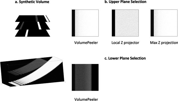
Results: We developed VolumePeeler, a versatile FIJI plugin for virtual 3D "peeling" of image stacks. The plugin implements spherical and spline surface projections. We applied VolumePeeler to perform peeling in 3D images of spherical embryos, as well as non-spherical tissue layers. The produced images improve the 3D volume visualization and enable analysis and quantification of geometrically challenging microscopy datasets.
Availability: ImageJ/FIJI software, source code, examples, and tutorials are openly available in https://cimt.uchile.cl/mcerda.
Keywords: 3D projections; Image processing; Microscopy; Virtual 3D peeling.
© 2023. The Author(s).
Conflict of interest statement
The authors declare that they have no competing interests.
Figures


References
-
- Herbert S, Valon L, Mancini L, Dray N, Caldarelli P, Gros J, Esposito E, Shorte SL, Bally-Cuif L, Aulner N, et al. LocalZProjector and DeProj: a toolbox for local 2D projection and accurate morphometrics of large 3D microscopy images. BMC Biol. 2021;19:1–13. doi: 10.1186/s12915-021-01037-w. - DOI - PMC - PubMed
-
- Schindelin J, Arganda-Carreras I, Frise E, Kaynig V, Longair M, Pietzsch T, Preibisch S, Rueden C, Saalfeld S, Schmid B, Tinevez J-Y, White DJ, Hartenstein V, Eliceiri K, Tomancak P, Cardona A. Fiji: an open-source platform for biological-image analysis. Nat Methods. 2012;9:676–682. doi: 10.1038/nmeth.2019. - DOI - PMC - PubMed
MeSH terms
LinkOut - more resources
Full Text Sources
