. 2023 Jul 13;24(1):186.
doi: 10.1186/s12931-023-02486-3.

Adenosine metabolized from extracellular ATP ameliorates organ injury by triggering A2BR signaling

Affiliations

Adenosine metabolized from extracellular ATP ameliorates organ injury by triggering A2BR signaling

Taha Kelestemur et al. Respir Res. .

Abstract

Background: Trauma and a subsequent hemorrhagic shock (T/HS) result in insufficient oxygen delivery to tissues and multiple organ failure. Extracellular adenosine, which is a product of the extracellular degradation of adenosine 5' triphosphate (ATP) by the membrane-embedded enzymes CD39 and CD73, is organ protective, as it participates in signaling pathways, which promote cell survival and suppress inflammation through adenosine receptors including the A2BR. The aim of this study was to evaluate the role of CD39 and CD73 delivering adenosine to A2BRs in regulating the host's response to T/HS.

Methods: T/HS shock was induced by blood withdrawal from the femoral artery in wild-type, global knockout (CD39, CD73, A2BR) and conditional knockout (intestinal epithelial cell-specific deficient VillinCre-A2BRfl/fl) mice. At 3 three hours after resuscitation, blood and tissue samples were collected to analyze organ injury.

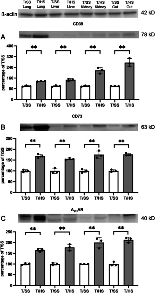
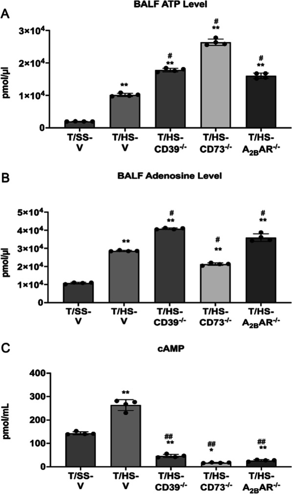
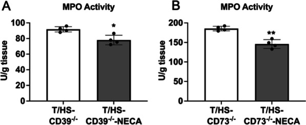
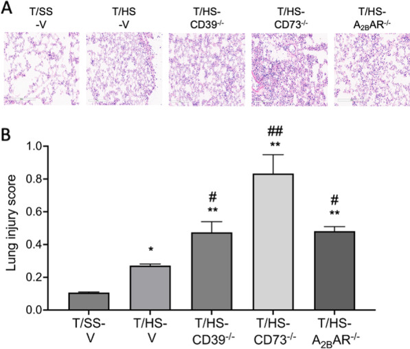
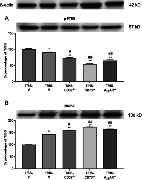
Results: T/HS upregulated the expression of CD39, CD73, and the A2BR in organs. ATP and adenosine levels increased after T/HS in bronchoalveolar lavage fluid. CD39, CD73, and A2BR mimics/agonists alleviated lung and liver injury. Antagonists or the CD39, CD73, and A2BR knockout (KO) exacerbated lung injury, inflammatory cytokines, and chemokines as well as macrophage and neutrophil infiltration and accumulation in the lung. Agonists reduced the levels of the liver enzymes aspartate transferase and alanine transaminase in the blood, whereas antagonist administration or CD39, CD73, and A2BR KO enhanced enzyme levels. In addition, intestinal epithelial cell-specific deficient VillinCre-A2BRfl/fl mice showed increased intestinal injury compared to their wild-type VillinCre controls.

Conclusion: In conclusion, the CD39-CD73-A2BR axis protects against T/HS-induced multiple organ failure.

Keywords: A2BR; Acute lung injury; Adenosine; CD39; CD73; Trauma hemorrhagic shock.


Conflict of interest statement

G.H. owns stock in Purine Pharmaceuticals, Inc. S.C.R. is the scientific cofounder of Purinomia and a consultant to SynLogic and eGenesis. The other authors have no financial conflict of interest.

Figures

Fig. 1
Fig. 1
T/HS increases CD39, CD73, and A2BAR expression in lung, liver, kidney, and gut. A–C CD39, CD73, and A2BAR expression were evaluated using western blotting of protein extracts. Data are mean ± S.D. (n = 4/group). **p < 0.01 compared with T/SS
Fig. 2
Fig. 2
ATP (A), adenosine (B), and cAMP (C) levels in BALF following T/HS in the CD39, CD73, and A2BAR KO samples. ATP and cAMP were measured colorimetrically and adenosine was measured by a fluorometric assay. Data are mean ± S.D. (n = 4/group) *p < 0.05 compared with T/SS-V, **p < 0.01 compared with T/SS-V, #p < 0.05 compared with T/HS-V, ##p < 0.01 compared with T/HS-V
Fig. 3
Fig. 3
CD39, CD73, and A2BR regulation of lung permeability and MPO activity. Lung permeability was determined using the EBD method (A, C, E, G) and MPO activity as a surrogate for neutrophil sequestration (B, D, F, H) was determined spectrophotometrically. Data are mean ± S.D. (n = 4/group). *p < 0.05 compared with corresponding T/SS, **p < 0.01 compared with corresponding T/SS, #p < 0.05 compared with corresponding T/HS-V, ##p < 0.01 compared with corresponding T/HS
Fig. 4
Fig. 4
Effect of NECA on lung MPO activity in CD39−/− and CD73−/− mice. MPO activity was determined from the lung spectrophotometrically (A, B). Data are mean ± S.D. (n = 4/group). *p < 0.05 compared with T/HS, **p < 0.05 compared with T/HS
Fig. 5
Fig. 5
Lung injury was induced by T/HS. Representative images of H&E-stained lung samples (A). Pulmonary injury was evaluated using 5 independent parameters (Neutrophils in the alveolar space, neutrophils in the interstitial space, hyaline membranes, proteinaceous debris filling the airspaces and alveolar septal thickness) (B). Data are mean ± S.D. (n = 4/group). *p < 0.05 compared with T/HS, **p < 0.05 compared with T/HS, #p < 0.05 compared with T/HS-V, ##p < 0.01 compared with T/HS-V
Fig. 6
Fig. 6
Quantification of pro- and anti-inflammatory cytokines and chemokines in the lung. Representative images of cytokine-chemokine arrays that were used to interrogate cytokine expression (A). Proteome Profiler Mouse Cytokine-Chemokine array was used to analyze cytokines in mice subjected to T/HS. Data were visualized by transforming them into a heat map B and also expressed as a bar graph (C). Data are mean ± S.D. (n = 4/group). *p < 0.05 compared with T/HS, **p < 0.05 compared with T/HS, #p < 0.05 compared with T/HS-V, ##p < 0.01 compared with T/HS-V
Fig. 7
Fig. 7
Macrophage accumulation in the lung after T/HS. Accumulated macrophages were determined by F4/80 immunostaining in the lung and representative images are shown as well as averages and means of macrophage counts in the various groups. Data are mean ± S.D. (n = 4/group). *p < 0.05 compared with T/HS, **p < 0.05 compared with T/HS, #p < 0.05 compared with corresponding T/HS-V, ##p < 0.01 compared with corresponding T/HS
Fig. 8
Fig. 8
Expression levels of survival-related kinases after T/HS. p-PTEN A and MMP-9 B expression levels were determined using western blot. Data are mean ± S.D. (n = 4/group). *p < 0.05 compared with T/HS, **p < 0.05 compared with T/HS, #p < 0.05 compared with corresponding T/HS-V, ##p < 0.01 compared with corresponding T/HS

References

    1. Cannon JW, Hemorrhagic Shock N Engl J Med. 2018;19:1852–3. - PubMed
    1. Huan Z, Tang Y, Xu C, Cai J, Yao H, Wang Y, Bu F, Ge X. PTPRO knockdown protects against inflammation in hemorrhage shock-induced lung injury involving the NF-kappaB signaling pathway. Respir Res. 2022;1:195. doi: 10.1186/s12931-022-02118-2. - DOI - PMC - PubMed
    1. Kao RL, Xu X, Xenocostas A, Parry N, Mele T, Martin CM, Rui T. Induction of acute lung inflammation in mice with hemorrhagic shock and resuscitation: role of HMGB1. J Inflamm (Lond) 2014;1:30. doi: 10.1186/s12950-014-0030-7. - DOI - PMC - PubMed
    1. Valade G, Libert N, Martinaud C, Vicaut E, Banzet S, Peltzer J. Therapeutic potential of mesenchymal stromal cell-derived extracellular vesicles in the Prevention of Organ Injuries Induced by Traumatic hemorrhagic shock. Front Immunol. 2021 doi: 10.3389/fimmu.2021.749659. - DOI - PMC - PubMed
    1. Hasko G, Xu DZ, Lu Q, Nemeth ZH, Jabush J, Berezina TL, Zaets SB, Csoka B, Deitch EA. Adenosine A2A receptor activation reduces lung injury in trauma/hemorrhagic shock. Crit Care Med. 2006;4:1119–25. doi: 10.1097/01.CCM.0000206467.19509.C6. - DOI - PubMed