. 2024 Jan 1;109(1):231-244.
doi: 10.3324/haematol.2023.283274.

DIS3 depletion in multiple myeloma causes extensive perturbation in cell cycle progression and centrosome amplification

Affiliations

DIS3 depletion in multiple myeloma causes extensive perturbation in cell cycle progression and centrosome amplification

Vanessa K Favasuli et al. Haematologica. .

Abstract

DIS3 gene mutations occur in approximately 10% of patients with multiple myeloma (MM); furthermore, DIS3 expression can be affected by monosomy 13 and del(13q), found in roughly 40% of MM cases. Despite the high incidence of DIS3 mutations and deletions, the biological significance of DIS3 and its contribution to MM pathogenesis remain poorly understood. In this study we investigated the functional role of DIS3 in MM, by exploiting a loss-of-function approach in human MM cell lines. We found that DIS3 knockdown inhibits proliferation in MM cell lines and largely affects cell cycle progression of MM plasma cells, ultimately inducing a significant increase in the percentage of cells in the G0/G1 phase and a decrease in the S and G2/M phases. DIS3 plays an important role not only in the control of the MM plasma cell cycle, but also in the centrosome duplication cycle, which are strictly co-regulated in physiological conditions in the G1 phase. Indeed, DIS3 silencing leads to the formation of supernumerary centrosomes accompanied by the assembly of multipolar spindles during mitosis. In MM, centrosome amplification is present in about a third of patients and may represent a mechanism leading to genomic instability. These findings strongly prompt further studies investigating the relevance of DIS3 in the centrosome duplication process. Indeed, a combination of DIS3 defects and deficient spindle-assembly checkpoint can allow cells to progress through the cell cycle without proper chromosome segregation, generating aneuploid cells which ultimately lead to the development of MM.


Figures

Figure 1.
Figure 1.
DIS3 expression analysis in plasma cell dyscrasias. (A) Box plot of DIS3 expression level in healthy donors (N) and plasma cell (PC) dyscrasias (proprietary dataset, GSE66293). The four N are RNA samples from bone marrow PC purified (>90%) from normal individuals and purchased from Voden (Medical Instruments IT). Total RNA samples from highly purified bone marrow CD138+ PC were profiled by Gene 1.0 ST array. (B) Box plot of DIS3 expression level in main IGH translocation groups in 774 cases from the CoMMpass cohort. The Kruskal-Wallis test was applied to assess differences in expression levels between groups. The pairwise comparisons were performed by the Dunn test; statistically significant results are marked in red, bold in the tables. MM: multiple myeloma; pPCL: primary plasma cell leukemia; sPCL: secondary plasma cell leukemia; trx.MAF: MAF translocation, trx.MYC: MYC translocation, double.trx: presense of two translocations, trx.neg: absence of the considered translocations.
Figure 2.
Figure 2.
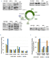
DIS3 expression analysis in DIS3-depleted cells. (A) Quantitative real-time polymerase chain reaction analysis of DIS3 mRNA in AMO-1, NCI-H929 and U266 cells at the indicated timepoints. DIS3 mRNA expression is represented as 2-ΔΔCt relative to the GAPDH housekeeping gene and the scrambled condition at the same timepoint used as a calibrator. *P<0.05, **P<0.01, Student t test. (B) Western blot analyses of DIS3-KD in AMO-1, NCI-H929 and U266 cells. Western blot results are quantified and plotted in a histogram. SCR: scrambled condition.
Figure 3.
Figure 3.
Functional impact of DIS3 depletion in multiple myeloma cells. (A) Growth curves of AMO-1, NCI-H929 and U266 cells following DIS3 silencing. (B) Flow cytometry analyses of apoptosis in AMO-1, NCI-H929 and U266 cells 6 days after treatment with gDIS3 (5 μM). (C) Colony formation assay performed on AMO-1, NCI-H929 and U266 cells treated for 21 days with gDIS3; representative pictures of colonies at day 21 are also shown, **P<0.01, ***P<0.001, Student t test. (D) Rappresentative images of May-Grünwald Giemsa staining of two CD138+ primary tumors treated for 6 days with gDIS3 (50x magnification). SCR: scrambled condition; 7-AAD: 7 amino actinomycin D.
Figure 4.
Figure 4.
Cell cycle analysis in DIS3-depleted cells. (A) Cell cycle progression in synchronized AMO-1, NCI-H929, and U266 cells was monitored by propidium iodide staining and fluorescence activated cell sorting analysis of the DNA content of cells at 6 h after release. (B) The percentage of cell cycle distribution is represented in the histograms; the standard deviations of three replicates are reported. *P<0.05, **P<0.01, ***P<0.001, Student t test. SCR: scrambled condition.
Figure 5.
Figure 5.
Western blot analyses of cell cycle checkpoint proteins. (A) Analysis performed in synchronized AMO-1, NCI-H929 and U266 cells 6 h after release. For NCI-H929 cells, pCDC20, CDC20 and CENP-A were evaluated on the same blot and thus with the same GAPDH control; to improve the overall symmetry of the figure and its comprehension, the same GAPDH panel has been reported twice. (B) Western blot results are quantified and plotted in a histogram. (C) Histogram of the modulation of the phosphorylated protein fraction upon gDIS3 treatment, calculated as a ratio of phosphorylated protein to total protein in gDIS3-treated cells compared to the ratio of phosphorylated protein to total protein in scramble-treated ones. SCR: scramble condition.
Figure 6.
Figure 6.
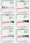
Transcriptomic analyses in DIS3-depleted cells. Enrichment plots of selected gene set enrichment analysis (GSEA) gene sets significantly modulated in DIS3-silenced cells in comparison to scrambled NCI-H929 cells. GSEA was performed on global annotated protein-coding gene expression profiles (19,048 genes) and the most significant gene sets were selected under stringent conditions (nominal P value <0.05 and false discovery rate q value <5%). The enrichment score and nominal P values are reported for each plot. NES: normalized enrichment score; FDR: false discovery rate.
Figure 7.
Figure 7.
Molecular validation of the transcriptomic analysis. (A) Quantitative real-time polymerase chain reaction validation of the indicated genes in DIS3-depleted AMO-1, NCI-H929, and U266 human multiple myeloma cell lines; gene expression is presented as 2-ΔΔCt relative to the GAPDH housekeeping gene and the scrambled condition used as a calibrator. *P<0.05, **P<0.01, ***P<0.001, Student t test. (B) Ex-vivo validation of the indicated genes in DIS3-depleted primary CD138+ tumor cells purified from four multiple myeloma patients. SCR: scrambled condition.
Figure 8.
Figure 8.
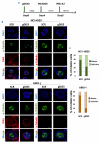
Images of metaphase spindles in NCI-H929 and AMO-1 cells treated with gDIS3 or scrambled gapmer. (A) Schematic representation of the experimental setup used to synchronize cells. (B) Representative images of multipolar spindles in both cell lines following DIS3-knockdown (right panel for each cell line). Scale bar, 5 μm. (C) Percentage of mitotic cells with defective spindles in NCI-H929 and AMO-1 cells treated with gDIS3 versus scrambled gapmer. IF: immmunofluorescence; SCR: scrambled condition.

Similar articles

Cited by

References

    1. Munshi NC, Anderson KC. New strategies in the treatment of multiple myeloma. Clin Cancer Res. 2013;19(13):3337-3344. - PMC - PubMed
    1. Morgan GJ, Walker BA, Davies FE. The genetic architecture of multiple myeloma. Nat Rev Cancer. 2012;12(5):335-348. - PubMed
    1. Todoerti K, Ronchetti D, Favasuli V, et al. . DIS3 mutations in multiple myeloma impact the transcriptional signature and clinical outcome. Haematologica. 2022;107(4):921-932. - PMC - PubMed
    1. Lionetti M, Barbieri M, Todoerti K, et al. . A compendium of DIS3 mutations and associated transcriptional signatures in plasma cell dyscrasias. Oncotarget. 2015;6(28):26129-26141. - PMC - PubMed
    1. Boyle EM, Ashby C, Tytarenko RG, et al. . BRAF and DIS3 mutations associate with adverse outcome in a long-term follow-up of patients with multiple myeloma. Clin Cancer Res. 2020;26(10):2422-2432. - PubMed

Publication types

Substances