

Intensive care unit sinks are persistently colonized with multidrug resistant bacteria and mobilizable, resistance-conferring plasmids
- PMID: 37439570
- PMCID: PMC10469867
- DOI: 10.1128/msystems.00206-23
Intensive care unit sinks are persistently colonized with multidrug resistant bacteria and mobilizable, resistance-conferring plasmids
Abstract
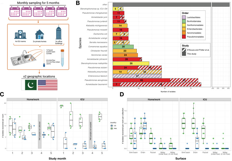
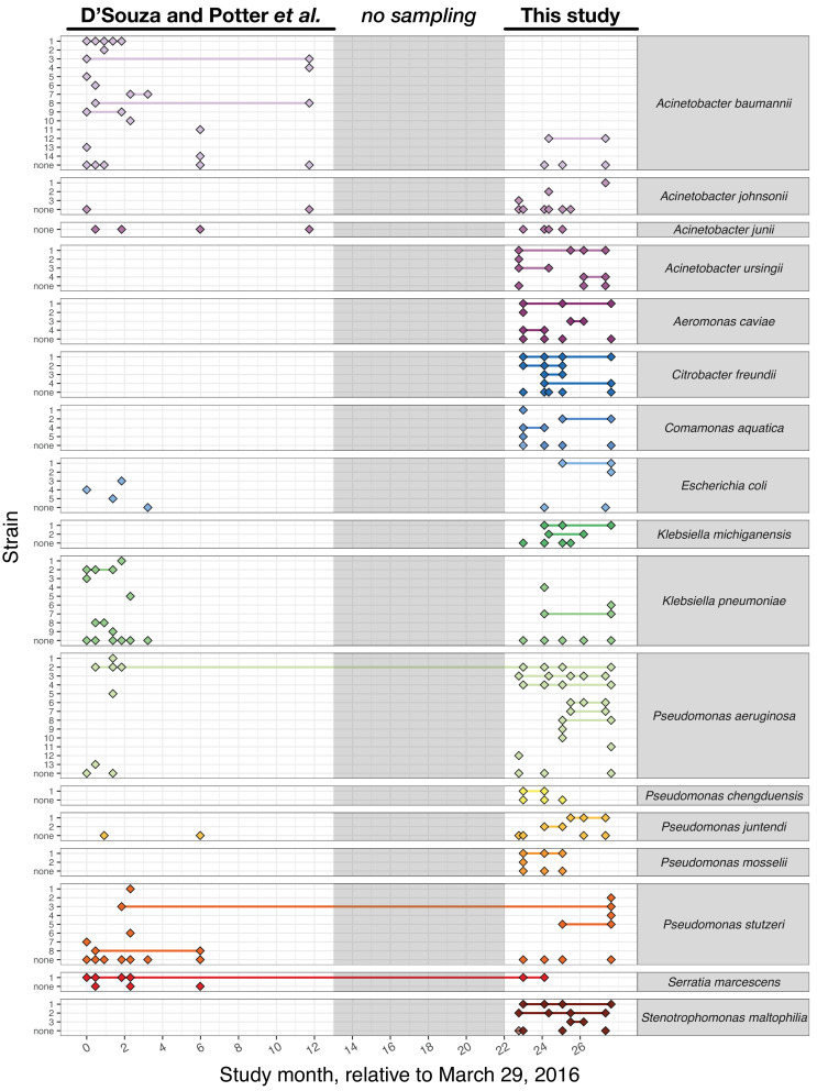
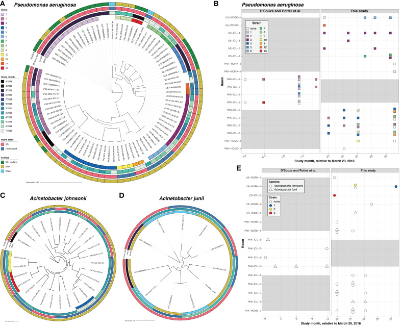
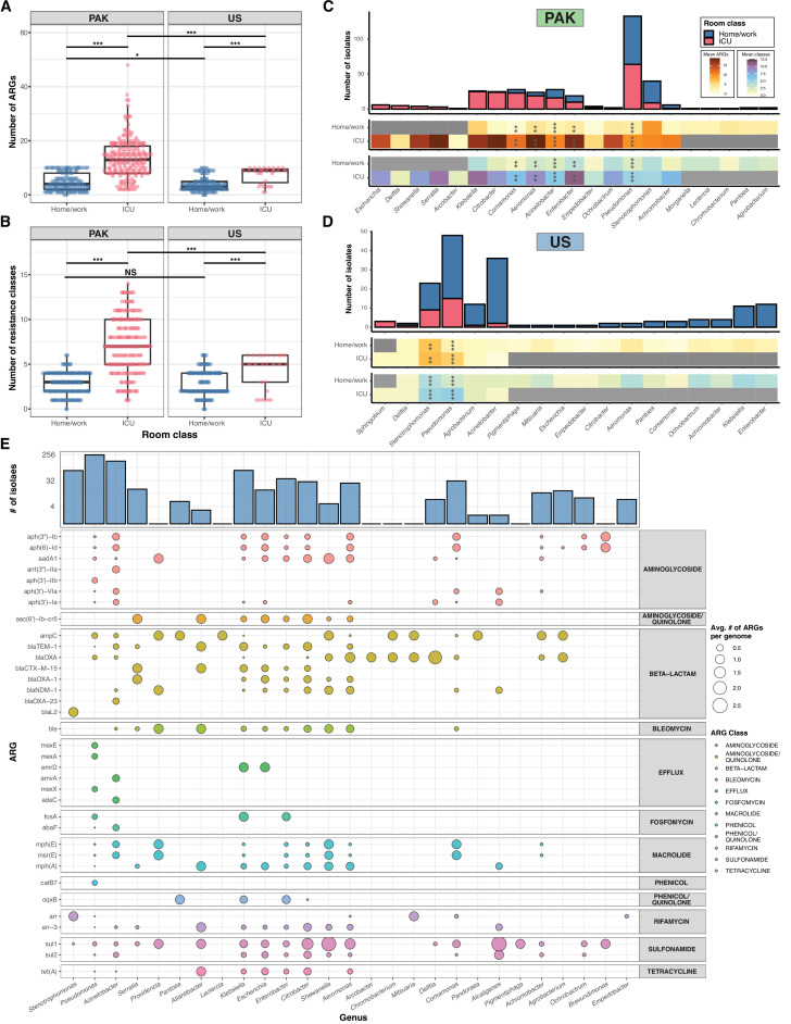
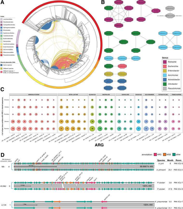
Contamination of hospital sinks with microbial pathogens presents a serious potential threat to patients, but our understanding of sink colonization dynamics is largely based on infection outbreaks. Here, we investigate the colonization patterns of multidrug-resistant organisms (MDROs) in intensive care unit sinks and water from two hospitals in the USA and Pakistan collected over 27 months of prospective sampling. Using culture-based methods, we recovered 822 bacterial isolates representing 104 unique species and genomospecies. Genomic analyses revealed long-term colonization by Pseudomonas spp. and Serratia marcescens strains across multiple rooms. Nanopore sequencing uncovered examples of long-term persistence of resistance-conferring plasmids in unrelated hosts. These data indicate that antibiotic resistance (AR) in Pseudomonas spp. is maintained both by strain colonization and horizontal gene transfer (HGT), while HGT maintains AR within Acinetobacter spp. and Enterobacterales, independent of colonization. These results emphasize the importance of proactive, genomic-focused surveillance of built environments to mitigate MDRO spread. IMPORTANCE Hospital sinks are frequently linked to outbreaks of antibiotic-resistant bacteria. Here, we used whole-genome sequencing to track the long-term colonization patterns in intensive care unit (ICU) sinks and water from two hospitals in the USA and Pakistan collected over 27 months of prospective sampling. We analyzed 822 bacterial genomes, representing over 100 different species. We identified long-term contamination by opportunistic pathogens, as well as transient appearance of other common pathogens. We found that bacteria recovered from the ICU had more antibiotic resistance genes (ARGs) in their genomes compared to matched community spaces. We also found that many of these ARGs are harbored on mobilizable plasmids, which were found shared in the genomes of unrelated bacteria. Overall, this study provides an in-depth view of contamination patterns for common nosocomial pathogens and identifies specific targets for surveillance.
Keywords: antimicrobial resistance; genomic epidemiology; horizontal gene transfer; hospital surveillance; plasmid ecology; whole-genome sequencing.
Conflict of interest statement
The authors declare no conflict of interest.
Figures
Similar articles
-
The ICU environment contributes to the endemicity of the "Serratia marcescens complex" in the hospital setting.mBio. 2024 May 8;15(5):e0305423. doi: 10.1128/mbio.03054-23. Epub 2024 Apr 2. mBio. 2024. PMID: 38564701 Free PMC article.
-
Tracking of Antibiotic Resistance Transfer and Rapid Plasmid Evolution in a Hospital Setting by Nanopore Sequencing.mSphere. 2020 Aug 19;5(4):e00525-20. doi: 10.1128/mSphere.00525-20. mSphere. 2020. PMID: 32817379 Free PMC article.
-
Systematic detection of horizontal gene transfer across genera among multidrug-resistant bacteria in a single hospital.Elife. 2020 Apr 14;9:e53886. doi: 10.7554/eLife.53886. Elife. 2020. PMID: 32285801 Free PMC article.
-
Approaches for characterizing and tracking hospital-associated multidrug-resistant bacteria.Cell Mol Life Sci. 2021 Mar;78(6):2585-2606. doi: 10.1007/s00018-020-03717-2. Epub 2021 Feb 13. Cell Mol Life Sci. 2021. PMID: 33582841 Free PMC article. Review.
-
Genome plasticity as a paradigm of antibiotic resistance spread in ESKAPE pathogens.Environ Sci Pollut Res Int. 2022 Jun;29(27):40507-40519. doi: 10.1007/s11356-022-19840-5. Epub 2022 Mar 29. Environ Sci Pollut Res Int. 2022. PMID: 35349073 Review.
Cited by
-
The hospital sink drain microbiome as a melting pot for AMR transmission to nosocomial pathogens.NPJ Antimicrob Resist. 2025 Jul 29;3(1):68. doi: 10.1038/s44259-025-00137-9. NPJ Antimicrob Resist. 2025. PMID: 40730864 Free PMC article. Review.
-
High genetic relatedness between multidrug resistant bacteria before and after the 2022 invasion of Ukraine.Genome Med. 2025 Jul 1;17(1):74. doi: 10.1186/s13073-025-01500-1. Genome Med. 2025. PMID: 40598567 Free PMC article.
-
High-Throughput Short Sequence Typing Schemes for Pseudomonas aeruginosa and Stenotrophomonas maltophilia Pure Culture and Environmental DNA.Microorganisms. 2023 Dec 27;12(1):48. doi: 10.3390/microorganisms12010048. Microorganisms. 2023. PMID: 38257875 Free PMC article.
-
Abundance and diversity of methicillin-resistant bacteria from bathroom surfaces at workplaces using CHROMagar media, 16S, and dnaJ gene sequence typing.Int J Mol Epidemiol Genet. 2024 Apr 15;15(2):12-21. doi: 10.62347/EJQK3362. eCollection 2024. Int J Mol Epidemiol Genet. 2024. PMID: 38736754 Free PMC article.
-
Characterization of vanA-harboring plasmids supports differentiation of outbreak-related and sporadic vancomycin-resistant Enterococcus faecium isolates in a tertiary care hospital.BMC Microbiol. 2025 May 28;25(1):337. doi: 10.1186/s12866-025-04058-5. BMC Microbiol. 2025. PMID: 40437365 Free PMC article.
References
-
- CDC . 2019. Antibiotic resistance threats in the United States 2019. Atlanta, GA: U.S. Department of Health and Human Services C. https://www.cdc.gov/drugresistance/biggest-threats.html.
-
- WHO . 2016. Global action plan on antimicrobial resistance
-
- Cassini A, Högberg LD, Plachouras D, Quattrocchi A, Hoxha A, Simonsen GS, Colomb-Cotinat M, Kretzschmar ME, Devleesschauwer B, Cecchini M, Ouakrim DA, Oliveira TC, Struelens MJ, Suetens C, Monnet DL, Burden of AMR Collaborative Group . 2019. Attributable deaths and disability-adjusted life-years caused by infections with antibiotic-resistant bacteria in the EU and the European Economic Area in 2015: a population-level modelling analysis. Lancet Infect Dis 19:56–66. doi:10.1016/S1473-3099(18)30605-4 - DOI - PMC - PubMed
-
- Mora M, Mahnert A, Koskinen K, Pausan MR, Oberauner-Wappis L, Krause R, Perras AK, Gorkiewicz G, Berg G, Moissl-Eichinger C. 2016. Microorganisms in confined habitats: microbial monitoring and control of intensive care units, operating rooms, Cleanrooms and the international space station. Front Microbiol 7:1573. doi:10.3389/fmicb.2016.01573 - DOI - PMC - PubMed
Publication types
MeSH terms
Substances
Grants and funding
LinkOut - more resources
Full Text Sources
Research Materials
Miscellaneous
