Review
. 2023 Aug 23;61(8):e0140322.
doi: 10.1128/jcm.01403-22. Epub 2023 Jul 13.

Testing for Human Papillomaviruses in Urine, Blood, and Oral Specimens: an Update for the Laboratory

Affiliations
Review

Testing for Human Papillomaviruses in Urine, Blood, and Oral Specimens: an Update for the Laboratory

Mario Poljak et al. J Clin Microbiol. .

Abstract

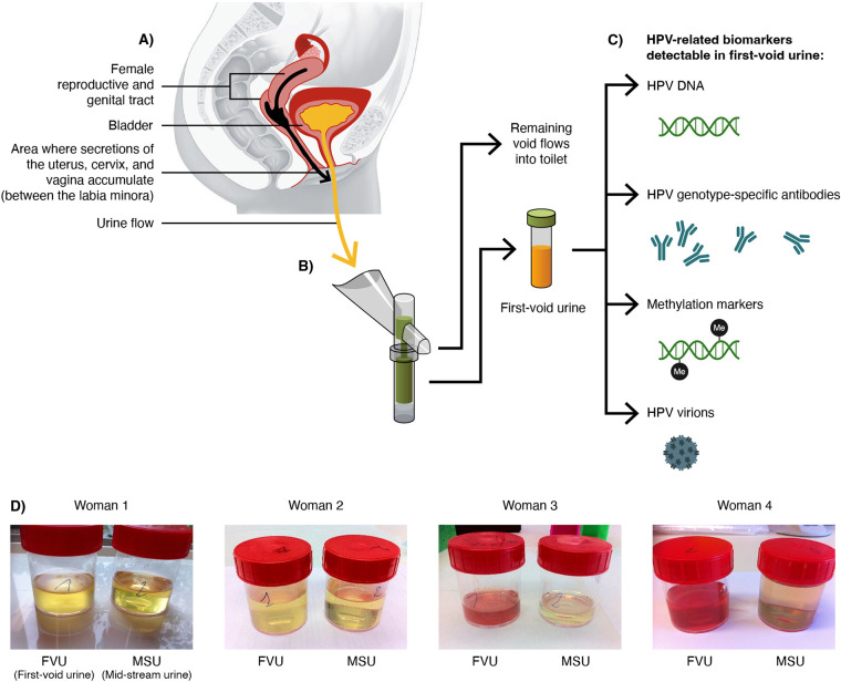
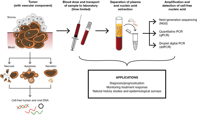
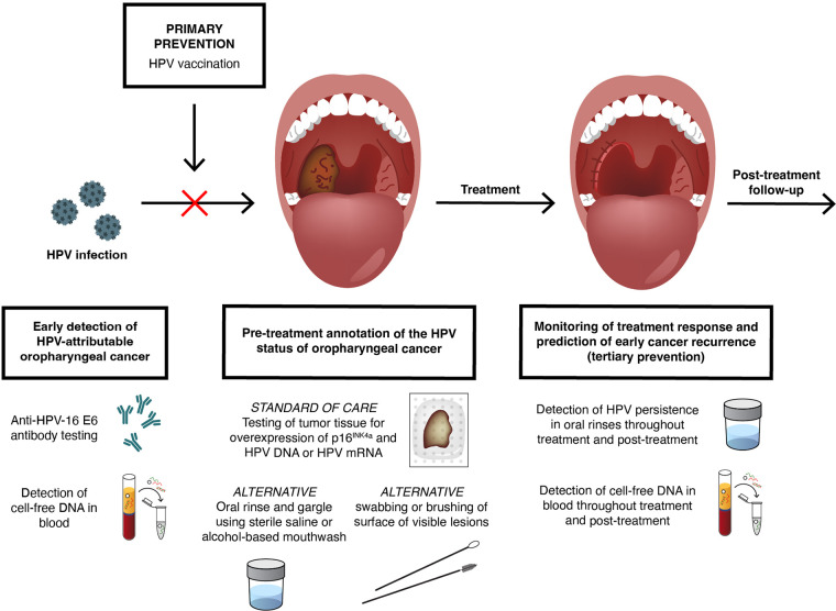
Twelve high-risk alpha human papillomavirus (HPV) genotypes cause approximately 690,000 cancer cases annually, with cervical and oropharyngeal cancer being the two most prominent types. HPV testing is performed in laboratory settings for various applications of a clinical, epidemiological, and research nature using a range of clinical specimens collected by clinicians or by individuals (self-collected specimens). Here, we reflect on the importance and justification of using the right test for the right application and provide practical updates for laboratories either participating in or anticipating involvement in HPV testing in three specimen types, namely, urine, blood, and oral specimens, which are considered "alternative" specimens by many. In addition to clinician-collected cervical samples and self-collected cervicovaginal samples, first-void urine is emerging as a credible specimen for HPV-based cervical cancer screening, triage of HPV screen-positive women, monitoring HPV vaccine impact, and HPV testing in groups for which a less invasive sample is preferred. Detection of cell-free DNA (including HPV DNA) in blood has great promise for the early detection of HPV-attributable oropharyngeal cancer (HPV-AOC) and potentially other HPV-driven cancers and as an adjunct prognostic marker in long-term tumor surveillance, including treatment response. The moderate sensitivity of HPV testing in oral rinses or swabs at HPV-AOC diagnosis prevents its use in HPV-AOC secondary prevention but represents a promising prognostic tool in HPV-AOC tertiary prevention, where the HPV persistence in oral rinses throughout treatment may predict early HPV-AOC recurrences and/or the development of secondary HPV-AOC. The increasing sophistication of specific collection devices designed for alternative samples and the enhanced precision of novel molecular technologies are likely to support the evolution of this field and catalyze potential translation into routine practice.

Keywords: HPV; blood; cervical cancer; oral specimens; oropharyngeal cancer; urine.


Conflict of interest statement

The authors declare a conflict of interest. M.P. institution received research funding, free-of-charge reagents, and consumables to support research in the last 3 years from Qiagen, Seegene, Abbott, and Roche, all paid to his employer. K.C. institution received research funding, reagents, and consumables to support research in the last 3 years from Cepheid, Euroimmun, GeneFirst, Self-screen, Hiantis, Seegene, Roche, Abbott, Hologic, and Vaccitech, all paid to her employer. K.C. also attended an advisory board meeting of Hologic where UK-based travel expenses were supported and is currently on the advisory board of Vaccitech (with any associated reimbursement paid to employer). L.A. institution received funding to support research in the last 3 years from Merck Sharp & Dohme, Roche, GSK, Vitro, Hologic, and Seegene, all paid to her employer. A.V. institution received funding to support research and setting up professional meetings in the last 3 years from Merck, Roche, GSK, Hologic, Abbott, Novosanis and Becton, Dickinson and Company, all paid to his employer. A.V. is the co-founder of Novosanis, a spin-off company of the University of Antwerp, Belgium, and a subsidiary of OraSure Technologies, Inc. since 2019, and he was a board member and minority shareholder until January 2019.

Figures

FIG 1
FIG 1
Rationale for the use of first-void urine as source of female reproductive and genital tract biomarkers and HPV-related biomarkers detectable in first-void urine. (A) Presents the anatomy of the female reproductive and genital tract and bladder, with the black arrow depicting the physiological movement of secretions of the uterus, cervix, and vagina subsequently mixed with debris of superficial cell layers of the epithelium covering the entire surface of the genital tract, exfoliated cells, and transudated or exudated immunoglobulins (mainly IgG). All secretions further drift and exit the vagina, where they are accumulated between the labia minora and around the urethra opening and flushed away by the urine originating from the bladder (yellow arrow). (B) Shows a first-void urine collection device (Colli-Pee; Novosanis, Wijnegem, Belgium) that can be prefilled with a urine preservative and is able to capture a prespecified volume of first-void or initial-stream urine without interrupting the urine void. Once the collection tube is filled, an outlet ensures that subsequent urine volume exits the device into the toilet. (C) Shows several relevant biomarkers detectable in first-void urine, as follows: (i) cell-free and cell-associated HPV DNA; (ii) transudated or excudated immunoglobulins, including HPV genotype-specific antibodies; (iii) methylated viral and human DNA; and (iv) HPV virions in the case of a productive HPV infection. (D) Shows selected examples of urine specimens collected by four out of eight women that captured their initial or first-void urine (FVU) and the subsequent void (often referred to as midstream urine [MSU]) directly into urine collection containers. In six of the eight women, macroscopically visible differences between FVU versus MSU samples due to substantially more debris derived from superficial layers of the epithelium, exfoliated cells, and genital tract secretions present in FVU were noticed. In the samples from women 3 and 4, who were menstruating at the time of sample collection, the FVU samples also contained menstrual blood visible with the naked eye compared with an almost transparent MSU sample(s).
FIG 2
FIG 2
Process and rationale for the detection of cell-free DNA (including HPV DNA). Abnormal lesions and cancers with a vascular component contain nuclear material that can be shed into the blood through various pathways, including necrosis, apoptosis, and secretion. This material, referred to as cell-free DNA (cfDNA), can be double or single stranded, and the concentration and stability depend on a number of factors, including lesion size, extent of proliferation, and extent of vascularization. cfDNA can be host or viral in origin, and it is feasible for a lesion to exude both. After a blood draw and centrifugation, nucleic acid that contains cfDNA can be extracted from the plasma component. Given the short half-life of some cfDNA targets, efficient sample transfer to the laboratory for processing and extraction is important. cfDNA can be used to detect viral and/or host sequences, specific mutations, integration hallmarks, methylation targets, and microsatellite alterations. Although a range of amplification technologies, such as quantitative PCR (qPCR), have been used for detecting cfDNA, those methods that can offer high analytical sensitivity are valuable, such as next-generation sequencing (NGS) and droplet digital PCR (ddPCR). The translation of cfDNA into routine diagnostic laboratory practice is at a relatively early stage for HPV-associated disease, but it holds promise for various applications, including diagnosis/prognostication and monitoring treatment response. In addition, cfDNA determination is valuable in natural history studies and epidemiological surveys.
FIG 3
FIG 3
Primary, secondary, and tertiary prevention of HPV-attributable oropharyngeal cancer (HPV-AOC). Primary prevention of HPV-AOC by HPV vaccination might work for HPV-AOC, pending the results of a postapproval confirmatory study, which is still ongoing. No validated screening strategy for the secondary prevention of HPV-AOC aiming for early disease detection in the healthy population is available at present, but the following two approaches have been extensively explored in the last decade: (i) the detection of antibodies against HPV-16 E6 protein in blood and (ii) “liquid biopsies,” or the detection of cell-free DNA (including HPV DNA) in blood samples. Differentiation of HPV-AOC from HPV-non-attributable oropharyngeal cancers is currently a major clinical application of HPV testing in the field. Annotation of the HPV status of oropharyngeal cancer is best ascertained by testing tumor tissue (formalin-fixed, paraffin-embedded or fresh-frozen tissues) for overexpression of p16INK4a using immunohistochemistry and the presence of HPV DNA or HPV mRNA by either in situ hybridization or PCR-based methods. Oral sample nontissue HPV-annotation alternatives, including (i) oral rinse and gargle using sterile saline or alcohol-based mouthwash and (ii) swabbing or brushing of surface of visible lesions, have been evaluated, and yet, the moderate sensitivity of these alternatives currently limits their use as diagnostics. In addition, nonoral alternatives, e.g., detection of cell-free DNA and antibodies against HPV-16 E6 protein in blood, have been also piloted for HPV-annotation purposes in selected clinical scenarios but with mixed and inconclusive results. The detection of HPV persistence in oral rinses and/or by detecting cell-free DNA in blood throughout treatment and posttreatment represent valid treatment response monitoring tools and promising predictive tools in the tertiary prevention of HPV-AOC (prevention of disease recurrences after definite therapy and/or prevention of the development of secondary HPV-AOC).

Similar articles

Cited by

References

    1. Kocjan BJ, Bzhalava D, Forslund O, Dillner J, Poljak M. 2015. Molecular methods for identification and characterization of novel papillomaviruses. Clin Microbiol Infect 21:808–816. doi:10.1016/j.cmi.2015.05.011. - DOI - PubMed
    1. Van Doorslaer K, Chen Z, Bernard H-U, Chan PKS, DeSalle R, Dillner J, Forslund O, Haga T, McBride AA, Villa LL, Burk RD, Ictv Report Consortium., ICTV Report Consortium . 2018. ICTV virus taxonomy profile: Papillomaviridae. J Gen Virol 99:989–990. doi:10.1099/jgv.0.001105. - DOI - PMC - PubMed
    1. Arroyo Mühr LS, Eklund C, Dillner J. 2021. Misclassifications in human papillomavirus databases. Virology 558:57–66. doi:10.1016/j.virol.2021.03.002. - DOI - PubMed
    1. Cubie HA. 2013. Diseases associated with human papillomavirus infection. Virology 445:21–34. doi:10.1016/j.virol.2013.06.007. - DOI - PubMed
    1. Burd EM. 2016. Human papillomavirus laboratory testing: the changing paradigm. Clin Microbiol Rev 29:291–319. doi:10.1128/CMR.00013-15. - DOI - PMC - PubMed

Publication types