. 2023 Aug 31;225(1):iyad129.
doi: 10.1093/genetics/iyad129.

Identification and characterization of protein interactions with the major Niemann-Pick type C disease protein in yeast reveals pathways of therapeutic potential

Affiliations

Identification and characterization of protein interactions with the major Niemann-Pick type C disease protein in yeast reveals pathways of therapeutic potential

Natalie Hammond et al. Genetics. .

Abstract

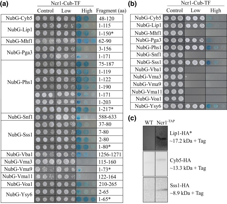
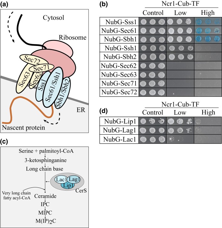
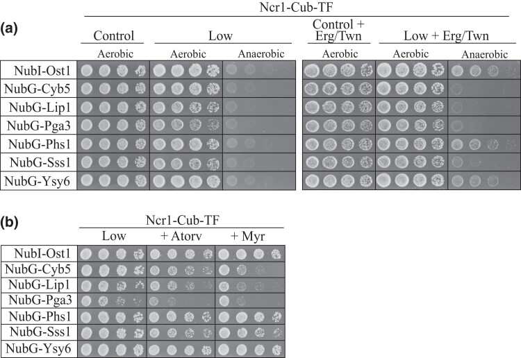
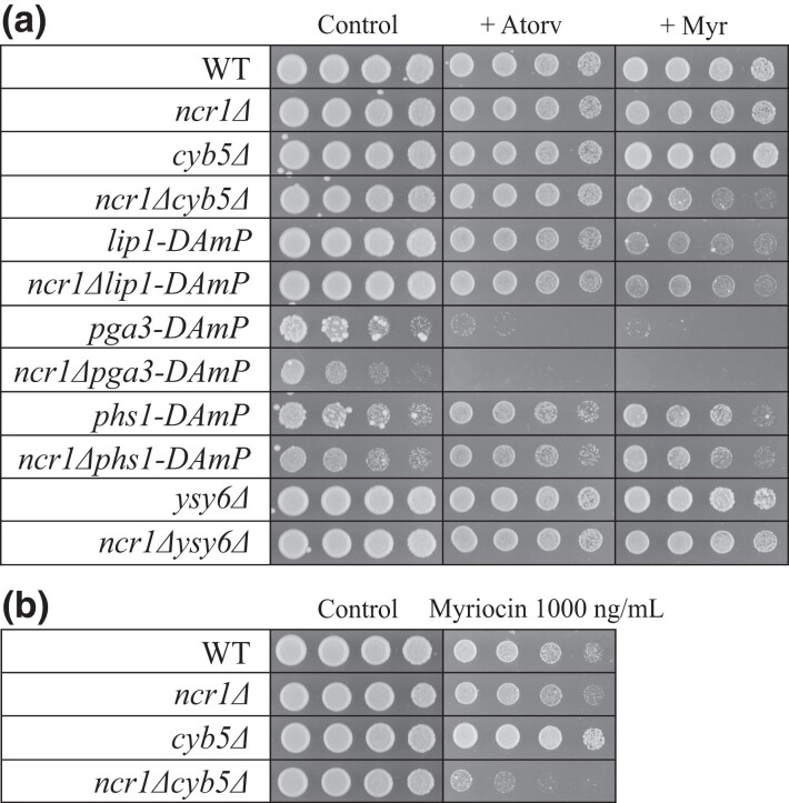
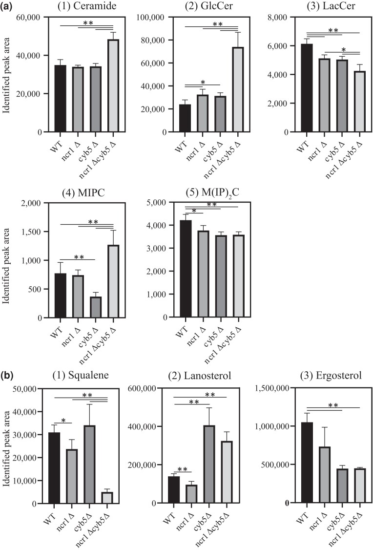
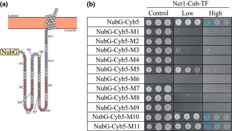
Niemann-Pick type C (NP-C) disease is a rare lysosomal storage disease caused by mutations in NPC1 (95% cases) or NPC2 (5% cases). These proteins function together in cholesterol egress from the lysosome, whereby upon mutation, cholesterol and other lipids accumulate causing major pathologies. However, it is not fully understood how cholesterol is transported from NPC1 residing at the lysosomal membrane to the endoplasmic reticulum (ER) and plasma membrane. The yeast ortholog of NPC1, Niemann-Pick type C-related protein-1 (Ncr1), functions similarly to NPC1; when transfected into a mammalian cell lacking NPC1, Ncr1 rescues the diagnostic hallmarks of cholesterol and sphingolipid accumulation. Here, we aimed to identify and characterize protein-protein interactions (PPIs) with the yeast Ncr1 protein. A genome-wide split-ubiquitin membrane yeast two-hybrid (MYTH) protein interaction screen identified 11 ER membrane-localized, full-length proteins interacting with Ncr1 at the lysosomal/vacuolar membrane. These highlight the importance of ER-vacuole membrane interface and include PPIs with the Cyb5/Cbr1 electron transfer system, the ceramide synthase complex, and the Sec61/Sbh1 protein translocation complex. These PPIs were not detected in a sterol auxotrophy condition and thus depend on normal sterol metabolism. To provide biological context for the Ncr1-Cyb5 PPI, a yeast strain lacking this PPI (via gene deletions) exhibited altered levels of sterols and sphingolipids including increased levels of glucosylceramide that mimic NP-C disease. Overall, the results herein provide new physical and genetic interaction models to further use the yeast model of NP-C disease to better understand human NP-C disease.

Keywords: lipid transport; lysosomal storage disease; neurodegenerative disease; rare disease; sphingolipid; sterol; yeast model.


Conflict of interest statement

Conflicts of interest: The authors declare no conflict of interest.

Figures

Fig. 1.
Fig. 1.
PPIs with Ncr1 using MYTH and co-IP analysis. MYTH was conducted where plasmids expressing prey proteins (NubG-Xxx) were transformed into an Ncr1-Cub-TF bait strain. Serial dilutions (1:5) of saturated transformants were plated on selection stringency agar [control (SD-W), low (SD-WH), high (SD-WH + X-Gal)] and incubated at 30°C for 3 days. Shown are cell dilutions 1:5, 1:25, and 1:125. a) Prey plasmids recovered from the prey plasmid library were retransformed into MYTH bait Ncr1-Cub-TF. b) Full-length prey proteins were constructed and transformed into Ncr1-Cub-TF. c) Co-IP analysis detected physical interactions between Cyb5-HA, Lip1-HA, and Sss1-HA with TAP-tagged Ncr1. Cyb5-HA, Lip1-HA, and Sss1-HA plasmids were transformed into WT and Ncr1-TAP tagged strains; co-IP analysis and subsequent western blot probed with anti-HA in WT and Ncr1-TAP tagged strains. *Digitonin was used instead of triton for the detergent. Expected sizes recovered on the western blot are indicated.
Fig. 2.
Fig. 2.
Members of the Sec61 protein translocation complex and the ceramide synthase complex physically interact with Ncr1. a) Simplified cartoon showing Sss1 functioning as part of the Sec61 protein translocation complex (Sss1, Sec61/Ssh1, Sbh1/Sbh2). Also functioning in protein translocation is the Sec63 complex (Sec62, Sec63, Sec71, and Sec72). Ribosome processing the mRNA binds to the Sec protein complexes on the ER membrane. Newly translated protein is transported into the ER through these complexes for further processing. Topology based on Itskanov and Park (2019). b) Full-length prey protein plasmids (NubG-Sss1, NubG-Sec61, NubG-Sbh1, NubG-Ssh1, NubG-Sbh2, NubG-Sec62, NubG-Sec63, NubG-Sec71, and NubG-Sec72) were transformed into Ncr1-Cub-TF. c) Simplified cartoon showing Lip1 functioning as part of the ceramide synthase complex (Lag1, Lac1, and Lip1) which converts sphingoid bases to ceramide in sphingolipid biosynthesis. d) Prey protein plasmids (NubG-Lip1, NubG-Lag1, and NubG-Lac1) were transformed into Ncr1-Cub-TF. For b) and d), serial dilutions (1:5) of saturated transformants were plated on selection stringency agar [control (SD-W), low (SD-WH), high (SD-WH + X-Gal)] and incubated at 30°C for 3 days. Shown are cell dilutions 1:5, 1:25, and 1:125.
Fig. 3.
Fig. 3.
MYTH analysis in conditions of obligatory ergosterol uptake (anaerobiosis) and treatment with lipid biosynthesis inhibitors. a) Prey constructs [NubI-Ost1 (control), NubG-Cyb5, NubG-Lip1, NubG-Pga3, NubG-Phs1, NubG-Sss1, and NubG-Ysy6] were transformed into Ncr1-Cub-TF. Resulting transformants were grown overnight, serial diluted (1:5), and plated on selection stringency agar: control (SD-W), control + 20 µg/mL in ergosterol in 0.5% Tween 80 and 0.5% ethanol (Erg/Twn) (SD-W + Erg/Twn), low (SD-WH), and low + Erg/Twn (SD-WH + Erg/Twn) under aerobic (de novo ergosterol biosynthesis) and anaerobic (obligatory ergosterol uptake) conditions. Plates were incubated at 30°C for 5 days. Shown are cell dilutions 1:5, 1:25, 1:125, and 1:625. b) Resulting transformants were grown overnight, serial diluted (1:5), plated on selection agar: low (SD-WH) with vehicle control or supplemented with 7 µg/mL atorvastatin or 100 ng/mL myriocin. Plates were incubated at 30°C for 3 days. Shown are cell dilutions 1:5, 1:25, 1:125, and 1:625.
Fig. 4.
Fig. 4.
Cell growth response of single and double gene mutants to lipid biosynthesis inhibitors. Cells were grown overnight, serial diluted (1:5), and plated onto SC agar with or without atorvastatin (14 µg/mL) or myriocin (700 ng/mL) a) or myriocin (1000 ng/mL) b). Plates were incubated at 30°C for 2 days. Shown are cell dilutions 1:5, 1:25, 1:125, and 1:625.
Fig. 5.
Fig. 5.
Mass spectrometry analysis of sphingolipids and sterols in ncr1Δcyb5Δ and single mutant strains. a) Polar lipids were extracted from 9.25 × 10⁷ cells (postdiauxic phase) using ethanol: ddH20: diethylether: pyridine: ammonium hydroxide (15:15:5:1:0.018) and subjected to UHPLC-MS to quantify sphingolipid species: (1) ceramide, (2) glucosylceramide (GlcCer), (3) lactosylceramide (LacCer), (4) MIPC, and (5) M(IP)2C. b) Neutral lipids were extracted from 1.85 × 10⁸ cells (early-log phase) using MTBE:MeOH (10:3) and subjected to SFC-MS to quantify sterol lipid species: (1) squalene, (2) lanosterol, and (3) ergosterol. Lipids were normalized to total lipid. Mean ± SD from 6 biological replicates. *P < 0.05; **P < 0.01, Brown–Forsythe and Welch ANOVA test and corrected for using a Games–Howell's multiple comparisons test.
Fig. 6.
Fig. 6.
Cell growth defect of ncr1Δcyb5Δ over a 48-h period. Strains were grown in selection media (SC, SC + clonNAT, SC + G418, or SC + clonNAT + G418) overnight and diluted to 0.128 × 10⁷ cells/mL before hourly reads for 48 h using the Envision 2102 Multilabel plate reader (Perkin Elmer) at 590 nm. Data shown are mean ± SEM.
Fig. 7.
Fig. 7.
Colocalization of Ncr1 and Cyb5 at the ER-vacuole membrane interface. Cells expressing Ncr1-mScarlet-I and Cyb5-GFP were grown in SC from a concentration of 0.255 × 10⁷ cells/mL to 1.26 × 10⁷ cells/mL and imaged using an IN Cell Analyzer 6500 with a 60 × objective lens.
Fig. 8.
Fig. 8.
Several Cyb5 mutants do not interact with Ncr1 via MYTH analysis, revealing possible sites of interaction. a) Predicted topology of Cyb5 with 11 mutants highlighted [mutant 1 (M1)–mutant 11 (M11)] (Omasits et al. 2014). Red circles, no MYTH interaction; blue circles, MYTH interaction; gray circles, no result. b) All Cyb5 constructs (NubG-Cyb5, NubG-Cyb5 M1, NubG-Cyb5 M2, NubG-Cyb5 M3, NubG-Cyb5 M4, NubG-Cyb5 M5, NubG-Cyb5 M6, NubG-Cyb5 M7, NubG-Cyb5 M8, NubG-Cyb5 M9, NubG-Cyb5 M10, and NubG-Cyb5 M11) were transformed into Ncr1-Cub-TF. Resulting transformants were grown overnight, serial diluted (1:5), and plated on selection stringency agar [control (SD-W), low (SD-WH), high (SD-WH + X-Gal)] and incubated at 30°C for 3 days. Shown are cell dilutions 1:5, 1:25, and 1:125.

Similar articles

Cited by

References

    1. Andreasen AA, Stier TJB. Anaerobic nutrition of Saccharomyces cerevisiae. I. Ergosterol requirement for growth in a defined medium. J Cell Comp Physiol. 1953;41(1):23–36. doi:10.1002/jcp.1030410103. - DOI - PubMed
    1. Babu M, Vlasblom J, Pu S, Guo X, Graham C, Bean BDM, Burston HE, Vizeacoumar FJ, Snider J, Phanse S, et al. Interaction landscape of membrane-protein complexes in Saccharomyces cerevisiae. Nature. 2012;489(7417):585–589. doi:10.1038/nature11354. - DOI - PubMed
    1. Bateman A, Martin MJ, Orchard S, Magrane M, Agivetova R, Ahmad Shadab, Alpi E, Bowler-Barnett EH, Britto R, Bursteinas B, et al. Uniprot: the universal protein knowledgebase in 2021. Nucleic Acids Res. 2021;49(D1):D480–D489. doi:10.1093/nar/gkaa1100. - DOI - PMC - PubMed
    1. Botstein D, Chervitz SA, Cherry JM. Yeast as a model organism. Science. 1997;277(5330):1259–1260. doi:10.1126/science.277.5330.1259. - DOI - PMC - PubMed
    1. Botstein D, Fink GR. Yeast: an experimental organism for 21st century biology. Genetics. 2011;189(3):695–704. doi:10.1534/genetics.111.130765. - DOI - PMC - PubMed

Publication types

MeSH terms