. 2023 Aug 11;381(6658):653-660.
doi: 10.1126/science.adh3694. Epub 2023 Jul 13.

RHINO directs MMEJ to repair DNA breaks in mitosis

Affiliations

RHINO directs MMEJ to repair DNA breaks in mitosis

Alessandra Brambati et al. Science. .

Abstract

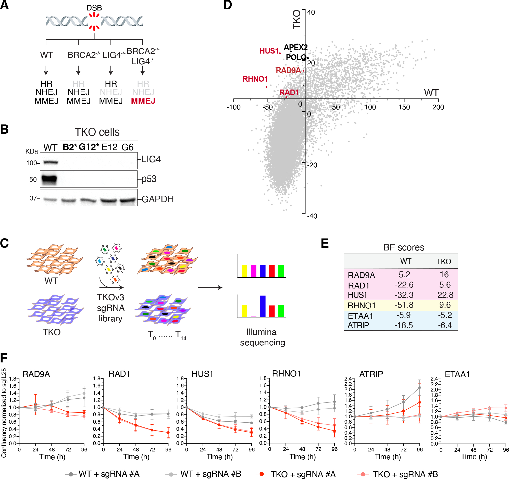
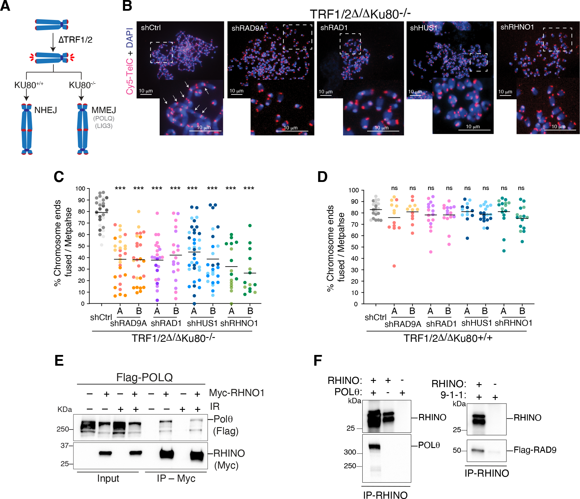
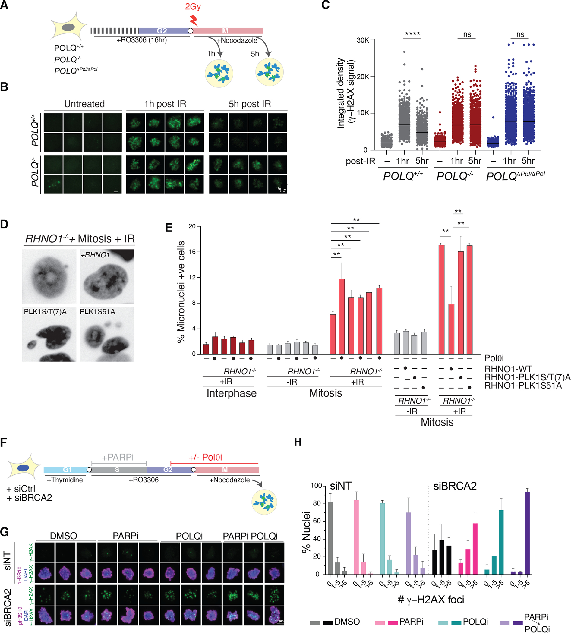
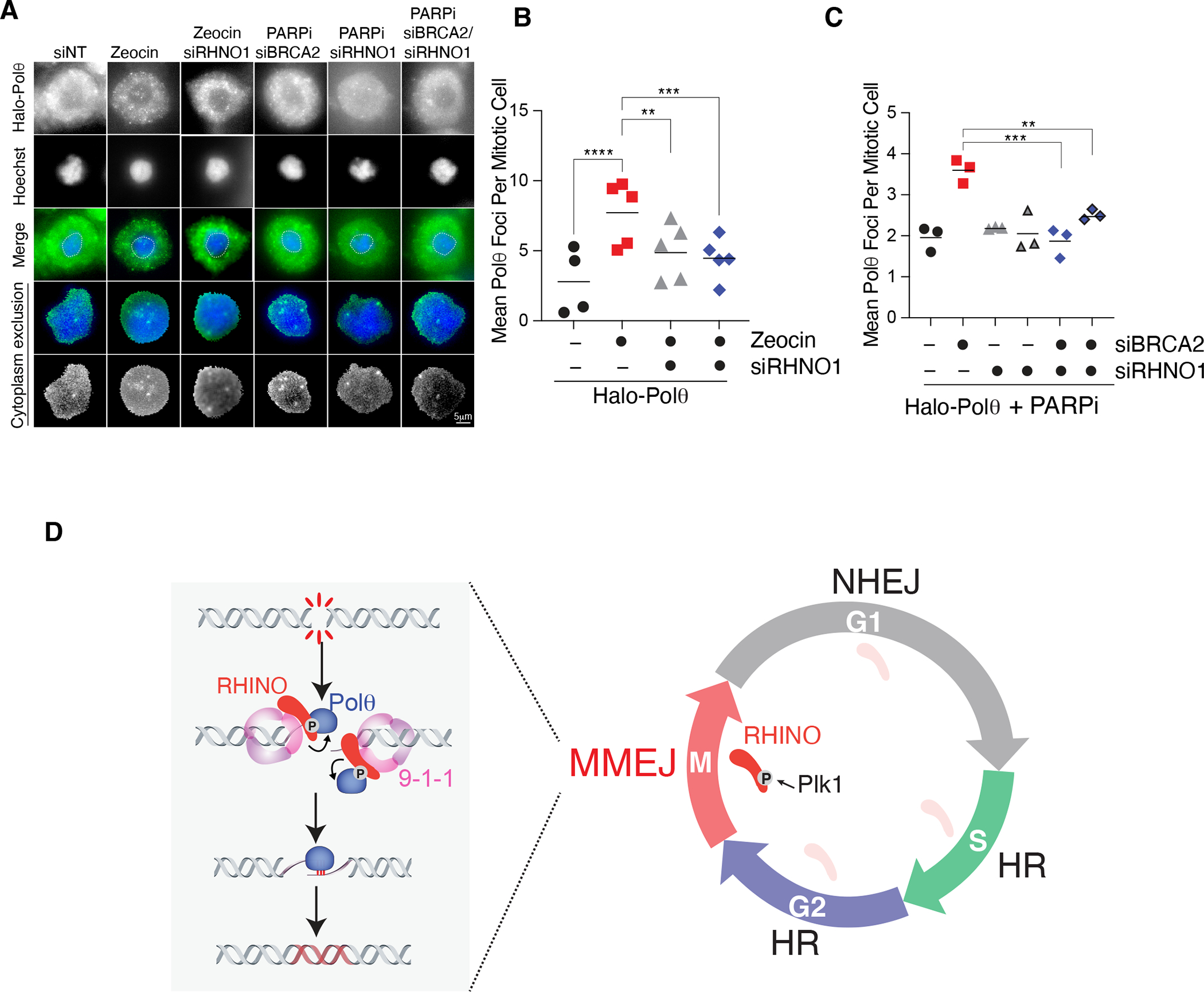
Nonhomologous end-joining (NHEJ) and homologous recombination (HR) are the primary pathways for repairing DNA double-strand breaks (DSBs) during interphase, whereas microhomology-mediated end-joining (MMEJ) has been regarded as a backup mechanism. Through CRISPR-Cas9-based synthetic lethal screens in cancer cells, we identified subunits of the 9-1-1 complex (RAD9A-RAD1-HUS1) and its interacting partner, RHINO, as crucial MMEJ factors. We uncovered an unexpected function for RHINO in restricting MMEJ to mitosis. RHINO accumulates in M phase, undergoes Polo-like kinase 1 (PLK1) phosphorylation, and interacts with polymerase θ (Polθ), enabling its recruitment to DSBs for subsequent repair. Additionally, we provide evidence that MMEJ activity in mitosis repairs persistent DSBs that originate in S phase. Our findings offer insights into the synthetic lethal relationship between the genes POLQ and BRCA1 and BRAC2 and the synergistic effect of Polθ and poly(ADP-ribose) polymerase (PARP) inhibitors.


Conflict of interest statement

Competing interest: Agnel Sfeir is a co-founder, consultant, and shareholder of Repare Therapeutics. All other authors have no competing interests

Figures

Fig. 1.
Fig. 1.. A genome-wide CRISPR-Cas9 screen uncovers an essential function for 9-1-1 and RHNO1 in cells lacking BRCA2 and LIG4.
(A) Schematic of the three major DSB repair pathways in mammalian cells. (B) Western blot analysis of LIG4 and p53 in clonally derived BRCA2−/− LIG4−/− TP53−/− DLD1 cells (TKO). The asterisk indicates clones used in the screen. (C) Schematic of the dropout CRISPR/Cas9 screen to identify synthetic lethal interactions. (D) Genome-wide CRISPR-Cas9 screen result in TKO cells. Genes with a Bayes factor (BF) score >5 (intersection and x- and y-axes) were considered essential. (E) BF scores for the indicated genes. (F) Growth curve of TKO and TP53−/− cells treated with the indicated sgRNAs. Data are mean ± s.d. of three independent experiments normalized to time point zero (one day after seeding) and a control sgRNA (sgIL25).
Fig. 2.
Fig. 2.. A non-canonical function for 9-1-1/RHINO in MMEJ.
(A) Schematic of the shelterin-free assay (37) to monitor MMEJ frequency at deprotected telomeres. (B) Representative images of metaphase spreads from TRF1/2Δ/ΔKu80−/− cells depleted for the subunits of the 9-1-1 complex and RHNO1 with two independent shRNAs. Telomeres are marked by FISH using a Cy5-[CCCTAA]3 PNA probe (red), and chromosomes are counterstained with DAPI (blue). White arrows indicate examples of telomeric fusions in the control sample. (C) Quantification of telomeric fusions mediated by MMEJ as shown in panel B. (D) Quantification of telomere fusions by NHEJ in TRF1/2Δ/ΔKu80+/+. Data in (C, D) are the mean of at least two independent experiments. One-way ANOVA (***p<0.001, **p<0.01). (E) Co-IP experiment depicting Polθ and RHINO interaction in whole-cell extracts from HEK293T cells co-transfected with plasmids expressing FLAG-Polθ and RHINO-MYC. Co-IPs were performed in cells treated with ionizing radiation (+IR, 20 Gy) and control cells. (F) Co-IP experiments showing Polθ/RHINO and 9-1-1/RHINO interaction with purified proteins. Polθ was purified from HEK293T cells, RHINO from E. coli, and 9-1-1 from S. cerevisiae.
Fig. 3.
Fig. 3.. RHINO is predominantly expressed in mitosis.
(A) Results from the CRISPR/Cas9 dropout screen in RHNO1−/− and isogenic RHNO1+/+ cells. Ranked z-scores of the difference in Bayes factor (BF) scores. (B) Reactome pathway overrepresentation analysis of synthetic lethal genes with RHNO1−/−. The fold enrichment of each pathway is plotted on the x-axis. The number of associated genes with each pathway is indicated by the size of the circle, while the color shade indicates the p-value. (C) Network analysis for POLQ and RHNO1 based on Pearson’s correlation of dependency scores derived from DepMap. (D) Representative immunofluorescence images of RHINO in interphase and mitotic cells. (E) Quantification of RHINO foci from panel D. (F) Western blot analysis of endogenous RHINO at different stages of the cell cycle. Extracts from RHNO1FLAG/FLAG cells at the indicated time points. pH3S10 antibody is used as a mitotic marker, and Lamin B1 as a loading control. (G) Control cells and ones expressing RHINO-MYC-FLAG were synchronized in mitosis and subjected to anti-FLAG immunoprecipitation followed by western blot for endogenous Polθ. I = interphase; M = mitosis. (H) Schematic of RHINO protein highlighting the binding domains for 9-1-1 and TOPBP1, D- and Ken-boxes (ΔDK), and the PLK1 phosphorylation sites (PLK1(S/T)7A and S51A). RHINO PLK1(S/T)A harbors alanine mutations in all 7 predicted PLK1 sites. RHINO S51A harbors a single mutation of serine 51 (conserved among primates and rodents) to alanine. (I) Western blot analysis of RHINO and RHINOΔDK during the cell cycle. (J) Co-IP experiments in HEK293T cells co-transfected with plasmids expressing FLAG-Polθ and RHINO-MYC mutants. RHINO mutants with a single S/T mutation to alanine are A through G.
Fig. 4
Fig. 4. MMEJ is the predominant DSB repair pathway during mitosis.
(A) Schematic of the experimental design to detect MMEJ in mitosis for panel B-C. (B) Representative images of γ-H2AX in cells treated as described in panel A. (C) Quantification of γ-H2AX intensity in mitotic cells with the indicated genotype. (n>450 cells; paired t-test.). (D) Representative images of micronuclei in cells with the indicated treatment and genotype. (E) Quantification of micronuclei formation after irradiation during interphase and mitosis as in panel D. Bars represent the mean of three independent experiments (n> 250 cells; paired t-test). (F) Schematic of the experimental pipeline for panels G-H. (G) Representative immunofluorescence images of mitotic cells treated as described in panel F and stained with anti-γ-H2AX. pH3S10 is used to mark mitotic chromosomes. DNA is stained with DAPI. (H) Quantification of γ-H2AX foci in mitotic cells with the indicated treatment. Bars represent the mean of three independent experiments (n> 50 cells).
Fig. 5
Fig. 5. RHINO recruits Polθ to damaged sites in mitosis.
(A) Representative images of Halo-Polθ foci in mitosis. Cells with the indicated siRNA treatment were synchronized according to the scheme in Fig. 4A. Cells in mitosis were treated with Zeocin for 1 hour. To monitor mitotic Polθ foci in cells with persistent damage from S phase, cells expressing siRNA against BRCA2 and RHNO1 were treated with Olaparib according to the schematic in Fig. 4F. (B) Quantification of mitotic Halo-Polθ foci in live-cells treated with Zeocin and depleted of RHNO1. Bars represent the mean of three independent experiments. n>40 nuclei (one-way ANOVA (***p<0.001, **p<0.01)). (C) Quantification of mitotic Halo-Polθ foci in live-cell imaging experiments in nocodazole-arrested cells treated with PARPi during S phase. Bars represent the mean of three independent experiments (n>40 nuclei; one-way ANOVA; ***p<0.001, **p<0.01). (D) NHEJ dominates in G1, and HR is preferred in S and G2. The confinement of MMEJ to mitosis occurs due to the accumulation of RHINO during M phase, PLK1- phosphorylation, and the recruitment of Polθ to DNA breaks

Update of

Comment in

References

    1. Mateos-Gomez PA et al., Mammalian polymerase theta promotes alternative NHEJ and suppresses recombination. Nature 518, 254–257 (2015). - PMC - PubMed
    1. Ceccaldi R et al., Homologous-recombination-deficient tumours are dependent on Poltheta-mediated repair. Nature 518, 258–262 (2015). - PMC - PubMed
    1. Wyatt DW et al., Essential Roles for Polymerase theta-Mediated End Joining in the Repair of Chromosome Breaks. Mol Cell 63, 662–673 (2016). - PMC - PubMed
    1. Zatreanu D et al., Poltheta inhibitors elicit BRCA-gene synthetic lethality and target PARP inhibitor resistance. Nat Commun 12, 3636 (2021). - PMC - PubMed
    1. Schrempf A, Slyskova J, Loizou JI, Targeting the DNA Repair Enzyme Polymerase theta in Cancer Therapy. Trends Cancer 7, 98–111 (2021). - PubMed