

Application of fluorescence endoscopy with methylene blue dye and indocyanine green dual-tracer method in sentinel lymph node biopsy for women with breast cancer
- PMID: 37441014
- PMCID: PMC10333774
- DOI: 10.21037/gs-22-469
Application of fluorescence endoscopy with methylene blue dye and indocyanine green dual-tracer method in sentinel lymph node biopsy for women with breast cancer
Abstract
Background: Indocyanine green (ICG) allows for the real-time visualization of lymphatic drainage and provides favorable performance for sentinel lymph node (SLN) mapping. However, the limited ability of tissue penetration of the near-infrared fluorescence of ICG may lead to the failure of lymph node detection in the traditional open approach of sentinel lymph node biopsy (SLNB) for breast cancer, especially in overweight or obese patients. To accurately and quickly detect SLNs, we applied fluorescence endoscopy with a dual-tracer method using ICG and methylene blue dye (MBD) in SLNB for breast cancer. We conducted this study to assess the feasibility and application value of this method in minimally invasive surgery.
Methods: A total of 117 patients who received dual-tracer injection of ICG and MBD prior to endoscopic SLNB from November 2020 to September 2021 were examined in this study. The number of SLNs identified, the SLN identification rate, the time to identify the first SLN, the procedure duration, and the postoperative morbidity were analyzed.
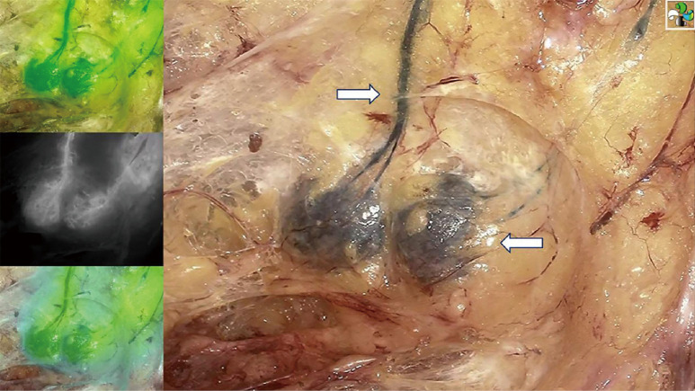
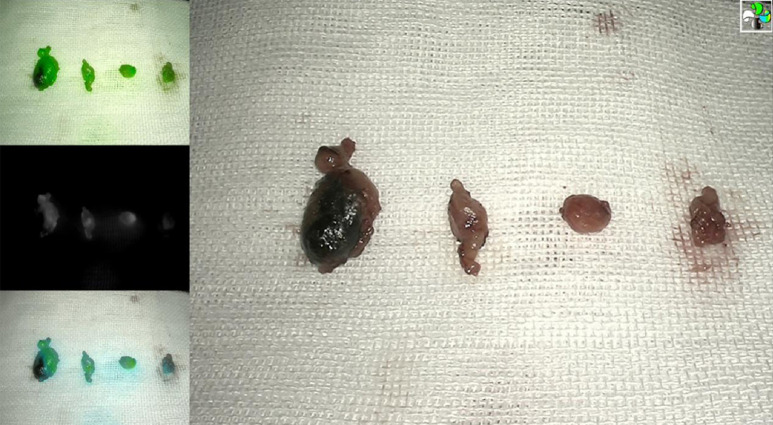
Results: Biopsied SLNs could be identified in 116 patients (99.15%) with an average number of 5.12±1.87 per patient. Blue-stained SLNs were found in 99 patients (84.62%) and fluorescent SLNs in 112 patients (95.73%). A total of 34 patients (29.06%) had positive SLNs. In 6 cases (5.13%), the positive SLNs were only stained with ICG fluorescence. In 1 case (0.85%), the positive SLNs were only blue-stained with no fluorescence staining. The mean durations for the identification of the first SLN and endoscopic SLNB were 7.14±6.31 and 37.75±16.94 min, respectively. Upper-limb lymphoedema was observed 5 cases (4.27%) during a median follow-up period of 10 months.
Conclusions: The fluorescence endoscopy method assisted by dual tracer facilitates SLN detection with a comparatively short procedure duration and low complication rate. This approach could serve as a new method for SLNB for patients with breast cancer.
Keywords: Indocyanine green (ICG); breast neoplasms; fluorescence endoscopy; methylene blue dye (MBD); sentinel lymph node biopsy (SLNB).
2023 Gland Surgery. All rights reserved.
Conflict of interest statement
Conflicts of Interest: All authors have completed the ICMJE uniform disclosure form (available at https://gs.amegroups.com/article/view/10.21037/gs-22-469/coif). The authors have no conflicts of interest to declare.
Figures
Similar articles
-
Can Low-cost Indo Cyanine Green Florescence Technique for Sentinel Lymph Node Biopsy Replace Dual Dye (Radio-colloid and Blue Dye) Technique in Early Breast Cancer: A Prospective Two-arm Comparative Study.Clin Breast Cancer. 2020 Oct;20(5):e576-e583. doi: 10.1016/j.clbc.2020.03.013. Epub 2020 Apr 6. Clin Breast Cancer. 2020. PMID: 32389561
-
Assessing the benefit of using indocyanine green in addition to methylene blue for breast cancer sentinel lymph node biopsy.Medicine (Baltimore). 2025 May 16;104(20):e42500. doi: 10.1097/MD.0000000000042500. Medicine (Baltimore). 2025. PMID: 40388739 Free PMC article.
-
Evaluation of indocyanine green combined with methylene blue staining in sentinel lymph node biopsy of breast cancer.Gland Surg. 2022 Sep;11(9):1489-1496. doi: 10.21037/gs-22-434. Gland Surg. 2022. PMID: 36221275 Free PMC article.
-
Indocyanine Green Fluorescence Versus Blue Dye or Radioisotope Regarding Detection Rate of Sentinel Lymph Node Biopsy and Nodes Removed in Breast Cancer: A Systematic Review and Meta-Analysis.Asian Pac J Cancer Prev. 2020 May 1;21(5):1187-1195. doi: 10.31557/APJCP.2020.21.5.1187. Asian Pac J Cancer Prev. 2020. PMID: 32458621 Free PMC article.
-
Detection of sentinel lymph node in vulvar cancer using 99mTc-labeled colloid lymphoscintigraphy, blue dye, and indocyanine-green fluorescence: a meta-analysis of studies published in 2010-2020.Arch Gynecol Obstet. 2023 Jun;307(6):1677-1686. doi: 10.1007/s00404-022-06605-1. Epub 2022 May 24. Arch Gynecol Obstet. 2023. PMID: 35608701 Review.
Cited by
-
The potential of indocyanine green fluorescence detection in surgical cut margin of breast conserving surgery.Gland Surg. 2024 Jun 30;13(6):1031-1044. doi: 10.21037/gs-24-195. Epub 2024 Jun 27. Gland Surg. 2024. PMID: 39015719 Free PMC article.
References
-
- International Agency for Research on Cancer. World Cancer Day: Breast cancer overtakes lung cancer in terms of number of new cancer cases worldwide. IARC showcases key research projects to address breast cancer. Lyon, France. Available online: www.iarc.who.int/pressrelease/
-
- Baeten IGT, Hoogendam JP, Braat AJAT, et al. Fluorescent Indocyanine Green versus Technetium-99m and Blue Dye for Bilateral SENTinel Lymph Node Detection in Stage I-IIA Cervical Cancer (FluoreSENT): protocol for a non-inferiority study. BMJ Open 2022;12:e061829. 10.1136/bmjopen-2022-061829 - DOI - PMC - PubMed
LinkOut - more resources
Full Text Sources