. 2023 Jun 30;12(6):834-852.
doi: 10.21037/gs-23-127. Epub 2023 Jun 27.

Silencing of AJAP1 expression by promoter methylation activates the Wnt/β-catenin signaling pathway to promote tumor proliferation and metastasis in salivary adenoid cystic carcinoma

Affiliations

Silencing of AJAP1 expression by promoter methylation activates the Wnt/β-catenin signaling pathway to promote tumor proliferation and metastasis in salivary adenoid cystic carcinoma

Liehao Jiang et al. Gland Surg. .

Abstract

Background: Salivary adenoid cystic carcinoma (SACC) is a unique malignant tumor of the salivary gland with poor prognosis, which is not effective with chemotherapy and targeted drugs. Therefore, it is important to explore the molecular mechanism underlying SACC invasion and metastasis to develop novel therapeutic strategies and targets in clinical research.

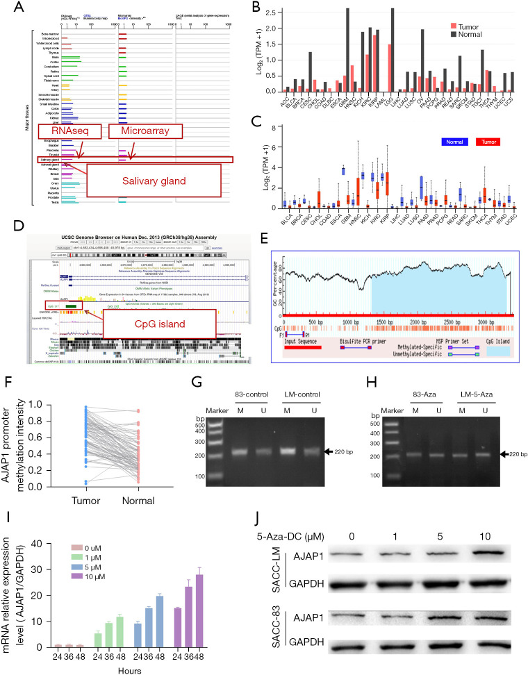
Methods: Real-time quantitative polymerase chain reaction (RT-qPCR) and western blot (WB) were performed to detect the expression of Adherens Junctions Associated Protein 1 (AJAP1). Methylation-specific PCR was used to evaluate the methylation of the AJAP1 promoter. AJAP1 was overexpressed or knocked down by lentivirus-mediated transfection. Kaplan-Meier analysis was conducted to create a survival curve and the log-rank test was used to analyze the overall survival (OS). The prognostic correlation was assessed using univariate and multivariate Cox regression analyses. Co-immunoprecipitation (Co-IP) was utilized to pull down the possible binding protein of AJAP1 and laser scanning confocal microscopy was applied to detect the subcellular localization of AJAP1, E-cadherin, and β-catenin. Cell viability, colony formation, wound healing, and Transwell invasion assays were performed to evaluate the function of AJAP1 in vitro. A subcutaneous xenograft assay in nude mice was performed to verify the function of AJAP1 in vivo.

Results: AJAP1 was downregulated in SACC tumors and was closely related to SACC lymph node/distant metastasis, which was an independent risk factor for SACC prognosis. Methylation-specific PCR confirmed that high methylation of the AJAP1 promoter was the main cause of its silencing. Overexpression or knockdown of AJAP1 in SACC cells could significantly inhibit or promote the proliferation, invasion, and metastasis of SACC cells, respectively, in both the in vitro and in vivo experiments. Mechanically, we found that AJAP1 binds to E-cadherin and β-catenin to form a complex in cytomembrane, reducing the nuclear translocation of β-catenin and blocking the Wingless/Integrated/β-catenin (Wnt/β-catenin) signaling pathway to play a suppressive role in cancer.

Conclusions: In conclusion, these results suggest that the downregulation of AJAP1 protein expression may play a certain role in progression and metastasis of SACC. Our study indicates that AJAP1 may be a potential prognostic molecular marker and therapeutic target for SACC.

Keywords: AJAP1; Salivary adenoid cystic carcinoma (SACC); metastasis; methylation; β-catenin.


Conflict of interest statement

Conflicts of Interest: All authors have completed the ICMJE uniform disclosure form (available at https://gs.amegroups.com/article/view/10.21037/gs-23-127/coif). The authors have no conflicts of interest to declare.

Figures

Figure 1
Figure 1
AJAP1 was lowly expressed in SACC tumors. (A) Heat map of differentially expressed genes in SACC tumor tissues and paracancerous tissues; (B) Volcano map of differentially expressed genes in SACC tumor tissues and paracancerous tissues; (C) pathway enrichment analysis showed that the differentially expressed genes were mainly enriched in extracellular matrix organization; (D) GO enrichment analysis showed that the differentially expressed genes were mainly enriched in biological adhesion; (E) immunohistochemical results showed that the expression level of the AJAP1 protein in paracancerous tumor tissues was significantly higher than that in tumor tissues; (F) Western blot results showed that the expression level of the AJAP1 protein (45 kDa) in paracancerous tumor tissues was significantly higher than that in tumor tissues; (G) immunohistochemical showed AJAP1 was highly expressed in the normal glandular tube (↑) but was poorly expressed in tumor tissues (×200) (★); (H) immunohistochemical showed the AJAP1 protein was highly expressed in some tumor tissues (×200); (I) immunohistochemical showed the expression of the AJAP1 protein was absent in tumors and highly expressed in tumor-associated nerves (↑) (×200). *, P<0.05. NC, normal control; GO, Gene Ontology; AJAP1, adherens junctions associated protein 1; IHC, immunohistochemistry; GAPDH, glyceraldehyde-3-phosphate dehydrogenase; T, tumor ; N, normal tissue; SACC, salivary adenoid cystic carcinoma.
Figure 2
Figure 2
OS and PFS of SACC patients. (A) The OS of patients in the AJAP1-negative group was significantly poorer than that in the positive group; (B) the OS of patients in the lymph node/distant metastasis-positive group was significantly poorer than that in the negative group; (C) the OS in the TNM stage I–II group was significantly better than that in the stage III–IV group; (D) among patients in the TNM stage I–II group, the OS of patients in AJAP1-negative group was significantly poorer than that of patients in the positive group; (E) among patients in the TNM stage III–IV group, the OS of patients in the AJAP1-negative group was significantly poorer than that of patients in the positive group; (F) the PFS in the AJAP1-negative group was significantly poorer than that in the positive group; (G) the PFS in the lymph node/distant metastasis-positive group was significantly poorer than that in the negative group; (H) the PFS in the TNM stage I–II group was significantly better than that in the stage III–IV group; (I) in the TNM stage I–II group, the PFS of the AJAP1-negative group was significantly poorer than that of the positive group; (J) in the TNM stage III–IV group, the PFS of patients in the AJAP1-negative group was significantly poorer than that of patients in the positive group. TNM, tumor nodes metastasis; AJAP1, adherens junctions associated protein 1; OS, overall survival; PFS, progression-free survival; SACC, salivary adenoid cystic carcinoma.
Figure 3
Figure 3
High methylation of the promoter CpG island leads to the silencing of AJAP1 expression in SACC tumors. (A) The GeneCards database showed that AJAP1 was highly expressed in normal salivary gland tissues; (B,C) the pan-cancer analysis showed that the expression of AJAP1 in various tumor tissues was significantly lower than that in normal tissues; (D) the UCSC database showed a large number of CpG islands in the promoter region of AJAP1 gene; (E) CpG analysis of the AJAP1 gene promoter; (F) methylation of the AJAP1 promoter in SACC tumor tissues was significantly higher than that in paracancerous tissues; (G) in the untreated group, methylation of the AJAP1 gene promoter was detected by MSP; (H) promoter methylation of the AJAP1 gene was detected by MSP in the 5-Aza treatment group; (I) the mRNA expression level of AJAP1 increased with the increase of 5-Aza treatment concentration in SACC cells; (J) the expression of AJAP1 protein (45 kDa) in SACC cells increased with the increase of 5-Aza treatment concentration. FPKM, Fragments Per Kilobase of exon model per Million mapped fragments; GTEx, genotype-tissue expression; TPM, transcripts per million; UCSC, University of California, Santa Cruz; AJAP1, adherens junctions associated protein 1; GAPDH, Glyceraldehyde-3-phosphate dehydrogenase; 5-Aza, 5-Azacytidine; M, methylation; U, unmethylation; SACC, salivary adenoid cystic carcinoma; CpG, cytosine-phosphoric acid-guanine.
Figure 4
Figure 4
AJAP1 overexpression or knockdown inhibited or promoted the invasion, metastasis, and proliferation of SACC cells. (A) The mRNA level of the AJAP1 gene was significantly increased after the introduction of overexpressed lentivirus into SACC-LM cells; (B) the mRNA level of AJAP1 gene was down-regulated after the introduction of knockdown lentivirus into SACC-83 cells; (C) the level of AJAP1 protein (45 kDa) was increased after the introduction of overexpressed lentivirus into SACC-LM cells; (D) the expression of AJAP1 protein (45 kDa) was significantly down-regulated in SACC-83 cells after the introduction of knockdown lentivirus; (E,F) the colony formation assay showed that AJAP1 overexpression in SACC-LM cells significantly down-regulated the colony formation ability (crystal violet staining, ×4); (G,H) the colony formation assay showed that the colony formation ability was significantly up-regulated after the knockdown of AJAP1 in SACC-83 cells (crystal violet staining, ×4); (I) the CCK8 proliferation assay showed that the overexpression of AJAP1 significantly down-regulated the proliferation of SACC-LM cells; (J) the CCK8 proliferation assay showed that the knockdown of AJAP1 significantly upregulated the proliferation of SACC-83 cells; (K,L) the scratch healing assay showed that the migration ability was significantly down-regulated after overexpression of AJAP1 in SACC-LM cells (white light, high contrast resolution, ×100); (M,N) the scratch healing assay showed that the migration ability was significantly up-regulated after AJAP1 knockdown in SSAC-83 cells (white light, high contrast resolution, ×100); (O,P) the Transwell assay showed that the invasion ability of SACC-LM cells was significantly down-regulated after the overexpression of AJAP1 (crystal violet staining, ×200); (Q,R) the Transwell assay showed that the invasion ability of SACC-83 cells was significantly upregulated after AJAP1 knockdown (crystal violet staining, ×200). *, P<0.05; **, P<0.01. AJAP1, adherens junctions associated protein 1; GAPDH, glyceraldehyde-3-phosphate dehydrogenase; SACC, salivary adenoid cystic carcinoma; LM, SACC-LM; NC, normal control.
Figure 5
Figure 5
AJAP1 reduces the nuclear localization of β-catenin by forming the AJAP1/E-cadherin/β-catenin complex. (A-C) The co-IP assays showed that endogenous protein of AJAP1 interacted with β-catenin and E-cadherin; (D) immunofluorescence displayed the co-localization among AJAP1 (45 kDa), E-cadherin (97 kDa), and β-catenin (85 kDa) (×200); (E) AJAP1 can bind to β-catenin in the cytoplasm, thereby reducing the content of β-catenin in the nucleus; (F) AJAP1 upregulation reduced the β-catenin/TCF/LEF-mediated transcription activity in SACC-LM cells; (G) AJAP1 depletion in SACC-83 cells increased the β-catenin/TCF/LEF-mediated transcription activity; (H-J) overexpression/knockdown of AJAP1 significantly up-regulated/down-regulated the expression of β-catenin downstream genes, respectively (AJAP1, 45 kDa; C-myc, 49 kDa; CyclinD1, 33 kDa; MMP1, 54 kDa; GAPDH, 36 kDa). *, P<0.05. IB, immunoblotting; IP, immunoprecipitation; IgG, immunoglobulin G; AJAP1, adherens junctions associated protein 1; DAPI, 4’,6-diamidino-2-phenylindole; SACC, salivary adenoid cystic carcinoma; LM, SACC-LM; NC, normal control; co-IP, co-immunoprecipitation; GAPDH, glyceraldehyde-3-phosphate dehydrogenase.
Figure 6
Figure 6
Regulation of Wnt/β-catenin signaling pathway could reverse the effect of AJAP1 on the function of SACC cells. (A,B) Wnt/β-catenin agonist 2 could reverse the downregulation of invasion ability of SACC-LM cells induced by AJAP1overexpression (crystal violet staining, ×200); (C,D) LF3 could reverse the upregulation of invasion ability of SACC-83 cells induced by the knockdown of AJAP1 (crystal violet staining, ×200); (E,F) Wnt/β-catenin agonist 2 could reverse the migration ability downregulation of SACC-LM cells induced by AJAP1 overexpression (white light, high contrast resolution, ×100); (G,H) LF3 could reverse the migration ability upregulation of SACC-83 cells induced by AJAP1 knockdown (white light, high contrast resolution, ×100); (I) Wnt/β-catenin agonist 2 could reverse the proliferative ability downregulation of SACC-LM cells induced by AJAP1 overexpression; (J) LF3 could reverse the proliferative ability upregulation of SACC-83 cells induced by AJAP1 knockdown. *, P<0.05. SACC, salivary adenoid cystic carcinoma; AJAP1, adherens junctions associated protein 1; Wnt/β-catenin signaling pathway, Wingless/Integrated/β-catenin signaling pathway; OD, optical density.
Figure 7
Figure 7
AJAP1 inhibited the growth and metastasis of SACC tumors in vivo. (A-C) AJAP1 overexpression in SACC-LM cells significantly reduced the subcutaneous tumorigenicity of SACC-LM cells in nude mice; (D-F) AJAP1 knockdown in SACC-83 cells significantly increased subcutaneous tumorigenesis of SACC-83 cells in nude mice; (G,H) immunohistochemical showed AJAP1 overexpression in SACC-LM cells significantly down-regulated the expression of β-catenin downstream genes (×200); (I,J) immunohistochemical showed AJAP1 knockdown significantly up-regulated the expression of β-catenin downstream genes in SACC-83 cells (×200). *, P<0.05. AJAP1, adherens junctions associated protein 1; SACC, salivary adenoid cystic carcinoma; NC, normal control.

Similar articles

Cited by

References

    1. Sahara S, Herzog AE, Nör JE. Systemic therapies for salivary gland adenoid cystic carcinoma. Am J Cancer Res 2021;11:4092-110. - PMC - PubMed
    1. Gentili F, Guerrini S, Mazzei FG, et al. Dual energy CT in gland tumors: a comprehensive narrative review and differential diagnosis. Gland Surg 2020;9:2269-82. 10.21037/gs-20-543 - DOI - PMC - PubMed
    1. Wang HF, Wang SS, Zheng M, et al. Hypoxia promotes vasculogenic mimicry formation by vascular endothelial growth factor A mediating epithelial-mesenchymal transition in salivary adenoid cystic carcinoma. Cell Prolif 2019;52:e12600. 10.1111/cpr.12600 - DOI - PMC - PubMed
    1. Lassche G, van Boxtel W, Ligtenberg MJL, et al. Advances and challenges in precision medicine in salivary gland cancer. Cancer Treat Rev 2019;80:101906. 10.1016/j.ctrv.2019.101906 - DOI - PubMed
    1. Tang YF, Wu WJ, Zhang JY, et al. Reconstruction and analysis of the aberrant lncRNA-miRNA-mRNA network based on competitive endogenous RNA in adenoid cystic carcinoma of the salivary gland. Transl Cancer Res 2021;10:5133-49. 10.21037/tcr-21-1771 - DOI - PMC - PubMed